New Facility for Bioengineering Research Opens in Los Angeles

In a world eager to solve the problem of rejection in organ transplantation, a young American scientist developed a breakthrough test in 1964 that would help establish the compatibility of tissue types between organ donors and patients in need of transplants. Even though, today, efforts to meet organ transplant demand are shifting toward the field of bioengineering, as researchers search for ways to recreate complex organs with patient-derived cells, the legacy of that scientist, Paul Ichiro Terasaki, continues to inspire discoveries in transplant medicine through his philanthropic ventures.

The Terasaki Institute for Biomedical Innovation (TIBI), a nonprofit research organization established by Terasaki, professor emeritus of surgery at the David Geffen School of Medicine at the University of California Los Angeles (UCLA), will open the doors to a new facility in 2022. The newly-acquired addition will house interdisciplinary research in bioengineering, micro- and nanoscale technologies to enable transformative biomedical innovation as part of continuing research to solve the biggest problems related to organ transplantation and beyond.

Earlier this month, the Terasaki Institute announced the revamping of a building in the Woodland Hills area of the city of Los Angeles. Once home to the Weider Health and Fitness Center, created by bodybuilder and entrepreneur Joe Weider, the two-story building will be custom-designed to house the latest technology in cutting-edge research and will provide 50,000 square feet of floor space for up to 200 employees.

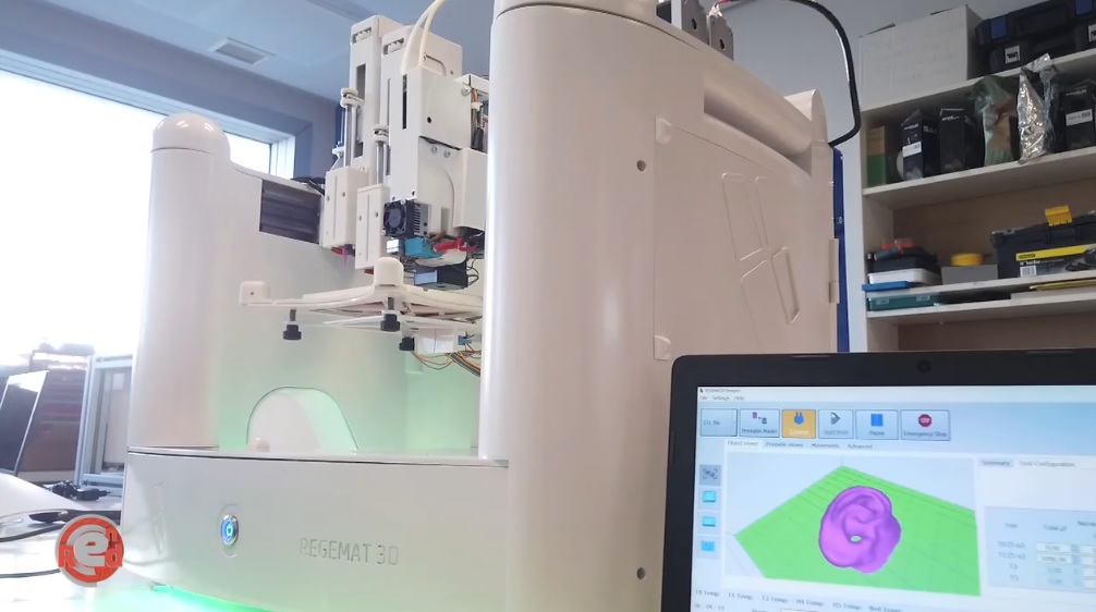
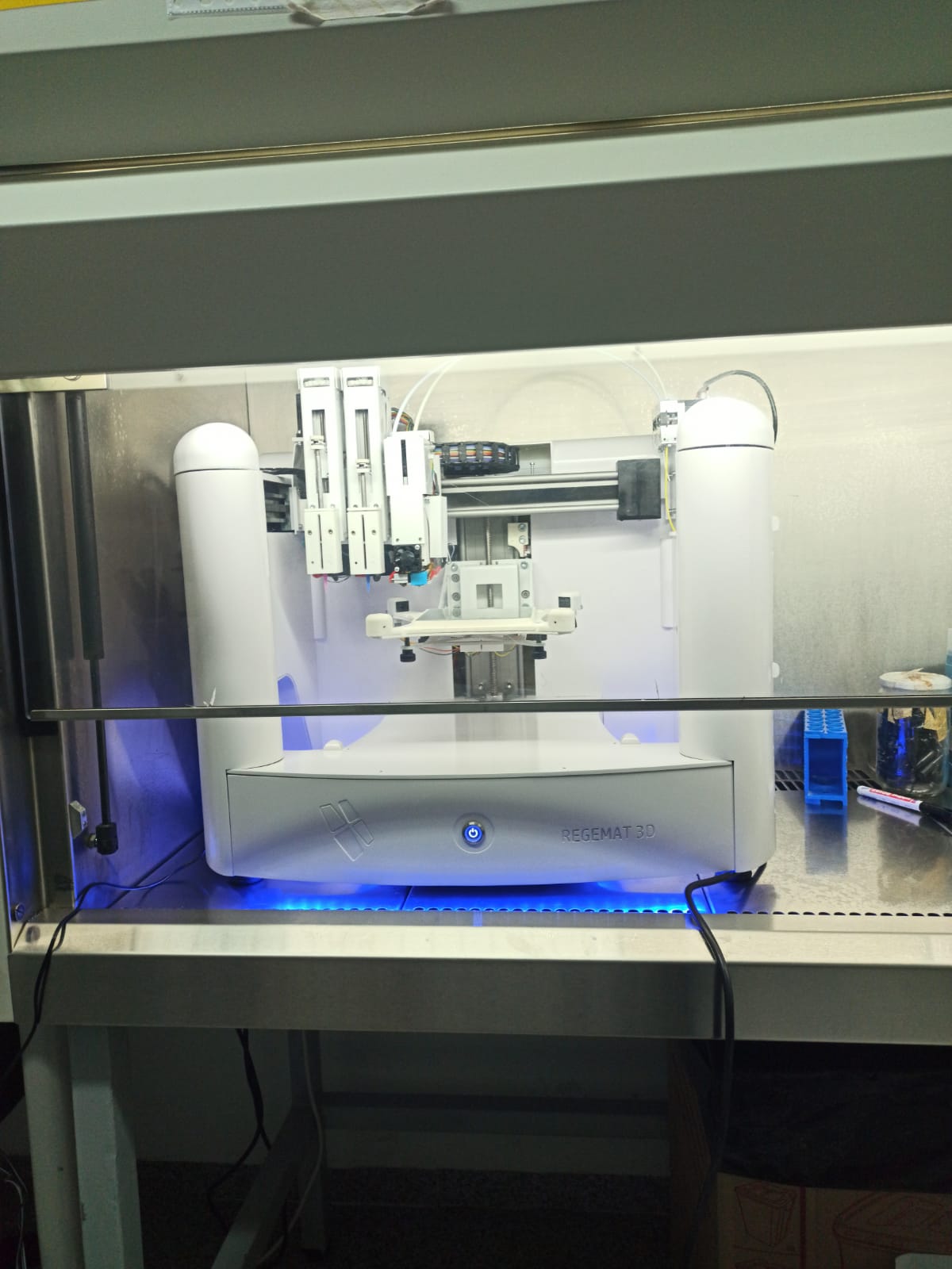
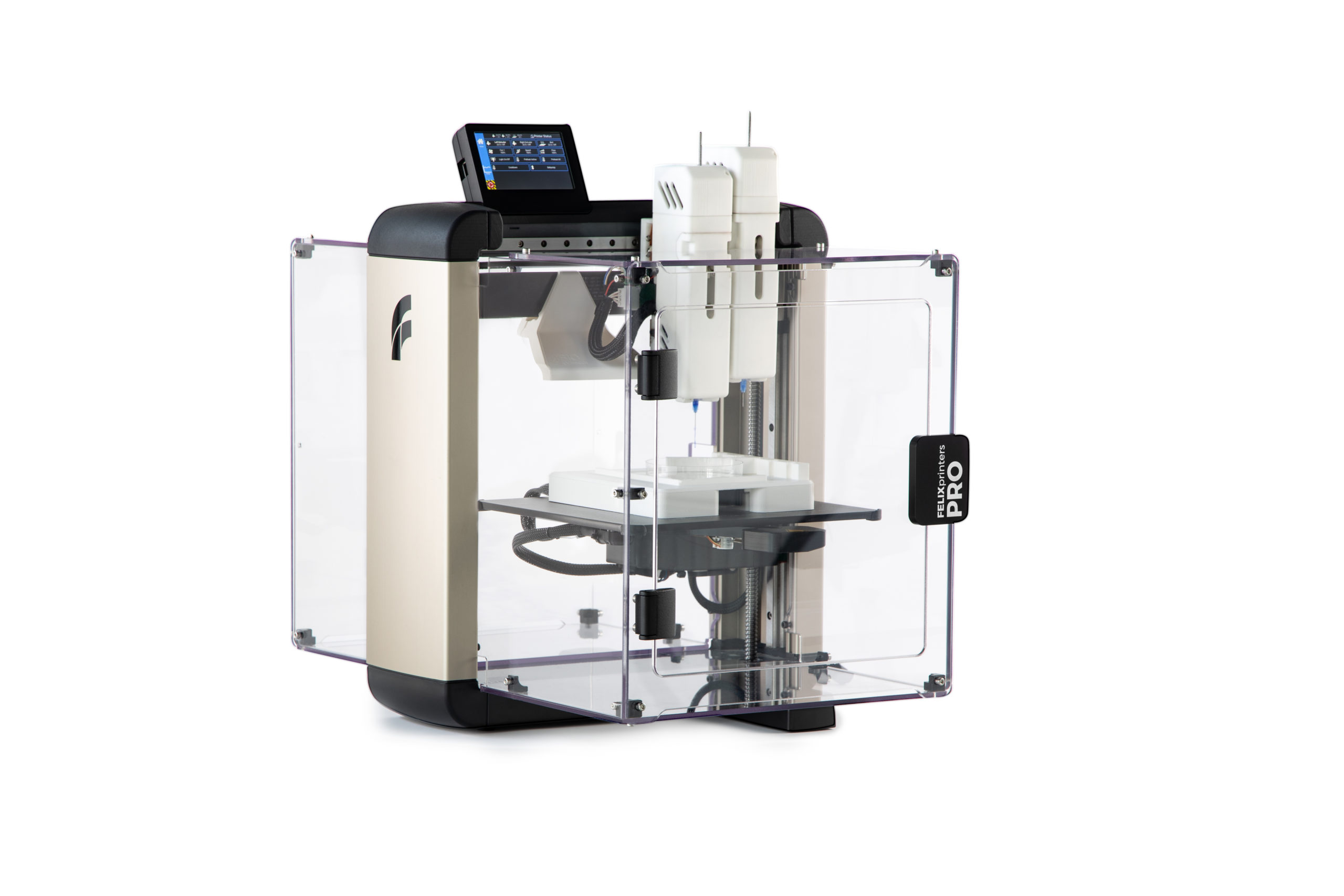
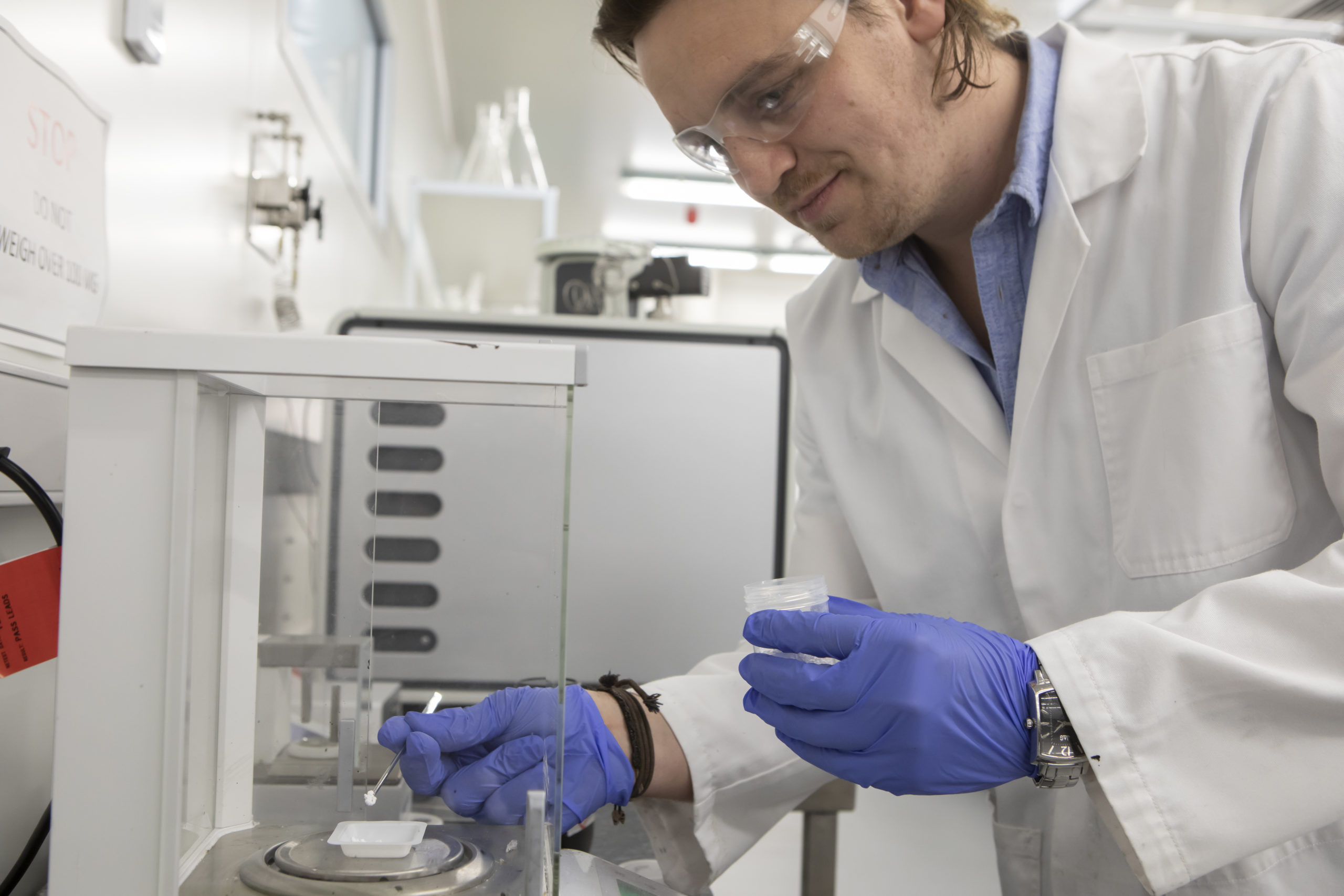
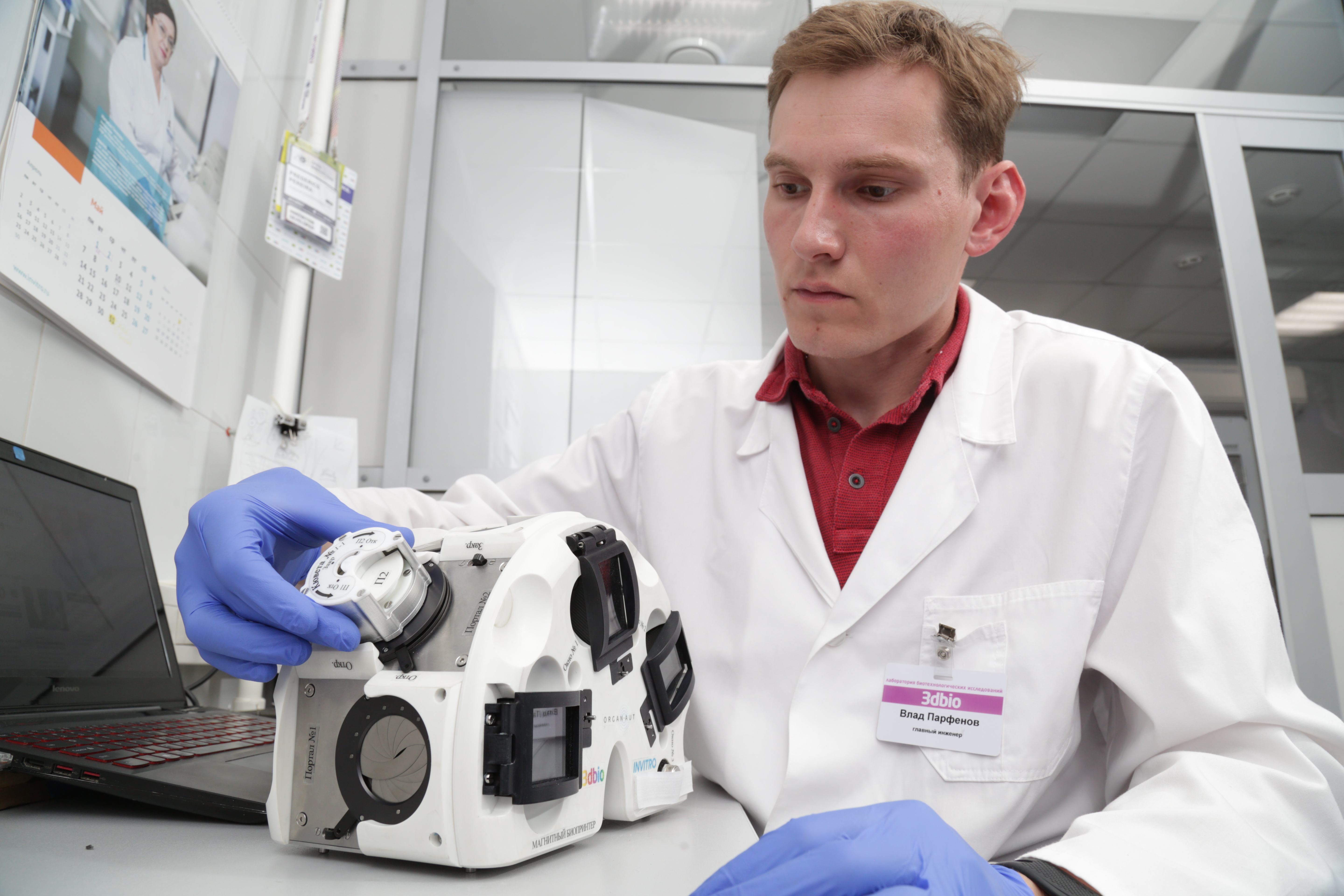
Located just 22 miles north of the original Terasaki Institute facilities in Westwood, the new space devoted to laboratory research will be designed to accommodate multiple teams of scientists, who will be developing bioengineered systems, devices, and other products with several biomedical applications. This new facility will be fully equipped to enable such technologies as tissue engineering and regeneration, biofabrication using 3D printing, nano- and micro-engineering, stem cell engineering, and the creation of human organs on chips.

When the new facility is inaugurated, with the renovation of the building set to begin in fall 2020, it will become the Terasaki Institute’s third research facility. In addition to the ample space and unique design features of the laboratory, the new facility will include in-house technology translation capabilities to be able to build prototypes and scale models of devices engineered by the institute. It will also be able to accommodate meetings, seminars, and conferences to further the education and exchange of ideas among its researchers and collaborators.

“I’m very excited about the addition of the new building to the Terasaki Institute. I believe that this addition will give us needed research space to bring together a number of leading scientists in our efforts to develop the next generation of biomedical innovations,” said Terasaki Institute’s new director and CEO, Ali Khademhosseini. “I’m particularly excited about furthering the great legacy of the Weider family and the building’s history in promoting health and fitness by focusing on individualized cures and diagnostics.”

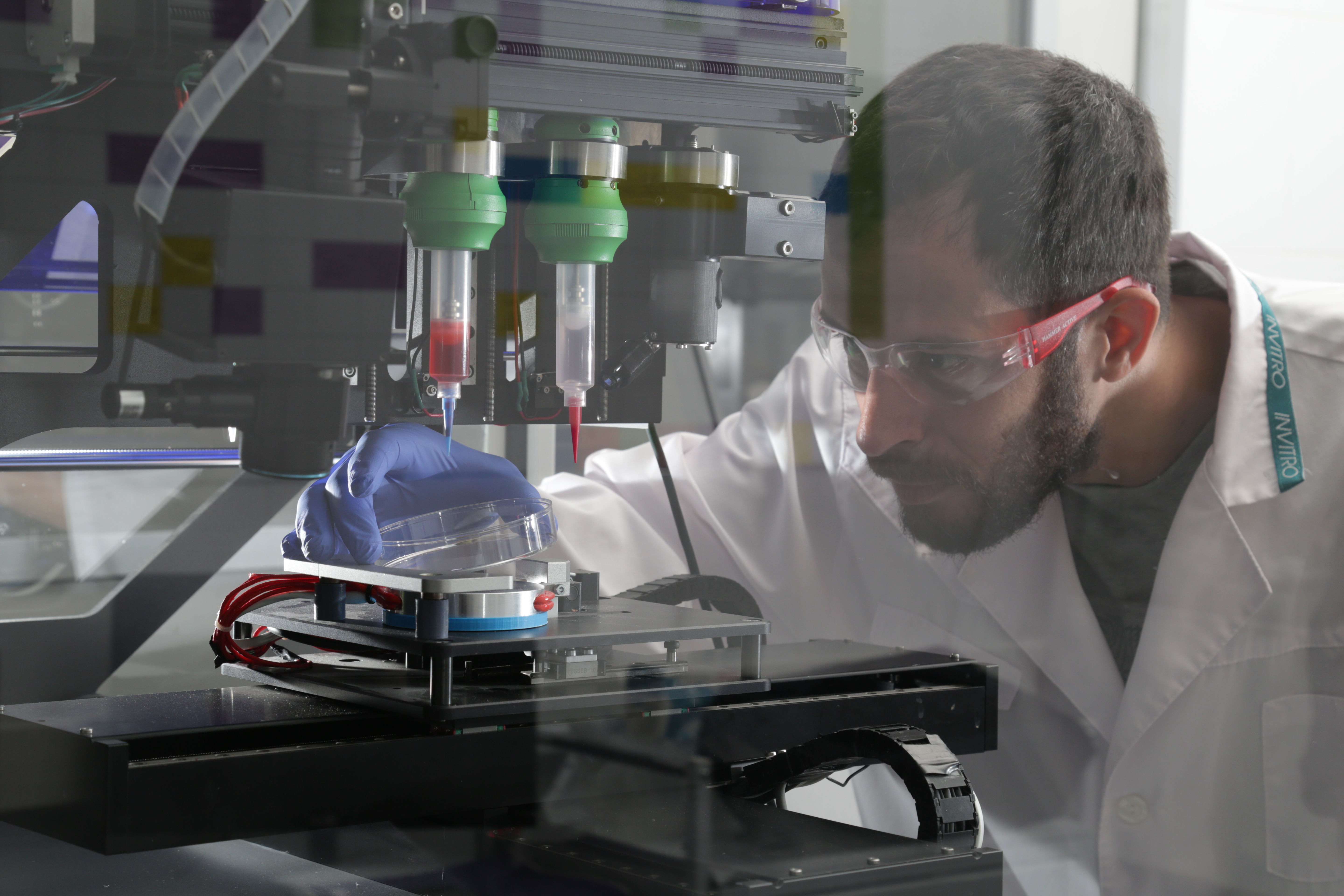
Previously at Harvard Medical School, the Wyss Institute for Biologically Inspired Engineering, and most recently at UCLA Bioengineering, Khademhosseini has been an influential figure in pushing bioengineering forward. His research in regenerative medicine, tissue engineering, and micro- and nanotechnologies for the treatment of diseases has been related to advancements that allow reprogramming of adult cells to become progenitors, as well as editing genes. The bioengineer has also created a technique that uses a specially adapted 3D printer that could help advance the field of regenerative medicine by making it possible to 3D print complex artificial tissues on demand. He has also established the Khademhosseini Lab, an industry-leading tissue engineering lab that is co-sponsored by both MIT and Harvard and acts as a strategic partner to 3D bioprinting startup BioBots.

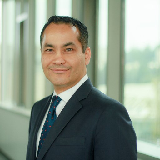
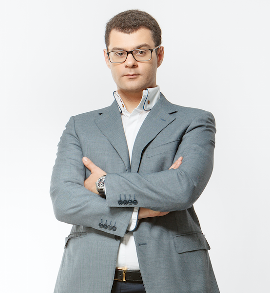
Ali Khademhosseini (Image: Ali Khademhosseini)

Stewart Han, president of the Terasaki Institute, has been working hard overseeing the planning and renovation of the new building: “It is exciting to be able to create a brand-new laboratory and research facility from the ground up, and it will greatly enhance our research capabilities when it’s completed. We also know that the new building will facilitate the future growth of our institute.

Founded in 2001, the Terasaki Institute was made possible through an endowment from the late Paul Terasaki, and it is expected to continue leveraging scientific advancements that enable an understanding of personalized medicine, from the macroscale of human tissues down to the microscale of genes, as well as to create technological solutions for some of the most pressing medical problems of our time.

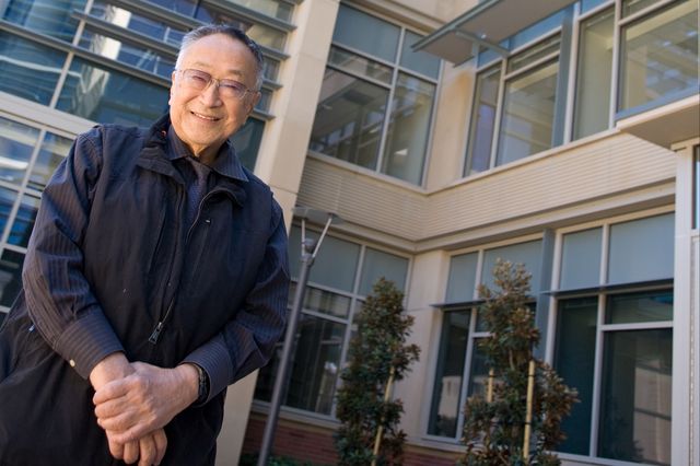
Paul Terasaki in front of the Terasaki Life Sciences Building UCLA. (Image: Leslie Barton/UCLA)

“The board of the Terasaki Institute is very excited about the purchase of the new building in Woodland Hills, and we look forward to developing it into a world-class biomedical research center,” said board chair and diagnostic radiology specialist Keith Terasaki. “My father, the late Paul I. Terasaki, started the Terasaki Institute in hopes that it will make impactful discoveries in medical research. This new research facility will enable us to do so.”

To the field of transplant surgery, transplant pioneer Paul Terasaki enabled a broad understanding of organ transplant outcomes around the world. More than 70 years after his original discovery, patients still rely on organ donor transplants and the fundamentals of Terasaki’s laboratory developed tissue typing tests are still used today for the determination of transplant compatibility. Nonetheless, the Terasaki Institute envisions a world where personalized medicine is available to all. So, as the researchers at the institute continue to address the challenges that can finally advance the field of organ transplants from human donors to bioengineered artificial organs, they might bridge the gap between sickness and health. With one of the most productive 3D printing researchers as director, Khademhosseini, and a new facility to further explore biofabrication technology, we can expect to hear much more from the Terasaki Institute in years to come.

The post New Facility for Bioengineering Research Opens in Los Angeles appeared first on 3DPrint.com | The Voice of 3D Printing / Additive Manufacturing.

Regemat3D Launches its New Bioreactors for Maturing Tissues

One of Spain’s leading biotech companies, Regemat3D, has been developing custom biofabrication systems and regenerative medicine solutions since 2011 to fulfill unique research requirements and offer customized solutions for patients’ needs. Now, the Granada-based startup has launched a new service to produce bioreactors for maturing tissues.

The bioreactors are called Bmap’s, which is short for bioreactors that mimic anatomy and physiology, and are expected to satisfy the demand of a large number of users that require them for growing organisms under controlled conditions. In fact, the demand for these devices has grown significantly in the past years, and Regemat3D plans to develop these mechanobiology devices to create functional tissues for the dynamic 3D culture that uses bioprinting methods, offering a favorable environment for increased growth and proliferation of cell cultures and extracellular matrix (ECM) production.

José Baena, Spanish entrepreneur and founder of Regemat3D said of the new initiative: “The potential of bioprinting is immense, but the industry is missing one part of the procedure, the maturation. A 3D printed scaffold with cells is not a tissue, we need a maturation procedure in a bioreactor in order to promote the tissue formation.”

Regemat3D’s premise has always been to “do not adapt your research to a device.” In fact, the company’s engineers will adapt the device to a customers’ particular research so that they can have better outcomes. The company claims that the selection of the right ingredients or bioinks and bioprinting procedure will be very important in the success of the creation of functional living tissues. However, Baena suggests that “if we think about bioprinting as a technology to recreate all the structures in the same form as shown in living tissue, we are going to fail.” Further highlighting that scientists need to think about bioprinting as a way of creating cell-laden 3D constructs as a precursor to functional tissue, while the maturation and tissue formation process will be as important or even more so than bioprinting.

According to the company, their environment-controlled bioreactors provide optimal nutrients and gases to growing cells and also trigger cellular mechanotransduction signaling pathways to stimulate tissue remodeling onto 3D scaffolding. The systems integrate sensors and actuators to control parameters, such as CO2, pH, humidity, and O2 to apply mechanical signals, like traction, compression, shear stresses, light, and ultrasound.

“The lack of tissue regeneration in human beings, the deficiency of allogeneic transplants and the higher mortality rate of people with organ dysfunction as we see these days with COVID-19, make the creation of functional tissues in the laboratory one of the most important problems for humanity right now,” indicated Baena. “We also need tissue samples that replicate human histology to develop new drugs faster, cheaper and without the use of animal models. However, the results obtained are still less than desired. Even though, the variety of commercial systems now available to researchers has increased, as well as the number of publications, the results obtained are still far from true clinical applications.”

Researchers trying out the bioreactor at the lab (Image: Regemat3D)

Moreover, Baena describes that “a common misconception that the industry has is the belief that we need to directly create functional tissue, but in reality, we are creating a matrix loaded with cells. The key here is to make these cells behave as they do in vivo and to promote the creation of functional tissues, which requires defining the right biofabrication and maturation strategy.”

Thereby, Regemat3D experts believe that in order to create living tissue, both the bioprinting process and the maturation of the construct are crucial. Recreating human adult conditions in the lab or the stimuli that occur during embryogenesis will move the results of tissue engineering closer to clinical applications.

The Bmap bioreactor (Image: Regemat3D)

The entrepreneur also pointed out that real-life experience helps researchers understand that mechanical stress distribution is crucial as a stimulus to create the right tissue. Thereby, he considers the selection of the right ingredients and the bioprinting procedure as a very important part of the success of creating functional tissues, and the maturation procedure applied to the 3D cell-laden constructs even more important.

“This approach will open a wide research area for tissue engineers to develop protocols with different stimuli to create functional tissues, either using direct or indirect bioprinting methods, such as using molds as temporal containers, fiber structure holding loads and a cell-friendly matrix, even adipose tissue containing blood vessels allowing the generation of functional, vascularized and ready to use tissues and organs.”

The new custom device provided by the company will address a broad range of tissue engineering processes and cell culture applications including that of single cells on microcarriers and slow-growing cell types with unsurpassed cell quality. Regemat3D expects their systems will accelerate cell growth, differentiation, and cell proliferation, mimicking native ECM in homogenous cell culture at the surface and core of the 3D scaffolds creating functional new living tissue.

The company also expects it will be used by research institutes, hospitals, biotechnology and pharmaceutical companies in a wide range of applications, such as bone regeneration, biomedical testing, adipose tissue for breast reconstruction, bone marrow stromal cells, cartilage regeneration, heart patch research, co-culture human fetal mesenchymal stem cells (hfMSC) and co-culturing with endothelial progenitor cells (EPC), and even stem cell expansion.

One of Regemat3D’s case studies involves a patented bioreactor, the Bmap Knee, that reproduces the in vivo conditions of the knee to generate functional cartilage, controlling the parameters, like the temperature. While another bioreactor, the Bmap Artery, mimicks in vivo conditions to generate functional arteries in vitro, controlling parameters such as flow and rotation for cell adhesion. Both of them are available via Regemat3D’s online shop, along with other customized bioreactors that the company is fully ready to develop. 

With so much work ahead for researchers in the field of biofabrication and enough pressure surmounting from the public to find novel solutions to common problems and diseases, perhaps devices like Regemat3D’s bioreactors could eventually help improve the lives of millions of people. Baena considers “it’s worth the time and effort.”

The post Regemat3D Launches its New Bioreactors for Maturing Tissues appeared first on 3DPrint.com | The Voice of 3D Printing / Additive Manufacturing.

FELIXprinters Launches Its First Bioprinter the FELIX BIOprinter

If we could visualize the future of medicine, drug testing, and artificial tissue and organ development, we would most certainly find bioprinters in the spotlight. Part of the vanguard vision of many companies and researchers alike is that the machines will become a familiar resource used in every bioengineering lab, university and even school around the globe. But building up to that momentum might take many years, even decades, yet this is becoming one of the most interesting times for the field, with a widening array of companies boosting bioprinting technology commercially, we can’t help but get excited when we hear about recent advances and newly launched machines.
Taking advantage of years of knowledge in 3D printing, Dutch manufacturer FELIXprinters announced today their latest venture, the commercial launch of a new bioprinter known as the FELIX BIOprinter. The company partnered with TRAINING4CRM and the Technical University of Denmark (DTU) to design a machine that works for all types of bioprinting research, equipped with strong motors that can extrude a wide range of material types and viscosities. According to the product site, the BIOprinter dispenses a wide range of viscous materials up to a viscosity of 64,000 Centipoise, with the ability to extrude materials and bioinks that range in consistency from liquids to pastes.
“The BIOprinter has been designed to be the ultimate bio research instrument in a cost-effective package, and has been developed alongside the brightest minds in the bioprinting sector,” suggested Wilgo Feliksdal, co-founder of FELIXprinters. “Uniquely, the BIOprinter combines dual sterilizable printheads which have a modular design for easy changeovers, and separate heads are available to print different bioinks at the same time. This integrates different material properties into a single scaffold structure.”

The new BIOprinter (Image: FELIXprinters)

Based on the existing and established FELIX products, the BIOprinter was developed on the chassis of the FELIXprinters product line. According to the company, the new printer is characterized by key features that are specifically designed for medical, scientific and research applications, including syringe cooling, print bed cooling and heating, a dual-head system, easy syringe positioning (ergonomic access to the machine innards supports researchers in their work), and automatic bed leveling.

It is also equipped with a touchscreen that has a user-friendly interface and embedded print server that allows remote print file monitoring, use in a multi-user environment, and print-file management.  A nozzle probing system enables automated bed leveling and calibration of the nozzles, plus a camera module that allows users to monitor prints remotely from their smartphone or computer complete the features of this machine. FELIX indicates that the BIOprinter also retracts with a highly precise motor for better dosage or materials and more accurate material flow versus alternative air pressure systems.

“The BIOprinter consists of an adaptable and flexible ecosystem to ensure that it can meet a wide range of researchers’ needs without generating unnecessary costs. One major advantage is the source control system which enables the user to use standard slicing software and make changes themselves if needed. Also, syringes are not restricted to expensive brand-specific or in-house produced products that essentially drive up operating costs. The machine instead has been designed to use a standard 5ml syringe, and standardized Petri dishes and culture plates, so there are no limitations on auxiliary parts and materials,” continued Feliksdal.

A big plus is that the machine uses familiar slicing software Simplify 3D, to allow fully in-control and customizable user experience. The BIOprinter is also WIFI and LAN enabled, comes with a one-year warranty, and lifetime customer support.

FELIXprinters officials claim that their new machine has been designed to be easily upgradeable, which means that its lifecycle can be extended without compromising quality, reliability, and productivity. While users can benefit from the fact that print heads are easy to sterilize, which eliminates the likelihood of contamination.
First introduced at Formnext‘s event in Frankfurt, Germany, last November, the machine is now commercially available with pre-orders already being processed. It was showcased alongside the company’s Tec 4, Pro 3 Touch, and Pro L and XL machines, which are used throughout an array of industry sectors for challenging AM production applications, and under the umbrella of their theme: “Going Dutch”, which displayed moving windmills, mini-clogs, and iconic colored tulips all created in FELIXprinter’s machines. It’s all part of the company’s Dutch heritage, which they are extremely proud of.
To develop the BIOprinter, which is handmade in the Netherlands, the company received funding from the European Union Horizon 2020 Programme, a funding program for research and innovation with nearly €80 billion of funding available over a seven-year period (from 2014 to 2020). While 13 research institutions participated in the development, including the University of Gothenburg, Universidad Autonoma de Madrid, Tufts University, Lund University and more.
New 3D bioprinters and bioinks bring so many opportunities to researchers with unique needs. And exploring new possibilities to work with different biomaterials and machines in the field of biofabrication helps them make new discoveries that can benefit everyone. For now, we will have to wait and see what FELIX BIOprinter users will create!

The post FELIXprinters Launches Its First Bioprinter the FELIX BIOprinter appeared first on 3DPrint.com | The Voice of 3D Printing / Additive Manufacturing.

West Point: Bioprinting for Soldiers in the Battlefield

Last summer, U.S. Army Lieutenant Colonel Jason Barnhill traveled to an undisclosed desert location in Africa with a ruggedized 3D printer and other basic supplies that could be used to biofabricate for field medical care, such as human mesenchymal stem/stromal cells (hMSCs). The aim was to discover how a 3D bioprinter could expedite healing and even replace damaged tissue for troops injured in combat.

Jason Barnhill with a 3D bioprinter that could replace damaged tissues for troops injured on the battlefield. (Image: Military Health System/West Point)

Barnhill, who is the life science program director of the United States Military Academy West Point Department of Chemistry and Life Sciences, is leading a project with a team of cadets working on experiments to advance bioprinting research in the field with an ultimate goal to develop technology for creating wound-healing biologics, bandages, and more for soldiers on-site or near the point-of-care. According to U.S. Army news, 26 first-class cadets at the United States Military Academy at West Point, in New York, are doing bioprinting research across seven different projects: two teams are working on biobandages for burn and field care; other two teams are working on how to bioengineer blood vessels to enable other bioprinted items that require a blood source, such as organs, to be viable; while one team is working on printing a viable meniscus, and another team is looking to print a liver.

“A lot of this has to do with the bioink that we want to use, exactly what material we’re using as our printer ink, if you will,” explained Class of 2020 cadet Allen Gong, a life science major conducting research for the meniscus project. “Once we have that 3D model where we want it, then it’s just a matter of being able to stack the ink on top of each other properly.”

Gong, along with his teammates, are researching how to use bioinks to create a meniscus that could be implanted into a soldier’s injured knee, while other cadets are seeking to print a liver that could be used to test medicine and maybe one day eliminate the shortage of transplantable organs. This is not the first time we hear the U.S. Army is using bioprinting for regenerative medicine, after all, they often suffer from trauma, resulting in loss of limbs, injuries to the face and severe burns. Deployed soldiers confront the risks of battle on a daily basis. However, being able to have immediate access to specialized bioprinters created to solve catastrophic medical injuries could be the dream-scenario solution many have been waiting for.

In 2014, scientists at the Armed Forces Institute of Regenerative Medicine (AFIRM), established by the Department of Defense, were using 3D bioprinters extensively for skin repair research; but the Army is also actively developing artificial 3D printed hearts, blood vessels, and other organs in a quest to develop customizable and 3D printed medicine. Barnhill’s pilot program in 2019, conducted by the Uniformed Services University of the Health Sciences (USU) in collaboration with the U.S. Military Academy at West Point, has shown that a 3D printer capable of biofabrication could potentially change the way deployed warfighters receive care also. Under his direction, the 3D printer successfully fabricated a number of products, including a scalpel capable of immediate use and a hemostat (a surgical tool used to control bleeding during surgery and capable of gripping objects) while locking them into place to hold a tissue or other medical implements. The tools were made of a material that could be sterilized on-site, reducing the chance of infection during practical use.

Common combat injuries include second and third-degree burns, broken bones, shrapnel wounds, brain injuries, spinal cord injuries, nerve damage, paralysis, loss of sight and hearing, post-traumatic stress disorder (PTSD), and limb loss. Many of these injuries could be tackled with customizable, on-site bioprinting machines, but for now, the cadets on each of the teams are in the beginning stages of their research before starting the actual printing process. This stage includes reading the research already available in their area of focus and learning how to use the printers, and after spring break, they will have their first chance to start printing with cells. The teams focusing on biobandage, meniscus, and liver will try to print a tangible product by the end of the semester as part of the initial research.

Another cadet and life science major working on the meniscus project, Thatcher Shepard, described in the U.S. Army article that “there are definitely some leaps before we can get to that point [of actually implanting what they print]. We have to make sure the body doesn’t reject the new bioprinted meniscus and also the emplacement. There can be difficulties with that. Right now, we’re trying to just make a viable meniscus, then, we’ll look into further research to be able to work on methods of actually placing it into the body.”

They claim that the meniscus team is starting with magnetic resonance images (MRI) of knees and working to build a 3D model of a meniscus, which they will eventually be able to print. A great deal of the team’s research will be figuring out how and when to implant those cells into the complex cellular structure they are printing.

Cadets at West Point Department of Chemistry and Life Sciences (Image: West Point)

According to Michael Deegan, another life science major and cadet working on one of the blood vessel projects, for now, it will involve a lot of research into what has already been done in the field and the questions that still need to be answered. He described the experience as “kind of like putting the cart before the horse.” Saying that “you’ve printed it, great, but what’s the point of printing it if it’s not going to survive inside your body? Being able to work on that fundamental step that’s actually going to make these organs viable is what drew me and my teammates to be able to do this.” Deegan and his colleagues will eventually decide on the scope and direction of their projects, knowing that their research will be key to allowing other areas of the field to move forward, since organs, such as livers and pancreases, have been printed, but so far, they can only be produced at the micro level because they have no blood flow.

While generating organs and blood vessels will be one of the great benefits of customized medicine in the future, the work behind the biobandage teams could have a direct use in the field during combat. The U.S. Army suggests that the goal is to be able to take cells from an injured soldier, specifically one who suffered burns and print a bandage with built-in biomaterial on it to jumpstart the healing process. Medical personnel could potentially be deployed with a 3D printer in their Forward Operating Base or it could be sent along in a column with a Humvee to enable bandages to be printed on-site.

“We’re researching how the body actually heals from burns,” said Channah Mills, a life science major working on one of the biobandage projects. “So, what are some things we can do to speed along that process? Introducing a bandage could kickstart that healing process. The faster you start healing, the less scarring and the more likely you’re going to recover.” “Being on the forefront of it and just seeing the potential in bioengineering, it’s pretty astounding,” Gong said. “But it has also been sobering just to see how much more complicated it is to 3D print biomaterials than plastic.”

At the moment, the projects are building on existing research on printing sterile bandages and then adding a bioengineering element. The bandages would be printed with specialized skin and stem cells necessary for the healing process.

More than half of the cadets working on the bioprinting projects plan to continue on to medical school following their graduation from West Point. This research, which will be presented during the academy’s annual Projects Day on April 30, is a great starting point for the future army doctors, as they begin to understand and work on some of the more complex technologies that could become their allies in the future, helping them heal soldiers in the field.

The post West Point: Bioprinting for Soldiers in the Battlefield appeared first on 3DPrint.com | The Voice of 3D Printing / Additive Manufacturing.

An Inside Look into the ACES Lab (Part II: TRICEP)

After peeking into some of the research labs at the ARC Centre of Excellence for Electromaterials Science (ACES), located at the Intelligent Polymer Research Institute (IPRI) in Australia’s University of Wollongong (UOW) Innovation Campus, thanks to a virtual tour by Gordon Wallace, Executive Director of ACES, audiences were able to get a first-hand look into another building at UOW. In the second part of the tour, Wallace showed the recently inaugurated Translational Research Initiative for Cell Engineering and Printing (TRICEP) and how it is leading the initiative for 3D bioprinting, encompassing bioinks, bioprinters, and bioprinting process developments, including the manufacturing of medical devices and the integration of living cells delivered using customized bioprinters to address specific medical challenges, and ultimately how they will scale up production of each of this innovations.

At TRICEP, scientists are using fundamental advances in materials science and engineering to solve real applications. The new facility was created last year to translate the advances into a manufacturable prototype or commercial product.

During this second part of the tour, Wallace explored the development of bioinks and bioprinters that will be commercially manufactured in the future, which is why the focus is more on material applications and protocol development for the manufacture of the bioinks and customized printers. A big part of what the researchers do involves exciting new developments to address very critical medical challenges, which in turn, create a commercial opportunity, according to Wallace.

“This is a great facility that incorporates the development of bioprinters, something you won’t find in a conventional university research environment, and we are taking all those 3D printing processes and making them ready for manufacture by implementing a quality management system that alows us to reallize real commercial opportunities,” suggested Wallace.

BIOINKS

The fundamental formulation for the bioinks developed come from the IPRI lab (as seen in the first part of the virtual tour), and TRICEP researchers need to use the new facility to scale it up and make it into a reliable product, but now without some challenges. Alexander Martyn, a Synthesis and Fabrication Chemist at TRICEP, is leading the bioink revolution and said: “Everything related to upscaling is a lot different here because we have to explore every single parameter of the experiments so that when we make the larger batches of bioink, they are reliable.” 

“We usually need to go back to the source of the material. For some of the research work, small scale is easy to achieve, but here you need to know the primary source of the material to ensure quality control. Before we start making a large scale production of the bioink we have to undergo a suite of characterization to determine a high purity, which basically means determining if it is a very good product for us to work with. So first we would characterize the material to ensure the qualities and then we turn it into the bioink,” said Martyn.

Alexander Martyn showing how the 10-liter reactor works

“We start with small scale reactions, that give us between 20 and 50 grams in scale, much more than the one gram that is used for research. And then we turn to our bioreactors, one of them is a 10-liter reactor, capable of generating quantities of upto 500 grams. It is controlled by a thermoregulation unit plummerd throug the external wallls, so that we can control the temperature and collect the material. We also have a 20 litre reactor, capable of producing kilogram batches, which is the big leap into an industrial process,” he went on.

PRINTING PRINTERS

At TRICEP, researchers are using conventional 3D printers to create a range of customized 3D printers which help them address important research challenges in the medical spheres and built new tools to train the next generation of biofabricators.

Stephen Beirne, Additive Fabrication Capabilities Head at TRICEP, explained that “one of our capabilities includes printing printers. We have a range of additive fabrication tools and have recently been able to expand, even more, acquiring a Mimaki 3D printer that allows us attain components in full-colour representations, that gives more detail so that our collaborators and commercial partners get real realizations on the parts they want to produce. Overall, these systems enable us to print new components for new printing systems.”

TRICEP is a 100 percent owned University of Wollongong initiative that draws on expertise and facilities available within ACES and the Australian National Fabrication Facility (ANFF) Materials Node, both based at the UOW Innovation Campus. It houses a range of additive manufacturing technologies, including the highest resolution metal printer in Australia and the country’s leading biofabrication capability to develop biomaterials, with teams producing specialized 3D printing devices and customized bioinks to treat specific medical conditions, such as wound healing and artificial skin for burn victims.

Scaling manufacture of 3D bioprinters (Image credit: TRICEP)

TRICEP can commercialize opportunities in 3D bioprinting including printer manufacturing, biomaterials, bioinks, and material-cellular combinations to address significant industry challenges that require an exclusive, tailored solution, bringing to life novel technology from concept stage through to prototyping and manufacture of hardware and the formulation of customized bioinks to accelerate product development and rapidly decrease time to market.

Professionals at the TRICEP facility are creating and developing prototypes, making sure that these are ready to be manufactured at scale. Instead of making devices and integrating those into commercial type platforms, they are developing their own customized platforms for clinical challenges at hand, that allow for additional functionalities, such as to incorporate additional extruder or deposition techniques simultaneously as well as cross-linking methods. 

For example, they are developing a series of handheld devices that are ideal for practical use in a clinical environment. Beirne described that “to characterize the device as we are going into our experimental protocols it is ideal to be able to transfer our mounting system into a tri-axis stage or even a five-axis stage, to have fine control over the scaffold and structures that we produce, to then be able to determine the repeatability and the consistency of the printing system we have developed.”

And the best part is, they have a whole suite of these, like the iFix pen, a cornea-correcting device created in collaboration with ACES at UOW. Beirne uses metal additive fabrication, basically, two selective laser melting systems, the first has a cylindrical build volume with a maximum built height of 74 mm (probably Trumpf or Sisma), while the newest and most recent system they added to the facility is a Concept Laser that allows printing volumes of 100mm x 100mm x 100 mm. They can also print in a range of different metals, like stainless steel and titanium, allowing for high resolution, low surface roughness components that are ideal for customized prototypes. He said that some of these structures are difficult to make with conventional manufacture devices.

“Some of the printers we created have also become educational printers and are an important part of our online teaching program. We have a graduate certificate on biofabrication which is available now, and that is the course work necessary to move to to the masters of biofabrication and it is exposure to this type of printers that give high level and state-of-the-art training in 3D printing. So that our prospective engineers and bioengineers have a range of tools and capabilities for them to learn, from the fundamentals of the 3D positioning systems all the way to the development of the mechanical extrusion mechanism and temperature management to allow them to see what happens to different materials and temperature conditions, printing conditions or extrusion parameters,” said Wallace.

Researcher working at the lab (Image credit: TRICEP)

One last customized printer to look at during the tour is 3D Alek, the bioprinter that replicates human ears for patients with microtia, built in collaboration with Payal Mukherjee, a nose and throat surgeon and Associate Professor at the University of Sydney School of Medicine. All of the components were built at the facility and it is able to print three different materials, each having their individual printing speed and condition. 

The in-house ability of TRICEP researchers to develop both customized hardware and bioinks, as well as the growing clinical network, makes them uniquely placed to help companies create a complete end product that is tailor-made to combat a specific medical challenge. In addition, their extensive medical network throughout Australia helps them develop clinically relevant systems and protocols.

“All the projects are driven by real clinical needs, we are very fond of working with clinicians around the country who are really defining the challenges with us, and implementing a plan with us, and then helping us in terms of translation,” said Wallace.

You can tune in to see the second part of the virtual lab tour here.

The post An Inside Look into the ACES Lab (Part II: TRICEP) appeared first on 3DPrint.com | The Voice of 3D Printing / Additive Manufacturing.

3D Bioprinting Solutions: The First Bioprinting Company in Russia

3Д Биопринтинг Солюшенс, also known as 3D Bioprinting Solutions, is the only company developing bioprinting technology and bioinks for commercial use in Russia. Back in the summer of 2014, they created the first Russian bioprinter: FABION. But there were more firsts for the company, late last year, their hardware, in the form of the OrganAut 3D bioprinter, made space history after being dispatched to the International Space Station (ISS) where Russian cosmonaut Oleg Kononenko represented his country as the first person to bioprint human cartilage tissue while in space. The Moscow-based firm is quick to become one of the big contenders of European biotechnologies, so 3DPrint.com spoke to co-founder Yusef Khesuani about how the company is preparing for the next steps in innovative biofabrication.

Scientists working at the 3D Bioprinting Solutions lab with the OrganAut

“Right now we are finishing our first set of space experiments with biofabrication of protein crystals, biofilms and cultured meat constructs onboard the ISS,” revealed Khesuani. “And by the end of this year, we plan to implement an in-situ bioprinter, which would combine a medical robotic arm and classic bioprinting extrusion technologies, allowing us to perform medical procedures and treat defects inside surgery rooms, and we plan to start with treating skin defects in mice.”

The ambitious resume is part of the company’s core purpose of advancing tissue engineering. Originally supported by the Moscow Healthcare Department and founded by INVITRO, the largest private medical lab chain in Eastern Europe, 3D Bioprinting Solutions has been delving into 3D printed organ transplants on mice for quite a while. Successfully bioprinting thyroid glands to transplant into living mice. The scientists, led by head researcher Vladimir Mironov, chose a thyroid gland as this organ is relatively simple, making it an uncomplicated subject for research work.

FABION 3D bioprinter


The bioprinting and successful transplantation of living functional mouse’s thyroid gland construct were done using FABION, a bioprinting method using nanotechnology that no longer requires the organic or artificial scaffold on which the cells are placed. But the company has since moved onto the FABION 2, a printhead, capable of bioprinting with single tissue spheroids.


“The FABION 2 bioprinter is part of the next generation, as it iterates on the same ideas [as FABION] and brings new functionality. It’s able to print complex structures using tissue spheroids and a wide range of hydrogels with different types of polymerization like thermosensitive hydrogels, multi-component hydrogels, photosensitive, Ph-sensitive and ionic-sensitive hydrogels,” explained Khesuani.

Specialist working with FABION 2 at the lab in Russia

“The key technological feature of the FABION 2 is high-speed printing with single tissue spheroids managed by a special device, developed by 3D Bioprinting Solutions. This proprietary device’s core element is a print head which operates as a “turnstile” controlling the injection and printing of single tissue spheroids. The competitive edge of this printing method is provided by the high cell density as well as the synthesis of extracellular matrix proteins within spheroids creating highly viable functional tissue constructs. Printing dispensers are calibrated using a unique laser positioning system with an unrivalled positioning accuracy of 5 μm, enabling FABION 2 to print highly complex structures,” went on the expert, who is Managing Partner of the Laboratory for Biotechnological Research of 3D Bioprinting Solutions.

The OrganAut

With FABION 2 being launched in 2016, the science and engineering team at 3D Bioprinting Solutions started focusing their efforts on a fundamentally new type of bioprinter, based on magnetic levitation in a controlled magnetic field, which would allow programmable self-assembly of tissue and organ constructs without solid scaffolds. The first prototype of this device saw the light in autumn of 2016, one year later the unique device became the OrganAut. After the first version of the printer was destroyed when the Russian spacecraft Soyuz MS-10 crashed due to a malfunction during liftoff, the company had already a second version prepped for liftoff. The second run was successful and on December 3, 2018, the bioprinter was delivered to the ISS onboard the Soyuz MS-11 manned spacecraft to perform formative biofabrication of 3D tissue and organ constructs in microgravity. For the first time in orbit, cosmonaut-researcher Kononenko printed human cartilage tissue and a rodent thyroid gland, opening up an opportunity to further expand 3D Bioprinting Solutions’ business to science (B2S) services.

Russian cosmonaut Oleg Kononenko using OrganAut in the ISS

According to Khesuani, the 3D printing community is pretty strong in Russia, particularly regarding materials for 3D printing, mainly for metal and SLA printing. With quite a few startups in the field of medical 3D printing, mainly dental, like 3DSmile. But in the realm of 3D bioprinting, they are the only company developing products in this field. Although he acknowledges that several research institutions have purchased commercially available bioprinters for their studies in the past few years.

Yusef Khesuani

Additionally, he claims that Russia has a wide range of STEM (science, technology, engineering, and mathematics) universities to collaborate with and to hire new interns and employees from. To this date, 3D Bioprinting Solutions has partnered with Moscow State University, First Moscow State Medical University, National Research Nuclear University, Moscow Institute of Physics and Technology, Stanford University, and also several undisclosed pharmaceutical and cellular agricultural companies. The company is beginning to adapt their existing technologies for cellular agriculture applications. Framework agreements have been signed with several leading startups in the field of clean meat and now they are carrying out several joint experiments using muscle cells of various species.

World Economic Forum report on STEM careers

The 3D bioprinting community in Russia could benefit from the professionals with science, technology, engineering, and mathematics degrees that are flying off the shelves since it is one of the six countries that are vigorously turning out more STEM graduates. The World Economic Forum reported that Russia comes in at fourth place, and not far behind from the United States, in producing STEM graduates, with 561,000 students. In fact, women in Russia are entering STEM  fields at a higher rate than many of their Western counterparts. If this trend keeps moving upward, bioprinting companies will benefit from the continuous inflow of professionals in the field.

3D Bioprinting Solutions is a wholly own subsidiary of VIVAX BIO, a biotech company focusing on 3D bioprinting and working on a range of hardware, materials, technologies, and products that are a part of this nascent industry. Based out of New York, VIVAX BIO  is more focused on sales and marketing, while their Russian counterpart is evolving through its research and development lab. Although, Khesuani revealed that in the very near future they are also planning to open a lab in New York.

The company continues working with human cells creating 3D bioprinted tissues and organoid models for drug discovery and disease modeling as a superior alternative to traditional 2D models. Along with world-renowned scientists with a lot of experience, the company hopes to use their intellectual and financial resources a strong background to coordinate successful projects that can revolutionize the world of regenerative medicine. They expect their engineering solutions to be part of the biotechnological research that will move the medical bioprinting industry forward. Much of their work weighs heavily on anticipating how their own technologies for the development of human organs from autologous stem cells will emerge within the next 25 years, and they hope to be part of the ultimate goal of their research, which is to bioprint a human kidney.

[Images: 3D Bioprinting Solutions]

The post 3D Bioprinting Solutions: The First Bioprinting Company in Russia appeared first on 3DPrint.com | The Voice of 3D Printing / Additive Manufacturing.

3DHeals Chicago Event

3DHEALS Chicago Event

It is important for us to engage with the world around us to learn in new ways daily. There is no better classroom than the real world. 3DHEALS is an organization that is building a global platform for people to learn and collaborate on healthcare 3D printing and bioprinting, and related technologies. This organization has been holding events across the globe for people to learn about bioprinting. Their events typically are held with a variety of local speakers from the community where the event is being held. It allows the local community within additive manufacturing and bioprinting to learn from each other. It is a great way to build community and allow people to collaborate and network. Outside of their events, 3DHEALS is focused on being a platform for emerging companies to have a network and possibly funding for their ideas.

3DHEALS has a focus on education as well. Jenny Chen, Founder and CEO of 3DHEALS who also has an MD from Harvard, briefly spoke on something that is important to her during a panel discussion I attended for 3DHEALS in Chicago recently. Jenny talked about the importance of decentralization of healthcare resources. She spoke on the fact that the traditional healthcare structure is weakening and it should be shifting towards a decentralization of resources and information, thus empowering people to be preventive within their own healthcare versus coming in for a problem after the fact. There is not a lot of info and resources out there for people to do such.

The Chicago 3DHEALS event was very informative. The event was technical enough to be challenging but had useful information for all levels of participants. From being around different people in the room, there was a sense of awe that a lot of people had not crossed paths until then. There were a lot of people who had significant overlap with their career fields, but never met before the event. That was nice to see the community of Chicago being able to mingle in that way.

The speakers for the event were great as well. These were the speakers at the Chicago event:

  • Adam Jakus PhD – CoFounder of Dimension Inx
  • Steven Morris – CEO of Biolife4D
  • Stephen Anderson PhD – Business Development Manager at Renishaw
  • Alejandro Espinoza – Spine Biomechanics Lab, Dept. of Orthopaedic Surgery – Biomechanics Researcher at Rush

Adam Jakus is a cofounder of the company Dimension Inx. Dimension Inx is a company that is focused on bioprinting materials, but more specifically a new technique that they believe will revolutionize 3D Printing. At Dimension Inx, they believe the key to unlocking and enabling revolutionary technologies and products lies in new, functional, advanced manufacturing compatible materials. With expansive and easy to implement material platform technologies, Dimension Inx want to enable the next revolution in medical and non-medical technologies across industries. Their technique is called 3D Painting. In a future article, we will do an interview with Dimension Inx and Adam to get a real sense of what their company and technology does.

Dimension Inx

Steven Morris is the CEO of BIOLIFE4D. BIOLIFE4D is committed to perfecting the technology to make viable organ replacement a safe, accessible and affordable reality. BIOLIFE4D, hopes to create a patient-specific, fully functioning heart through 3D bioprinting and the patient’s own cells, eliminating the challenges of organ rejection and long donor waiting lists that plague existing organ transplant methods. The biggest problem with transplants in the medical scene is that we are using a foreign object and placing it into a system that does not recognize the transplant instantly. Biolife4D is leveraging stem cells within our own bodies to create tissue scaffolds that will lead to the creation of organs. We will outreach to them soon to get a more in-depth look at what they are doing as well.

BIOLIFE4D

Stephen Anderson is Business Development Manager at Renishaw. Renishaw is a global company with core skills in measurement, motion control, healthcare, spectroscopy and manufacturing. The company supplies products and services used in applications as diverse as jet engine and wind turbine manufacture, through to dentistry and brain surgery. It is also produces metal 3D printing machines, where it is the only UK business that designs and makes industrial machines which ‘print’ parts from metal powder. Stephen spoke about interesting initiatives the team at Renishaw is exploring within bioprinting, and specifically, outsourcing their expertise to people looking to build new polymers. The most applicable usage of their technology within the world of bioprinting is related to dentistry implants as well as spinal implants.

Renishaw

Alejandro Espinoza is a Biomechanics Researcher for the Spine Biomechanics Lab at Rush University. The Spine Biomechanics Laboratory is housed in the Department of Orthopedic Surgery. As part of a program that is consistently ranked one of the nation’s top orthopedics programs by U.S. News & World Report, the laboratory has access to world-class orthopedic care, driven by research. They study the effects of aging, tissue degeneration and altered biomechanics in the cervical and lumbar regions of the spine. Their main goal is to find an explanation to the elusive question of why back pain happens and how can one best help patients overcome it. He spoke in-depth about the need for the universities to adopt this type of technology more readily.

Rush

All of these speakers were able to give 10 minute summaries of their work for an audience of additive manufacturing professionals in Chicago. It was a great event with a lot of learning and networking done. 3DHEALS is really leveraging social media well and it is amplifying the connection to others within the newer space of bioprinting. I would highly suggest anyone who wants to learn more about bioprinting to check out an event by 3DHEALS and see if they are coming to your city soon.


Interview With Jay Hoying and Michael Golway of Bioprinting Company Advanced Solutions Life Sciences

Some of the biggest impacts 3D printing will have on the world are still quite far away. In labs around the world, people are taking the initial baby steps in bioprinting, tissue printing, using 3D printing in regenerative medicine and making things such as drug loaded implants. We can scarcely conceive of the impacts that bioprinting will have on medicine. We should also especially in this area be careful in distinguishing from the possible to the probable. While researchers see 3D printed organs in a clinical setting to be something like twenty years out most regular consumers see it a something that is bound to happen in a few years. In the middle of this exciting development sits the Advanced Solutions LifeSciences which makes bioprinters, bioprinting software and bioinks and is a part of the larger firm, Kentucky based Advanced Solutions Inc.(ASI). We interviewed Michael Golway the CEO of ASI and the company’s scientific advisor James Hoying to find out what they’re doing in bioprintig.

What is Advanced Solutions LifeSciences?

Michael: “Advanced Solutions Life Sciences (ASLS) exists to democratize and continually improve its BioAssembly 3D Bioprinting Platform, resulting in curative therapies that deliver improved longevity and quality of health while reducing global healthcare costs.”

What does the BioBot Basic do? And how much is it? Who is it intended for?

Michael: “The BioBot Basic is our entry-level bioprinter offering for $4,995. This bundle includes our tissue modeling software (TSIM) and 3D bioprinter which enables research institutions and private companies to rapid prototype with biomaterials.”

BioBot Basic

Can I adapt the unit for filament, other materials, heating materials etc.? 

Michael: “The BioBot Basic is an ambient dispense unit with the ability to 3D print up to 5 different materials in a single print.  Our BioAssemblyBot platform enables users the flexibility to adapt temperature control, UV Cure, material movement, etc.”

What is TSIM? 

Michael: “TSIM stands for Tissue Structure Information Modeling – it is a 3D tissue modeling software program that enables the user to view both DICOM and 3D solid model constructs within the same workspace to precisely design and prototype simple to highly complex tissue structures.”

TSIM Screenshot

Why do researchers need Tissue Modeling software? 

Michael: TSIM integrates many of the software-related tasks needed to generate living 3D tissues into a single workspace. Once a DICOM file is imported, the user can seamlessly navigate and edit the “digital tissue” generated by TSIM from the file to identify regions of interest for design and fabrication. Users can also create tissue models de novo, using whatever 3D design and segmentation tools they prefer. Once created, the digital prototype is sent to our biofabrication platform for production. No other software is required. Upcoming expansions to the TSIM platform will also enable the user to develop automated fabrication and manufacturing processes for tissue production; leveraging the broad manufacturing capabilities of the BioAssemblyBot.

What is the BioAssemblyBot?

Michael: The patented BioAssemblyBot is the world’s first 6-axis robotic arm that 3D prints human tissue structures. The BioAssemblyBot can perform both ‘Additive’ and ‘Contour’ 3D printing.  In addition to the 3D printing tasks, the BioAssemblyBot has the flexibility to attach different tools to robotically control the assembly and material movement workflow within the workstation while also interfacing to other agile bioprocessing equipment.

BioAssemblyBot

Why a Six-Axis arm? 

Michael: The first industrial robot was invented in 1954.  In 60 years, the 6-axis robot has proliferated manufacturing plants across the planet resulting in exponential improvements in assembly and workflow tasks.  Today, the technology offers high throughput, exceptional quality, low cost and extreme precision that enables us to now realize manufacturing for patient-matched human tissues.  The workflow required to 3D bioprint and assemble complex human tissues are well-suited for a 6-Axis robotic arm and the BioAssemblyBot workstation.     

What do you mean with contour 3D printing?

Michael: Contour 3D printing allows a continuous deposition of material along a path in the X, Y and Z plane. Traditional 3D printing is only in the X and Y plane with incremental Z movement.  Since the 6-Axis Robot Arm moves with the freedom of a human arm, we are able to 3D print directly onto complex geometric surfaces.  These surfaces can pre-exist (e.g. an existing object in the print space) or the result of printing from the 3D Model.

Contour Printing

How does it assemble as well as print? 

Jay: While many of these individual subassemblies may be printed, it’s unlikely that an entire organ, with all of its different components will be printed in a single run. Thus, in building complex tissues and organs, we envision the fabrication of sub-assemblies (e.g. valves, vessels, muscle sheets, etc. that make up the heart) which are then assembled into the larger, final product. The robotic arm is designed to perform a variety of manufacturing tasks to enable not just fabrication, but also assembly and bioprocessing. Our BioAssemblyBot does this by automatically switching tool-heads at the end of the arm from fabrication to pick-n-place, to imaging/scanning, and so on. The range of motion of the arm also enables the addition to an existing construct or organ part. In this way, our BioAssembly® Platform enables true tissue and eventually organ manufacturing.

For whom is this intended? 

“Michael: Our first suite of products is targeted for research and pharmaceutical applications.  We are beginning to release products specifically targeted for clinical applications.”

Why is tool head and motion stage temperature control important? What other tool heads can I add? 

Jay: Many of the materials used in regenerative medicine exhibit complex temperature-dependent behaviors that can be leveraged in a tissue fabrication strategy.

  • For example, a common preparation of collagen, a native material present in nearly all tissues, requires it to be maintained at cold temperatures. However, at warm temperatures (such as body temperature) the collagen will gel – helping to form the tissue structure. Thanks to our cold bioprinting tool, BioAssemblyBot users can keep the collagen throughout the preparation and entire fabrication process. Our build platform can be heated such that as the cold collagen is printed, it begins to gel immediately as it is added to the structure. The independent temperature control possible with the different aspects of the platform enables flexibility and customization of fabrication protocols.
  • The universal adapter at the end of the robotic arm in the BioAssemblyBot® passes power, pneumatics, and data to and from whatever tool head that can be deployed. Thus, the types of tool heads, and therefore manufacturing functionality, is limited by the imagination of our engineering team and users. Everything from 3D scanners to specialized gripper tools to multi-material mixing tool heads are being deployed. We are constantly developing new and custom tool-heads for our customers depending on their applications.

For what kind of an application would I use all eight bioinks on the bot plus pick and place?

Jay: The BAB is capable of working with 8 different tool heads in a single fabrication operation. These could represent 8 different bioinks or a few bioinks plus a pick-n-place, as suggested. Or the operation might include the same bioink in tools fitted with different print nozzle diameters or shapes. One example application involving multi-tool head involves building 3D tissue models in multi-well plates, commonly used in throughput assays and screens. The breadth of tool use depends on how many different cell types, materials, and fabrication approaches are involved. For example, in one application we are developing, the pick-n-place tool is used to move tissue culture plates (before and after fabrication of the tissue) into the work envelope, 3 different tool heads fitted with different caliber nozzles are used to pattern a sacrificial structure, and an additional temperature-control tool is used to dispense cells in matrix. This example highlights the multi-tool use capability and process workflow control of our platform in automating tissue assay production at a high-throughput-like scale.

Can I use other materials than bioinks? 

Jay: The BioAssembly Platform can utilize a spectrum of soft materials ranging in operational temperatures from 4oC up to 110oC. As most bioinks fit within this range, the platform is ideal for biomanufacturing. However, nearly any soft material that can be extruded can be employed with the system including silicones, ceramic pastes, glues, paints, biological extracts, food materials, etc. Coupled with our automation controls, the platform is promising great utility in a variety of industries beyond tissue fabrication. Related to this, and reflecting the flexibility of the platform, as our customers identify and develop next generation (bio)materials, we design and create novel printhead technologies for those materials.

Is the pick and place meant for manufacturing or could it be used for mechanized testing? 

Michael: Today it is meant to move a tissue through our precision bioprinting workflow. Currently in our development pipeline is a mechanical analytics tool that captures mechanical performance and reliability of a bioprinted tissue or material. Such measurements are critical for assessing the stiffness, elasticity, and viscosity of tissues often employed in load-bearing applications such as menisci, vertebral discs, and bone.

What kind of things have people made with your machines?

Jay: Our Innovations Laboratory, customers, and partners are using our BioAssembly Platform to fabricate a variety of structures, devices, and objects. These include 3D tissue models for research and informative assays, microfluidic platforms for drug discovery and development, tissue patches, small caliber guide tubes, tissue microenvironments for device development, implant systems, organ models, and much more.

Why should I work with you?

Michael: Our organization is founded on the principle of innovating on behalf of the customer to advance the science, and our products are designed so that we can constantly improve them – it likely comes from our roots as a software company. We are obsessed with the promise of regenerative medicine and are working on key partnerships to unlock bioprinting at a therapeutic level, not just research. Not only do our customers get access to the world’s most advanced bioprinter technology platform, but they gain access to a dedicated team of professionals whose sole focus is our customer’s research, commercial and clinical success.