3D Printed Medicine Uses Fish Gelatin to Deliver Cancer Treatment

Japanese researchers Jin Liu, Tatsuaki Tagami, and Tetsuya Ozeki have completed a recent study in nanomedicine, releasing their findings in “Fabrication of 3D Printed Fish-Gelatin-Based Polymer Hydrogel Patches for Local Delivery of PEGylated Liposomal Doxorubicin.” Experimenting with a new drug delivery system, the authors report on new potential for patient-specific cancer treatment.

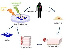
The study of materials science continues to expand in a wide range of applications; however, bioprinting is one of the most exciting techniques as tissue engineering is expected to lead to the fabrication of human organs in the next decade or so. Such research has also proven that bioprinting may yield much more powerful drug delivery whether in using hybrid systems, multi-drug delivery systems, or improved scaffolds.

Here, the materials chosen for drug delivery are more unique as the researchers combined printer ink with semi-synthesized fish gelatin methacryloyl (F-GelMA)—a cold fish gelatin derivative.

In providing aggressive cancer treatment to patients, the use of doxorubicin (DOX) is common as an anti-carcinogen for the treatment of the following diseases:

  • Breast cancer
  • Bladder cancer
  • Kaposi’s sarcoma
  • Lymphoma
  • Acute lymphocytic leukemia

DOX may also cause serious cardiotoxicity, however, despite its use as a broad-spectrum drug. As a solution, PEGylated liposomal DOX, Doxil has been in use for treatment of cancer with much lower cardiotoxity. The nanomedicine has also been approved by the FDA, and is used for targeting local tumors; for instance, this type of drug delivery system could be suitable for treating a brain tumor.

“PEGylating liposomes can prolong their circulation time in blood, resulting in their passive accumulation in cancer tissue, called the enhanced permeability and retention effect,” state the authors.

Using a 3D bioprinter, the authors developed liposomal patches to be directly implanted into cancerous cells.

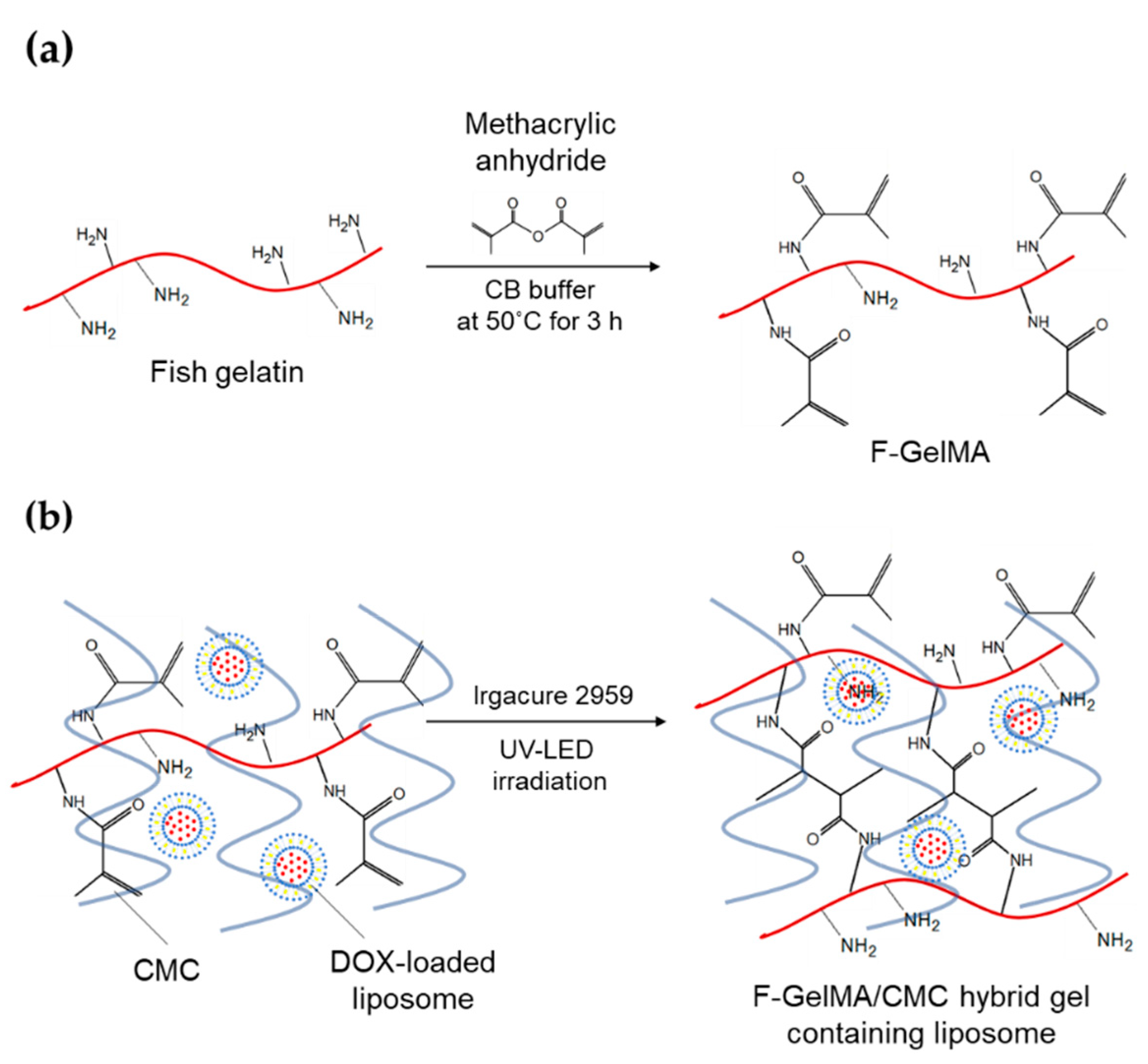
(a) Synthesis of fish gelatin methacryloyl (F-GelMA). (b) Hybrid gel of cross-linked F-GelMA and carboxymethyl cellulose sodium (CMC) containing PEGylated liposome. The reaction scheme was prepared in previous studies

“We used a hydrogel containing semi-synthetic fish-gelatin polymer (fish gelatin methacryloyl, F-GelMA) to entrap DOX-loaded PEGylated liposomes. Fish gelatin is inexpensive and faces few personal or religious restrictions,” stated the authors.

Fish gelatin has not been used widely in bioprinting, however, due to low viscosity and rapid polymerization. To solve that problem, the authors created a bioink composite with elevated viscosity.

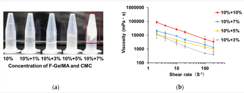
Viscous properties of drug formulations used as printer inks. (a) The appearance of F-GelMA hydrogels containing different concentrations of CMC. (b) The viscosity profiles of F-GelMA hydrogels containing different concentrations of CMC. The data represent the mean ± SD (n = 3).

And while hydrogels are generally attractive for use due to their ability to swell, for this study, the researchers fabricated a variety of different materials—with the combination of 10% F-GelMA and 7% carboxymethyl cellulose sodium (a thickening agent) showing the highest swelling ratio.

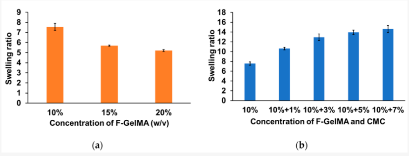
Swelling properties of hydrogels after photopolymerization. (a) Swelling ratio of different concentrations of F-GelMA. (b) Swelling ratio of mixed hydrogel (10% F-GelMA with different concentrations of CMC). The data represent the mean ± SD (n = 3).

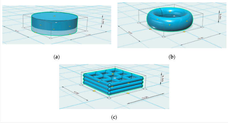
Design of the different 3D geometries: (a) cylinder, (b) torus, and (c) gridlines.

Patches were printed in three different sample shapes, using a CELLINK bioprinter syringe as the authors tested drug release potential in vivo. Realizing that surface area, crosslinks density, temperature, and shaker speed would play a role, the team relied on a larger surface volume for more rapid release of drugs.

Printing conditions of patches.

While experimenting with the torus, gridline, and cylindrical sample patches, the researchers observed gridline-style patches as offering the greatest potential for sustained release.

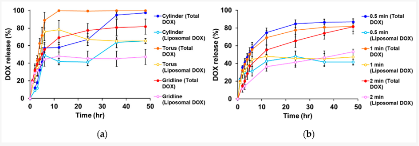
Drug release profiles of liposomal doxorubicin (DOX). (a) Influence of shape on drug release. The UV exposure time was set to 1 min. (b) Influence of UV exposure time on drug release. The gridline object was used for this experiment. The data represent the mean ± SD (n = 3).

“These results indicate that CMC is useful for adjusting the properties of printer ink and is a useful and safe pharmaceutical excipient in drug formulations. We also showed that drug release from 3D-printed patches was dependent on the patch shapes and UV exposure time, and that drug release can be controlled. Taken together, the present results provide useful information for the preparation of 3D printed objects containing liposomes and other nanoparticle-based nanomedicines,” concluded the authors.

[Source / Images: ‘Fabrication of 3D Printed Fish-Gelatin-Based Polymer Hydrogel Patches for Local Delivery of PEGylated Liposomal Doxorubicin’]

The post 3D Printed Medicine Uses Fish Gelatin to Deliver Cancer Treatment appeared first on 3DPrint.com | The Voice of 3D Printing / Additive Manufacturing.

Biodiscoveries: CELLINK is bioprinting its way into the future

Back in 2015, Erik Gatenholm realized there was no place to purchase bioink for 3D bioprinting. So, blown away by this gap in the market, he quickly worked with co-founder Hector Martinez to create a universal bioink that anyone working with bioprinting could use. It was quite a high stakes bet, and at the beginning they set up a webshop to see if they got any bites. It only took 24 hours for the first sale. With more orders quickly coming in, they realized the enormous potential of the product they developed, and CELLINK was born, becoming the first company to commercialize a universal bioink for bioprinting of human tissues and organs.

CELLINK co-founders Erik Gatenholm and Hector Martinez

In the United States alone, every 10 minutes another person is added to the growing waiting list for organ transplants, most of them (60%) in need of a kidney, and with over 130,000 organs transplanted every year worldwide, is no wonder how demand certainly outweighs supply almost everywhere. In some countries the wait can take years, making 3D printing of organs one of the most sought after technologies out there. Bioprinting in the future could allow patients and doctors to reduce waiting times, increase compatibility and decrease immunological failure. For this to happen medical researchers will need to design organs using modeling software, and then print them with biomaterials such as polymers and hydrogels, in addition to the patient’s own cells. Although currently focused on growing cartilage and skin cells suitable for testing drugs and cosmetics, the Swedish company founded in Gothenburg in 2016, hopes to progress the technology far enough to create replacement organs for transplant in humans in the next 15 years.

“In the coming decade we would like to continue to push the boundaries of 3D bioprinting until it becomes an established technology in the medical field. We have the vision of becoming the first and number one provider of bioprinters, bioinks, software and technical know-how for the next generation of medical device manufacturers,” co-founder and CTO Hector Martinez told 3DPrint.com.

Their unique bioink is a biomaterial innovation that allows human cells to grow and thrive as they would in circumstances close to their natural environment. The startup has already managed to print human skin and is also working on producing liver tissues, as well as the beta cells that produce the insulin we need to survive. In 2018 it began printing tumors to combat cancer as part of a research project that doesn’t endanger human lives, and just a few weeks ago, it teamed up with Volumetric to develop Lumen X, a digital light processing bioprinter, designed to enhance inventions in creating more substantial vascular structures. Skin care products, topological drugs and medical treatments are all in need of enhanced testing procedures that can increase the transability from in vitro testing to in vivo usage of products. With tissue engineering and 3D bioprinting more representative in vitro models can be constructed, limiting the use of testing in animals.

CELLINK’s bioinks

Actually, academic labs and companies worldwide are trying to bioengineer all kinds of sophisticated creations for regenerative medicine, drug testing, screening, and tissue engineering. So it’s no wonder CELLINK has their research team focused on creating the next generation of bioinks. Their top selling product is making bioprinting much easier than it used to be some 10 years ago, with 30 different types of bioink available, with prices that go from 99 to 900 dollars. So, what makes one bioink more expensive than the other? It’s all about the components. Collagen and laminin are more expensive to produce than gelatin, raising the price of the end-product. According to CELLINK, scientists mix their live cells into the company’s bioink, a kind of gel designed to allow cells to survive and multiply. The ink is then loaded into a 3D printer by the customer, which forms the desired shape layer by layer as the gel solidifies. By the time the lights inside CELLINK’s box turn green, researchers have an object that acts like human tissue, and can then apply their drug and see how the living cells inside respond.

CELLINK team printing liver models at the lab

“Today we are taking the necessary steps to build and expand our technology offering and exploring new methods for bioprinting tissues. Such technologies include multiple contact-less dispensing methods and light-based bioprinting techniques that enable the bioprinting of high resolution tissue constructs. Refining such technologies will take a close collaboration with our customers as we define the best practices for bioprinting different tissues and specific functions. We can already anticipate that the integration of different bioprinting technologies with post-bioprinting, real-time monitoring systems will be of utmost importance as the bioprinted tissue matures and attains a specific function through an active and precise manipulation of its environment.” Chief IT Officer Jockum Svanberg explained to 3DPrint.com.

Creating the raw material for bioprinting processes is no easy task. Cellink has been focusing on process-compatible soft biomaterials loaded with living cells to create its bioinks since September 2015. The process of bioprinting requires a delivery medium for cells which can be deposited into designed shapes acquired from computer-aided design (CAD) models, which can be generated using 3D medical images obtained through MRIs or CT scans. Some important features of an ideal bioink material are bioprintability, high mechanical integrity and stability, insolubility in cell culture medium, biodegradability at a rate appropriate to the regenerating tissue, non-toxicity and non-immunogenicity, and the ability to promote cell adhesion. Some bioink types, like hydorgels, are not always suitable as construction materials which is why CELLINK is working on a study to provide an upgraded version of the current CELLINK BONE bioink by incorporating collagen and hydroxyapatite. The bioink currently offered does not get close to the real stiffness of the natural bone tissue, but finely resembles its chemical composition. The advantage of such a soft material is to be able to incorporate cells and, during the bioprinting process, to locate them at a precise position throughout the scaffold. This is still for research use only and might take a few years until it is compatible for human use.

With CELLINK bioinks, 3D bioprinting of tissues will help hasten bone fracture healing

Since its start the technology firm has grown to become one of the big competitors in the industry. CELLINK had only been in existence for ten months before they decided to pursue their IPO in November of 2016, listing on Nasdaq First North after a 1070% oversubscribed IPO, which means that demand for their shares was ten times what they expected. Since then, shares have risen over 400%, giving the company a present-day market cap of around $257 million. CELLINK’s affordable printers have already been bought by customers in 25 countries around the world, mostly universities, like Stanford, Harvard, Yale, Princeton and MIT, and some private customers, including Shiseido, Roche, Merck, Johnson and Johnson, and Toyota

But it’s not just about bioprinting it’s way into the future of medicine, CELLINK is also working with other disruptive technologies, such as machine learning. CELLINK told 3DPrint.com that “they want to empower our users with better tools to simplify the bioprinting learning process and broaden its adoption”. One example of this is by developing algorithms that analyse printed structures and based on the results can recommend printing parameters to the users. Using this tool in the development, has helped them speed up the bioink development process. They have just launched a new product: CELLCYTE X, a live cell imaging microscope with live monitoring and analysis of cells in the cloud. Traditionally cell studies have involved manual labor and relied on analysis of the images from an expert, but using deep learning models they are automating this process to provide better and more reliable analysis to their users. The system relies on the latest in serverless system architecture to provide the most scalable, reliable and most intuitive system on the market.

What do you think, will CELLINK continue its upward trajectory? Will it become superseded by other larger firms or get passed by newer start ups? Find out more through our series of articles exploring bioprinting, Biodiscoveries.

Bioprinting 101 Part 9 – Decellularized Extracellular Matrices


Decellularization Process

Today we will be learning about one of the more complex sounding materials within bioprinting. Decellularized extracellular matrices are important within bioprinting processes. That term itself seems like a mouthful. Do not fret. We will break it down into simple parts, and it will be manageable to understand.

Firstly let us define what it means to be decellularized. Decellularization is the process used in biomedical engineering to isolate the extracellular matrix (ECM) of tissue from its inhabiting cells, leaving an ECM scaffold of the original tissue, which can be used in artificial organ and tissue regeneration.

The extracellular matrix is a three-dimensional network of extracellular macromolecules, such as collagen, enzymes, and glycoproteins, that provide structural and biochemical support of surrounding cells. The composition of an ECM varies between multicellular structures. Typically cell to cell communication occurs within an ECM, as well as cell differentiation. Cell differentiation refers to the process of how a cell changes from one cell type to another.

Most connective tissues within animals consist of a type of ECM. Collagen fibers and bone mineral comprise the ECM of bone tissue; reticular fibers and ground substance comprise the ECM of loose connective tissue; and blood plasma is the ECM of blood. Reticular fibers refer to a type of fiber in connective tissue composed of type III collagen secreted by reticular cells. Reticular fibers crosslink and form a fine network. This network is leveraged as a mesh in soft tissues such as liver, bone marrow, and the tissues and organs of the lymphatic system. Ground substance refers to an amorphous gel-like substance in the extracellular space that contains all components of the extracellular matrix except for fibrous materials such as collagen and elastin.

dECM Tissue Structure

When we are within the scope of bioengineering, we are using decellularized extracellular matrices for the purpose of organ and tissue creation. An ECM has great physical properties due to its ability to vary in terms of stiffness and elasticity. This range allows an ECM to be used for soft brain tissues as well as hard bones. The elasticity and stiffness of an ECM is dependant on collagen and elastin concentration within the substance. ECM’s have the ability to regulate cellular processes extensively due to their flexibility in biomechanical properties.

An important note to understand is that decellularized extra matrices are used because they are separated from cells within the body. An ECM is naturally within the body, but a dECM is what we use outside of the body to create bioprinting materials.

The benefit of using a decellularized extracurricular matrix (dECM) is the fact that it is abundant within the body. This means that it is likely to not be rejected when repurposing this material for other biological usages in the body (this refers to us using ECM’s for bioprinting purposes for tissue creation and or organ creation). We can then increase or decrease levels of collagen or elastin within this material to repurpose it quickly for a different usage within the body. So we can effectively take some ECM from a pig and repurpose it for brain tissue. Also one must understand that ECM’s can be combined with stem cells to regrow organs as well. It also allows a smooth immune system response due to it being a substance already found within the body.

The biggest problem with ECM’s are that they have poor mechanical strength. These are typically soft tissue based materials on their own. To create a strong load bearing tissue, it is important to strengthen this material as well as combine it with synthetic biology methods to get the best results. DECM’s can be combined with synthetic polymers through electrospinning to create hybrid scaffolds. Electrospinning is a fiber production method which uses electric force to draw charged threads of polymer solutions or polymer melts up to fiber diameters in the order of some hundred nanometers. Hybrid scaffolds refer to synthetic biology and dECM’s to create dual property scaffolds.

Electrospinning Process

dECM’s are useful for in vivo and in vitro purposes. dECM is particularly of interest for regenerative tissue in vivo because of how it most effectively captures the complex array of proteins and many other matrix components that are found in native tissue, providing ideal cues for regeneration and repair. In vitro, dECM’s are used for general tissue growth and experimentation. This is the best material we have discussed thus far in the series in terms of in vivo practicality for bioprinting.

To create a scaffold through bioprinting we would apply many of the techniques we have already discussed within this series. We can convert a dECM into a bioink or hydrogel which may be extruded for 3D bioprinting purposes. We can also apply different common 3D printing technologies such as SLA printing with this type of material. It is most common to use extrusion as this material is readily made in an aqueous state. Something to ponder in the future is turning this material into a non aqueous material. This can lead to possible developments within how we can use the material as a whole.

As we have taken an indepth look at dECM’s, we understand that it is a burgeoning material within the bioprinting industry. It is important to understand how we can leverage dECM’s in the future with other polymers in order to go more in depth with the uses. We currently focus on soft tissue applications for dECM’s. More research needs to be done in order for us to unlock the potential of this material. We will have a follow up article as well on electrospinning and its relevance for bioprinting as a whole.

This aticle is part of a series that wishes to make bioprinting more accessible. It starts with bioprinting 101, Hydrogels, 3D Industrial Bioprinters, Alginate, Bioinks, Pluronics, Applications and Gelatin.

Interview with Dr. Vahid Serpooshan, Who Created a ‘Heart Attack Patch’ With the Help of 3D Printing

According to the World Health Organization, cardiovascular disease is the number one cause of death globally. An estimated 17.9 million people died from cardiovascular disease in 2016, representing 31% of all global deaths. Treatments for all manner of cardiovascular impairments are therefore a high priority. A very prevalent ailment in that category is a heart attack which happens to over 735,000 Americans a year

A team of researchers led by Dr. Vahid Serpooshan, an assistant professor of Biomedical Engineering and Pediatrics at Georgia Institute of Technology and Emory University School of Medicine, have created a patch with a regenerative protein to treat the infarcted myocardium (cardiac muscle, a part of the heart that gets damaged during a heart attack). According to Georgia Institute of Technology the patch will cover the infarcted tissue with a collagen patch infused with a  protein called FSTL1, which is tailored to fit an area a little larger than the dead tissue. Dr. Serpooshan used bioprinters to develop the patch using a collagen gel. The patch is about to start its pre-clinical trial phase at Emory University’s School of Medicine in Atlanta, and a clinical trial is already ongoing in Europe for a version of this patch device. So it could be a couple more years before they are available for patient treatment. Dr. Serpooshan’s Lab (which has two locations at both Emory University and Georgia Tech) uses a multidisciplinary approach to design and develop micro/nano-scale tissue engineering technologies with the ultimate goal of generating functional tissues and organs. 3DPrint.com reached out to talk to Dr. Serpooshan about his team’s groundbreaking work. 

Professor Vahid Serpooshan sets up the medical 3D printer that will print a patch engineered to strengthen heart muscle damaged in a heart attack. (Photo by Rob Felt, Georgia Institute of Technology)

How has 3D printing helped to engineer the patch?

3D printing has been a huge help in manufacturing the patch in so many ways. It enabled us to incorporate vasculature inside the patch, something that would not have been available with traditional tissue engineering techniques, since they offer really limited potential to create complex, functional vasculature in thick, 3D tissue constructs. Additionally, it allowed us to have a controlled deposition of different cells, biomaterials, and molecules to form heterogenous, complex 3D structures that closely mimic native tissue structure. Also it provided us with patient -and disease- specificity: we utilize MR or CT imaging data obtained from patients to create 3D printed tissue constructs that precisely match the patient’s diseased or damaged tissue. Finally, bioprinting technologies have enabled tissue engineers to markedly enhance resolution and precision in various tissue manufacturing endeavors, making precision medicine even more promising.

What 3D printer do you use to create the patch?

We are mainly using two types of 3D bioprinters for this project. A BioAssemblyBot bioprinting system, made by Advanced Solutions Inc. (an American company in Louisville, Kentucky). This is the ONLY six-axis robotic arm printer in the market, with a resolution down to approximately 20 to 50 μm (microns). The second bioprinter is a BIO-X from Swedish biotech company Cellink, a pioneer in bioprinting.

An illustration of the heart with the cardiac patch already in place (Serpooshan Lab)

How long will it take to manufacture the patch?

Biomanufacturing of a cardiac patch at a clinically-relevant scale would take about 15 to 30 minutes, depending on the number of cell types involved, the vasculature design, and other factors. This enables the clinics, hopefully in the near future, to have heart attack patients come to the clinic, conduct CT or MR imaging, prepare 3D digital model of the damaged tissue, and sending it to the bioprinter to manufacture a patch device customized for that patient.

Vahid Serpooshan designing the myocardial infarct patch (Photo by Rob Felt, Georgia Institute of Technology)

Is there already a procedure in place to apply the patch?

Actually, there already is a procedure to apply the patch. We use a left thoracotomy approach to get access to the surface of the heart and suture the patch onto the epicardial layer of the heart, covering the myocardial infarction (also called a heart attack) area. There are currently some ongoing works trying to minimize the aggressiveness of this surgical procedure, potentially using catheter-based techniques.

What are the benefits of using collagen in the patch?

Collagen is the most abundant protein of the human body. This protein highly supports cell viability, proliferation, and function. It is a biodegradable protein which is a major advantage for in vivo applications, facilitating timely integration of the implant with the host tissue and avoiding long-term immunogenic complications. For our 3D bioprinting works, we use gelatin methacrylate (gelMA). Gelatin is derived from collagen (a hydrolyzed collagen) and offers similar advantages. The main advantages of gelMA for bioprinting of cardiac patches include: acceptable compatibility with cardiac cells (cardiomyocytes), tunable biomimetic mechanical properties (stiffness), controllable degradation rate, and of course, great printability (rheology properties).   

The collagen gel used to develop the 3D printed patch. (Photo by Rob Felt, Georgia Institute of Technology)

What percentage of heart disease patients will benefit from this device?

This therapy is designed for a specific group of heart attack patients, approximately one third of patients who either arrive too late to the clinics or are resistant to current catheter-based reperfusion methods and pharmacologic therapies.

If approved, Dr. Serpooshan’s patch could help over 200,000 patients per year.

Zero Gravity Bioprinter is Ready for Delivery to the International Space Station

An amazing thing happened two years ago, when Techshot and nScrypt worked together to 3D print a human heart – in zero gravity. Not a working heart, yet, but the two companies were the first to 3D print cardiac and vascular structures in zero gravity using adult human stem cells. Why would they do such a thing, you might ask? The reasoning is much more than “because they can,” as 3D bioprinting in zero gravity is actually easier and more effective than it is on Earth.

Earth-based bioprinting requires thick, viscous bioinks that can contain chemicals or other materials necessary for providing structural support. The lack of gravity in space, however, means that thinner, purer bioinks can be used, as well as thinner print nozzles, allowing for more precision and control. When the first viable human organ is 3D printed, it may very well happen in space.

After nScrypt and Techshot pulled off their successful zero-G bioprinting feat, they began work on the 3D BioFabrication Facility, or BFF, a 3D bioprinting system for the International Space Station. The BFF can 3D print thick tissue and organs using adult stem cells. The printing will happen on an nScrypt 3D Bio Assembly Tool, or BAT; the bioink will be printed into a specialized cell culturing bioreactor cassette designed by TechShot and conditioned in the TechShot ADvanced Space Experiment Processor (ADSEP). The BFF and ADSEP are scheduled to launch on their way to the ISS in February 2019.

The nScrypt BAT 3D printer features high-precision motion and extreme dispensing control, and will use nScrypt’s patented SmartPump, which has 100 picoliter volumetric control and uses super-fine nozzles, down to 10 microns, to dispense biomaterials. This enables the highly controlled and repeatable placement of bioink, which is necessary for printing the fine details of tissues and organs.

“Especially when dealing with something as important as tissue, it is vital to place the correct amount of material in the correct position every time,” said nScrypt CEO Ken Church. “This is what our machines offer and what has contributed to our success in bioprinting as well as other applications. This is an exciting time for discovery and more importantly a time of impact for those that are seriously seeking solutions to grow thick vascularized tissue, which is the basis for a fully printed organ.”

The first complete print, after the initial test prints, will be a cardiac patch for damaged hearts. Cells will be printed into the bioreactor cassette, and the bioreactor will then provide media perfusion to deliver nutrients and remove toxins from the tissue, keeping it alive while providing electrical and mechanical stimulus to encourage the cells to become beating heart tissue.

Rendering of the BFF in an EXPRESS rack [Image: nScrypt]

The BFF may truly be an astronaut’s BFF; in addition to 3D printing tissue for people on Earth, it can print pharmaceuticals and even food on demand for people on the International Space Station.

“We are very excited to see this project, and all that it can provide, come to life,” said Techshot President and CEO John C. Vellinger. “With the goal of producing everything from organs, to pharmaceuticals, to perhaps even food, the BFF has the ability to improve the lives of people on earth and help enable deep space exploration.”

Discuss this and other 3D printing topics at 3DPrintBoard.com or share your thoughts below.