Aalto University Develops a Novel Bioink for Cardiac Tissue Applications

Finland is one of Europe’s most forested nations. Over 70 percent of the country’s boreal forest is covered with spruce, pine, downy birch, and silver birch. But beyond the splendor of the Finnish woodlands, all these trees have one thing in common, and that is nanocellulose. A light solid substance obtained from plant matter which comprises cellulose nanofibrils (CNF) and is considered a pseudo-plastic that possesses the property of specific kinds of gels that are generally thick in normal conditions. Overall, it is a very environmentally friendly and non-toxic substance that is compatible with the human body and has the potential to be used for a range of medical applications.

In 2018, the Department of Bioproducts and Biosystems at Aalto University, located just outside Helsinki, began searching for new ideas to revitalize one of the country’s traditional economic engines, forests (which are handled sustainably thanks to renewable forest resources). At the time, they noticed that one of the possible applications could be working with nanocellulose. Forward two years and the researchers have come up with a new bioink formulation praising nanocellulose at its basis.

Thanks to the structural similarity to extracellular matrices and excellent biocompatibility of supporting crucial cellular activities, nanocellulose-based bioprinting has clearly emerged for its potential in tissue engineering and regenerative medicine. The qualities of the generally thick and fluid light substance make it an excellent match to develop bioinks that are both suitable and scalable in their production, but also have consistent properties. However, there have been major challenges in processing nanocellulose.

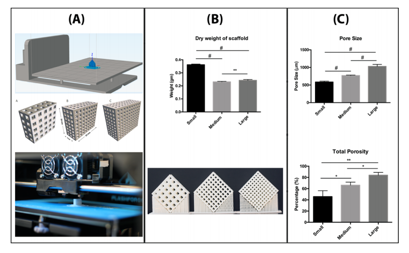
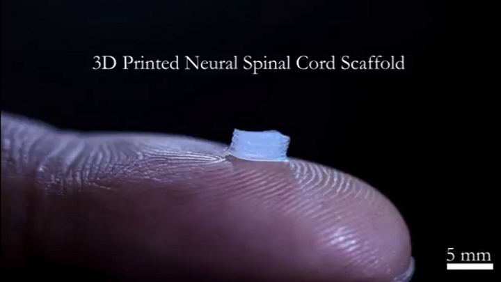
As described by Aalto University researchers in a recently published paper in the science journal ACS Publication, the unresolved challenges of bioink formulations based on nanocelluloses are what stops the substance from becoming one of the preferred components for 3D bioprinting structures. This is why Finnish researchers focused on developing a single-component bioink that could be used to create scaffolds with potential applications in cardiac biomedical devices, while fundamentally dealing with some of the limitations of using nanocellulose-based bioinks.

A co-author of the paper and a doctoral candidate at Aalto’s Department of Bioproducts and Biosystems, Rubina Ajdary, told 3DPrint.com that “other than natural abundance and as a renewable resource, nanocellulose has demonstrated to have an outstanding performance in tissue engineering.” She also suggested that “recent efforts usually consider the use of nanocellulose in combination with other biopolymers, for example, in multicomponent ink formulations or to encapsulate nanoparticles. But we were interested in investigating the potential of monocomponent nanocellulose 3D printed scaffolds that did not require crosslinking to develop the strength or solidity.”

In fact, the Biobased Colloids and Materials (BiCMat) research group at Aalto University, led by Orlando Rojas, proposed heterogeneous acetylation of wood fibers to ease their deconstruction into acetylated nanocellulose (AceCNF). As a unique biomaterial opportunity in 3D scaffold applications, the team considered using nanocelluloses due to the natural, easy to sterilize, and high stability porosity of the substance, and chose to introduce AceCNF for the generation of 3D printed scaffolds for implantation in the human body. The team then went on to evaluate the interactions of the scaffolds with cardiac myoblast cells.
“Most modifications make the hydrogels susceptible to dimensional instability after 3D printing, for instance, upon drying or wetting. This is exacerbated if the inks are highly diluted, which is typical of nanocellulose suspensions, forming gels at low concentrations,” went on Ajdary. “This instability is one of the main reasons why nanocellulose is mainly combined with other compounds. Instead, in this research, we propose heterogeneous acetylation of wood fibers to ease their deconstruction into acetylated nanocellulose for direct ink writing. A higher surface charge of acetylated nanocellulose, compared to native nanocellulose, reduces aggregation and favors the retention of the structure after extrusion even in significantly less concentration.”
This is exactly why it was important for the researches to develop a single component bioink. Nanocellulose has shown promises when combined with other biopolymers and particles. However, Ajdary insists that benefits including similarity to the extracellular matrix, high porosity, high swelling capacity, ease of surface modification, and shear thinning behavior of cellulose, encouraged them to study the potential of monocomponent surface-modified nanocelluloses.

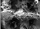
Acetylated nanocellulose (Credit: Aalto University School of Chemical Engineering)

The team at Aalto University used the sustainable and widely available nanocelluloses to make several formulations of bioinks and evaluate them, including unmodified nanocellulose CNF, Acetylated CNF (AceCNF), and TEMPO-oxidized CNF.
To 3D bioprint the hydrogels, researchers used Cellink bioprinters, something Ajdary attributed to the user-friendliness of the device and because it provided a lot of flexibility to test different types of hydrogels and emulsions produced in the research group.
In this new process, the single-component nanocellulose inks were first 3D printed into scaffolds using Cellink’s BIO X bioprinter, which is equipped with a pneumatic print head was used to extrude single filaments and form the 3D structures. Then freeze-dried to avoid extensive shrinkage, and sterilized under UV light. After sterilization the scaffold was ready and cells seeded on the samples.
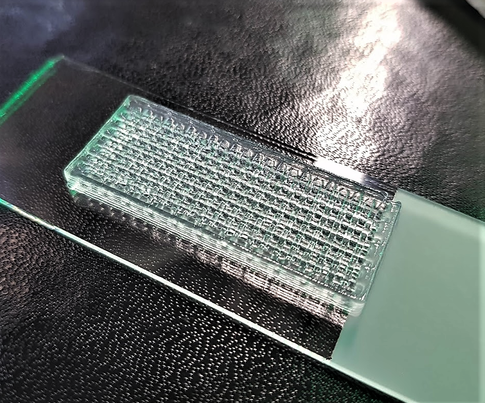
“3D structures of acetylated nanocellulose are highly stable after extrusion in far less concentrations. The lower concentration in wet condition facilitates the scaffold with higher porosity after dehydration which can improve the cell penetration in the structure and assist in nutrient transport to the cells as well as in the transport of metabolic waste,” specified Ajdary.
The researchers claim that the method was successful as the 3D printed scaffolds were compatible with the cardiomyoblast cells, enabling their proliferation and attachment, and revealing that the constructs are not toxic. Although still in research stages, these bioinks and technique can be used for the inexpensive, consistent fabrication and storage of constructs that can be applied as base materials for cardiac regeneration.
What is novel in this study is the particular focus on single-component nanocellulose-based bioinks that open up a possibility for the reliable and scale-up fabrication of scaffolds appropriate for studies on cellular processes and for tissue engineering. Since this is an ongoing research, we can expect to read more published material from Aalto University researchers as they continue testing their unique technique even further.

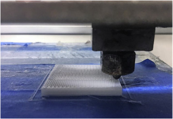
Scaffolds corresponding to 3D printed AceCNF (Credit: Aalto University)

The post Aalto University Develops a Novel Bioink for Cardiac Tissue Applications appeared first on 3DPrint.com | The Voice of 3D Printing / Additive Manufacturing.

Beijing University of Chemical Technology: 3D Printed HA/PCL Tissue Engineering Scaffolds

3D printed bone scaffolds used for tissue engineering purposes need to have a good amount of mechanical strength, since the scaffold needs to be able to provide support for the tissue. As bone scaffolds also require the correct pore structure to help provide a good environment for the differentiation, proliferation, and repairing of damaged tissue cells, bioactive materials, such as polycaprolactone (PCL) and hydroxyapatite (HA), are needed.

Researchers Zhiwei Jiao, Bin Luo, Shengyi Xiang, Haopeng Ma, Yuan Yu, and Weimin Yang, from the Beijing University of Chemical Technology (BUCT), published a paper, titled “3D printing of HA / PCL composite tissue engineering scaffolds,” about their work constructing nano-HA/PCL and micro-HA/PCL tissue engineering scaffolds using the melt differential FDM 3D printer they developed.

The abstract reads, “Here, the internal structure and mechanical properties of the hydroxyapatite/polycaprolactone scaffolds, prepared by fused deposition modeling (FDM) technique, were explored. Using hydroxyapatite (HA) and polycaprolactone (PCL) as raw materials, nano-HA/PCL and micro-HA/PCL that composite with 20 wt% HA were prepared by melt blending technology, and HA/PCL composite tissue engineering scaffolds were prepared by self-developed melt differential FDM 3D printer. From the observation under microscope, it was found that the prepared nano-HA/PCL and micro-HA/PCL tissue engineering scaffolds have uniformly distributed and interconnected nearly rectangular pores. By observing the cross-sectional view of the nano-HA/PCL scaffold and the micro-HA/PCL scaffold, it is known that the HA particles in the nano-HA/PCL scaffold are evenly distributed and the HA particles in the micro-HA/PCL scaffold are agglomerated, which attribute nano-HA/PCL scaffolds with higher tensile strength and flexural strength than the micro-HA/PCL scaffolds. The tensile strength and flexural strength of the nano-HA/PCL specimens were 23.29 MPa and 21.39 MPa, respectively, which were 26.0% and 33.1% higher than those of the pure PCL specimens. Therefore, the bioactive nano-HA/PCL composite scaffolds prepared by melt differential FDM 3D printers should have broader application prospects in bone tissue engineering.”

Melt differential 3D printer.

PCL is biocompatible, biodegradable, and has shape retention properties, which is why it’s often used to fabricate stents. But on the other hand, due to an insufficient amount of bioactivity, the material is not great for use in bone tissue engineering. HA, which has been used successfully as a bone substitute material, has plenty of bioactivity, which is why combining it with PCL can work for bone tissue engineering scaffolds.

“On the whole, the existing tissue engineering scaffolds preparation process have problems of low HA content, easy agglomeration, low stent strength, and single printing material,” the researchers explained.

“The HA/PCL composite particles are used as printing materials, and the mechanical properties and structural characteristics of the two tissue engineering scaffolds are compared and analyzed. The raw material of the melt differential 3D printer is pellets, which eliminates the step of drawing compared to a conventional FDM type 3D printer. The 3D printer is melt-extruded with a screw, and a micro-screw is used for conveying and building pressure. At the same time, precise measurement is performed by a valve control system. This printing method shows advantages in simple preparation process of the composite material, higher degree of freedom in material selection, simple printing process, and shorter preparation cycle of tissue engineering scaffolds.”

The team mixed PCL particles and HA powder together to make the scaffolds. Their melt differential 3D printer uses pellets, and features a fixed nozzle with a platform that moves in three directions. A twin-screw extrusion granulator was used to prepare the PCL material, and the melt differential 3D printer fabricated the tissue engineering scaffolds out of the nano-HA/PCL and micro-HA/PCL composite particles.

The working principle diagram of the polymer melt differential 3D printer.

A microcomputer-controlled electronic universal testing machine was used to test the scaffolds’ bending and tensile properties. A scanning electron microscope was used to observe the micro-HA particle size, as well as the scaffolds’ cross section, while an optical microscope was used to observe their surface structure and a transmission microscope was used to look at the nano-HA particles’ particle diameter and morphology. The scaffold material’s crystallization properties were analyzed using a differential thermal analyzer.

3D printing tissue engineering scaffolds.

Testing showed that the micro-HA was spherical, with a 5–40 μm diameter, and contained some irregularly-shaped debris. The nano-HA was rod-shaped, with a 20–150 nm length.

The crystallization peak temperature of the HA/PCL composites was higher than pure PCL material, because adding HA caused its molecular chain to form a nucleate after absorbing on the HA’s surface. Additionally, adding HA to pure PCL increased the material’s melting temperature, as the latter material had crystals “of varying degrees of perfection.”

The nano-HA/PCL and micro-HA/PCL tissue engineering scaffolds “could form a pre-designed pore structure and the pores were connected to each other,” which is seen in the image below.

“…the micro-HA/PCL and the nano-HA/PCL composite tissue engineering scaffolds can form a three-dimensional pore structure with uniform distribution and approximately rectangular shape.”

External views of micro-HA/PCL and nano-HA/PCL composite tissue engineering scaffolds.

These rectangular pores, with a 100-500 μm length and width, are good news for cell adhesion and proliferation, and the fact that they’re interconnected is positive for nutrient supply.

As for mechanical properties, the nano-HA/PCL specimens had the highest tensile and bending strengths – between 25 and 35% higher than the pure PCL. The micro-HA/PCL specimens had higher tensile and flexural strengths than the PCL, but the nano-HA/PCL was stronger than the micro-HA/PCL, because the HA’s modulus is higher than the PCL’s.

“In addition, nano-HA was more evenly distributed in the composite, while micro-HA had obvious agglomeration in the composite, so the tensile strength and flexural strength of nano-HA/PCL specimens were higher than that of micro-HA/PCL specimens,” the researchers wrote.

Finally, the pore structure of the nano-HA/PCL and micro-HA/PCL tissue engineering scaffolds offered a favorable environment for the discharge of cellular metabolic waste, in addition to facilitating nutrient transport and blood vessel growth. The researchers concluded that their 3D printed composite scaffolds had more potential applications in bone tissue engineering.

Discuss this and other 3D printing topics at 3DPrintBoard.com or share your thoughts below.

The post Beijing University of Chemical Technology: 3D Printed HA/PCL Tissue Engineering Scaffolds appeared first on 3DPrint.com | The Voice of 3D Printing / Additive Manufacturing.

Bioprinting Hot Dogs with Hierarchical Structures

Researchers around the world continually surprise us with innovation, but rarely do they reach into the roots of popular culture—and the food that accompanies it—applying it to the world of bioprinting…and with hot dogs in mind, no less. However, that’s exactly what has inspired German and Chinese scientists to build bioprinting structures, with their study outlined in the recently published ‘3D Printing of Hot Dog-Like Biomaterials with Hierarchical Architecture and Distinct Bioactivity.’

Fabrication and morphology of hot dog‐like scaffolds (HD‐AKT). a) The schemata of preparation of HD‐AKT, combining 3D printing and bidirectional freezing to realize 3D printed scaffolds containing rods with aligned lamellar microstructure. b–e) 3D micro‐CT images and f,g) 2D micro‐CT images of HD‐AKT from different views showing the scaffold structure, which is hollow tube macrospores embedded by bioceramic rods with the uniformly aligned lamellar structure of rods.

The researchers fabricated hierarchical structures using direct ink writing (DIW), with the hot dog structure figuring in with the use of tubes:

“The scaffolds are composed of hollow bioceramic tubes (mimicking the “bread” in hot dogs, pore size: ≈1 mm) embedded by bioceramic rods (mimicking the “sausage” in hot dogs, diameter: ≈500 µm) and the sausage‐like bioceramic rods possess uniformly aligned lamellar micropores (lamellar pore size: ≈30 µm),” said the researchers.

While hierarchical structures are often used in bioprinting, it can be challenging to find suitable materials in creating micro/nanostructures, and especially with DIW. And while challenges have continued in implanting 3D printed scaffolds to regenerate bone growth, there has been some success. The researchers contend, however, that hierarchy in structure is needed to promote better tissue growth, with the ‘hot-dog like structure’ lending itself to better cell adhesion and supply of nutrients, with the rods enhancing delivery of osteogenic drugs.

“By mimicking the function of nutrition supply for sausages in hot dog, the potential of the scaffolds for loading icariin (Ica, a model osteogenic drug), was investigated in this study,” explained the researchers.

“However, the Ica loading efficiency and capacity of S‐AKT were much lower than that of HD‐AKT, indicating the excellent loading capacity of the hierarchical hot dog‐like scaffolds. The thermogravimetric analysis further verified the significantly improved loading efficiency of HD‐AKT scaffolds as compared to those without hot dog microstructure.”

Hot dog‐like scaffolds are an excellent carrier for drug and protein. The drug Ica and protein BSA loading and release properties of the HD‐AKT. a) The Ica loading efficiency, and b) the loading capacity of traditional solid struts scaffolds(S‐AKT), H‐AKT, and different kinds of HD‐AKT. c) Thermogravimetric analysis of the scaffolds after loading the Ica. d) The Ica release of different kinds of scaffolds after loading Ica (S‐AKT/Ica, H‐AKT/Ica, HD‐20AKT/Ica, HD‐30AKT/Ica, HD‐40AKT/Ica, and HD‐50AKT/Ica). e) The BSA loading efficiency. f) The BSA release of the scaffolds. g–i) SEM images of the hot dog rod of scaffolds g) after 90 d Ica release. h) Surface images and i) the cross‐section image show lots of mineralized hierarchical structures on the surface of hot dog rod of scaffolds. Compared with the S‐AKT and H‐AKT, the HD‐AKT have higher loading efficiency and capacity. Meantime, HD‐AKT possess longer Ica/BSA release time.

The research team also discovered that other drugs could be distributed too, such as the large molecule protein bull serum albumin (BSA). Even more encouraging, the researchers found that the scaffolds exhibited ‘excellent bioactivity,’ proven through in vivo processes for bone regeneration as they implanted drugs into ‘rabbit femoral defects’ for eight weeks with no inflammation, and proof of bone tissue growth.

Hot dog‐like scaffolds are an excellent platform for cell delivery and differentiation. The proliferation, morphology, and relative genes expression of rBMSCs cultured on different scaffolds. a‐d) Confocal and SEM images of rBMSCs cultured on a) S‐AKT, b) H‐AKT, c) HD‐30AKT, and d) HD‐30AKT/Ica. e) The rBMSCs (red color) adhesion on the rod of HD‐AKT. f) The proliferation of rBMSCs after seeding in different kinds of scaffolds, showing hot dog‐like scaffolds are beneficial for cell proliferation. g) The relative osteogenic genes expression (OCN, Runx2, OPN, and ALP) of rBMSCs in scaffolds, indicating that the Ica release from the scaffolds promotes the relative osteogenic genes expression of rBMSCs (n = 6, *P < 0.05, and **P < 0.01.).

“Our study suggests that the hot dog‐like scaffolds can be used for the multifunctional biomaterials for drug delivery, tissue engineering, and regenerative medicine. The combined strategy of DIW 3D printing with bidirectional freezing is a promising method to prepare biomimetic and hierarchical biomaterials,” concluded the researchers.

Hot dog‐like scaffolds possess excellent bone‐forming bioactivity after implanted in the femoral defects of rabbits. The characterizations of hot dog‐like scaffolds for osteogenesis in vivo. a1–d1) Digital, a2–d2) 2D micro‐CT, and 3D micro‐CT images (a3–d3: transverse view, and a4–d4: sagittal view) of the defects at week 8. In 3D micro‐CT images, green, red and white represents new bone, scaffold, and primary bone, respectively. e) Micro‐CT reconstruction analysis exhibits the volume ratio of the new bone to the original defect regions (BV/TV) at week 8. HD‐30AKT and HD‐30AKT/Ica indicate significantly. f) The newly formed bones (red) grow into the lamellar microstructures (green arrows) of hot dog‐like scaffold. g–j) Hard histological sections stained with Van Gieson’s picrofuchsin of g) Blank, h) H‐AKT, i) HD‐AKT, and j) HD‐AKT/Ica, red color stands newly formed bone and black color represents scaffolds. Newly formed bone can grow into the hierarchical rods of the scaffolds (blue circle point to the new bone in the hierarchical rods). improvement in new bone regeneration as compared to Blank control. In addition, Ica can also promote osteogenesis, suggesting that both hierarchical structure and the Ica delivery of the hot dog‐like scaffolds contribute to the bone regeneration (n = 6, *P < 0.05, **P < 0.01, and ***P < 0.001.).

While man-made processed foods are certainly inspiring to many, undoubtedly nature continues to propel scientists and designers forward from creating conductive parts to custom-made shoes, and liquid polymers for 3D printing. What do you think of this news? Let us know your thoughts! Join the discussion of this and other 3D printing topics at 3DPrintBoard.com.

[Source / Images: ‘3D Printing of Hot Dog-Like Biomaterials with Hierarchical Architecture and Distinct Bioactivity’]


The post Bioprinting Hot Dogs with Hierarchical Structures appeared first on 3DPrint.com | The Voice of 3D Printing / Additive Manufacturing.

McGill University Researchers: Can We Use PLA for Desktop Bioprinting?

Bioprinting has proven to be useful for bone regeneration, as researchers learn to create more stable structures that mimic human tissue. In ‘Three-Dimensional Printed Polylactic Acid Scaffolds Promote Bonelike Matrix Deposition in Vitro,’ authors Rayan Fairag, Derek H. Rosenzweig, Jose L. Ramirez-Garcialuna, Michael H. Weber, and Lisbet Haglund explore the uses of desktop bioprinting with PLA.

Even in conventional medicine today, surgeons find difficulty in repairing bones that have undergone trauma, whether due to an accident, tumor, or other serious issue. Grafting can still be challenging to complete, and then problematic later in terms of pain, infection, and the need for multiple procedures. Materials such as calcium phosphate bone cement (a synthetic graft) have become more popular for repairing bone defects, but there are also limitations due to lack of mechanical strength. While poly-cements have been used also, they can cause stress around the ‘target area,’ and lead to secondary fracture, which defeats the purpose of healing altogether.

Here, the researchers have investigated the use of tissue engineering for bone repair in growing cells, scaffolds, and using numerous bioactive factors. 3D printing has been successful in fabricating scaffolds using different polymers like PLA.

“The ideal material for scaffold development should fulfill specific criteria,” state the researchers. “The material must be biocompatible and must be capable of being generated with an interconnected network to mimic the natural tissue architecture.”

Cell sustainability is the greatest challenge, along with creating stable structures. The researchers sought to create scaffolds that would allow for complete cell sustainability, along with the best environment for encouraging tissue to form. They must also allow for the following:

  • Fabrication in different, complex shapes
  • Resistance to inflammation and toxicity
  • Strong mechanical properties
  • Appropriate porosity
  • Affordability

In previous studies, the researchers were aware that PLA 3D printed from the desktop was suitable for both chondrocyte and nucleus pulposus tissue engineering applications. Here, they tested PLA scaffolds with pore sizes of 500, 750, and 1000 μm, fabricating accurate structures with good porosity; in fact, all scaffolds reflected pores in line with the initial designs, leaving the authors to conclude that this ‘suggested accuracy’ with desktop 3D printer—in this case, the Flashforge Creator Pro.

Pore size results were as follows:

  • Small pore scaffolds – 585.61 μm ± 26.40
  • Medium pore scaffolds – 769.94 μm ± 12.98
  • Large pore scaffolds – 1028.85 μm ± 57.54, p < 0.0001

“The scaffold fabrication and replication process manifests high accuracy and precision as evidenced by μCT analysis, which proves the value of low-cost printing in tissue engineering applications,” stated the researchers.

The authors reported the following for mechanical properties:

“Significant differences in stiffness were observed between the three sizes (p < 0.05, p < 0.0001) in which Young’s modulus for the small pore size was 206.7 MPa ± 0.17 SD, medium size scaffold was 137.5 MPa ± 6.98 SD, and 116.4 MPa ± 5.97 SD for the large size PLA scaffold.”

Mechanical properties of 3D-printed scaffolds. (A) Young’s modulus representing 5−10% compressive stress/strain curves of printed PLA scaffolds. For each set, (n = 3), error bars represent ±SD and (* = P value < 0.05), (# = P value < 0.0001). (B) Stress/strain curves of 500, 750, and 1000 μm showing the amount of deformation, elastic (proportionality) limit, and plastic region. For each set, (n = 3).

“The failure point of each scaffold was determined from the stress/strain curves in which the small-size failure point was around 21.63 MPa, around 11.86 MPa for the medium size, and around 8.53 MPa for the large-pore scaffold. Our results demonstrated an overall higher compressive modulus with smaller pores because of the addition of bulk material (smallest pore size has the highest amount of material and is the stiffest).”

The use of PLA was successful, indicating both accuracy and reproducibility, and the scaffolds presented properties like native bone. The authors stated that the data reflected structures stable enough for an environment recruiting host stem cells and repairing bone.

Morphological characterization of 3D printed scaffolds. (A) Representative images of the 3D models with dimensions and printing process. (B) Quantification of scaffold weight, (n = 6), error bars represent ±SD (** = P value < 0.005), (# = P value < 0.0001), with a representative image of printed scaffolds (Canon EOS 350d Camera). (C) Pore size was calculated by scanning electron microscopy, and porosity was determined by μ-CT. For each set, (n = 3), error bars represent ±SD and (* = P value < 0.05), (** = P value < 0.005), (# = P value < 0.0001).

“In vivo studies will be necessary to determine potential adverse effects, bone repair, and scaffold resorption rates,” stated the researchers. “It comes without surprise that 3D printing has been strongly adopted by orthopedic surgery clinical practice, medical education, patient education, and orthopedic-related basic science.

“Whereas 3D printing has been used for some time to generate patient models of defects for presurgical planning, there is a growing shift in using this technology in actual bone or tissue repair. One major focus in orthopedic and reconstructive surgery is to use 3D printed constructs for filling bone defects, substituting current standard therapies as an innovative approach for bone repair. Several studies have shown applicability and clinical relevance of using different types of 3D-printed polymers as a graft substitute.”

From 3D printing in hospitals to bioprinting in outer space and bringing forth materials which may eventually yield fabricated human organs, researchers are driven to create what used to be considered impossible, with a wide range of innovations already in use around the world. Find out more about desktop bioprinting here. What do you think of this news? Let us know your thoughts! Join the discussion of this and other 3D printing topics at 3DPrintBoard.com.

SEM of acellular and cell-seeded scaffolds. Representative SEM images of acellular, osteoblasts, MSC−OST seeded scaffolds at 80×,450×,1500×, and 22 000× magnifications and scale bars represent 1 mm, 200, 50, and 5 μm with the rectangular marker indicating the region of the scan (n = 3).

[Source / Images: ‘3D-Printed Polylactic Acid (PLA) Scaffolds Promote Bone-like Matrix Deposition In-vitro’]

UPC Researchers Develop New Method of Designing Porous Scaffolds for FDM 3D Printing

Trabecular structure with thin walls.

From manufacturing customized prosthetics and implants to surgical planning and bioprinting organs and tissues, 3D printing has many medical applications. In terms of bone tissue engineering and 3D bioprinting, 3D printed scaffolds are used as templates to help with tissue formation and initial cell attachment, as well as to fix prostheses through osseointegration. We’ve seen scaffolds made with all sorts of materials, like a compound in turmeric, sugar, and plastic, but the best are those with good porosity that can simulate tissue.

It’s not always easy to fabricate porous structures with specific pore sizes using FDM technology. A trio of researchers from the Polytechnic University of Catalonia (UPC) in Barcelona published a paper, titled “3D Printing of Porous Scaffolds with Controlled Porosity and Pore Size Values,” explaining how they developed a new method of designing porous scaffolds for FDM 3D printing.

The abstract reads, “The present paper provides a methodology to design porous structures to be printed. First, a model is defined with some theoretical parallel planes, which are bounded within a geometrical figure, for example a disk. Each plane has randomly distributed points on it. Then, the points are joined with lines. Finally, the lines are given a certain volume and the structure is obtained. The porosity of the structure depends on three geometrical variables: the distance between parallel layers, the number of columns on each layer and the radius of the columns. In order to obtain mathematical models to relate the variables with three responses, the porosity, the mean of pore diameter and the variance of pore diameter of the structures, design of experiments with three-level factorial analysis was used. Finally, multiobjective optimization was carried out by means of the desirability function method. In order to favour fixation of the structures by osseointegration, porosity range between 0.5 and 0.75, mean of pore size between 0.1 and 0.3 mm, and variance of pore size between 0.000 and 0.010 mm2 were selected. Results showed that the optimal solution consists of a structure with a height between layers of 0.72 mm, 3.65 points per mm2 and a radius of 0.15 mm. It was observed that, given fixed height and radius values, the three responses decrease with the number of points per surface unit. The increase of the radius of the columns implies the decrease of the porosity and of the mean of pore size. The decrease of the height between layers leads to a sharper decrease of both the porosity and the mean of pore size. In order to compare calculated and experimental values, scaffolds were printed in polylactic acid (PLA) with FDM technology. Porosity and pore size were measured with X-ray tomography. Average value of measured porosity was 0.594, while calculated porosity was 0.537. Average value of measured mean of pore size was 0.372 mm, while calculated value was 0.434 mm. Average value of variance of pore size was 0.048 mm2, higher than the calculated one of 0.008 mm2. In addition, both round and elongated pores were observed in the printed structures. The current methodology allows designing structures with different requirements for porosity and pore size. In addition, it can be applied to other responses. It will be very useful in medical applications such as the simulation of body tissues or the manufacture of prostheses.”

Cross-section of specimen 1: (a) 3D view, and (b) 2D view.

In order to design 3D printed porous scaffolds that can simulate tissues, they need mechanical strength, which helps with protection and support; permeability, which can direct the transport of nutrients; and surface area and interconnectivity, both of which relate to good cell growth. Other researchers have tried to achieve the necessary porosity in scaffolds by using hierarchical design and topology optimization. But the UPC team went a different way.

“Unlike other methods that are based on truss structures, the present model allows obtaining irregular porous structures from random location of columns in the space, which leave voids among them. Specifically, the structure was modelled with parallel planes joined by columns, with a certain number of columns on each plane,” the researchers wrote.

They applied their model to a disc shape and defined three different variables:

  1. Distance between parallel planes
  2. Number of base points for columns on each plane
  3. Radius of each column

Then, the team used dimensional analysis to lower the number of process variables to just two, and so defined their requirements “for a specific application case: the use of a porous structure in external layers of hemispherical hip prostheses.”

Printed porous structure (rescaling of the designed scaffold by scaling factor of five).

To compare the results of their experiment with computationally calculated results, the researchers used a dual-extruder Sigma 3D printer from BCN3D to fabricate three sample scaffolds out of PLA, then measured their pore size and total porosity. The researchers found that the measured results were not dissimilar to the calculated results.

“In future work, other requirements for structures, related to either mechanical strength or mass transport, will be addressed. In addition, improvement of the FDM printing process is required in order to obtain more accurate and smooth parts,” the researchers concluded.

Co-authors of the paper are Irene Buj-CorralAli Bagheri, and Oriol Petit-Rojo.

Discuss this research and other 3D printing topics at 3DPrintBoard.com or share your thoughts below.

Breakthrough 3D Printed Neural Scaffold Could Help Patients with Spinal Cord Injuries Regain Some Functions

Right now, 285,000 people in the US suffer from spinal cord injuries, with roughly 17,000 new injuries each year. 3D printed spinal implants have been shown to help patients recover more easily, and a team of engineers and medical researchers from the University of Minnesota (UMN) have spent the last two years developing an innovative new 3D printed medical device that could help long-term spinal cord injury patients regain some function in the future.

“This is a very exciting first step in developing a treatment to help people with spinal cord injuries. Currently, there aren’t any good, precise treatments for those with long-term spinal cord injuries,” said Ann Parr, MD, PhD, a UMN Medical School Assistant Professor in the Department of Neurosurgery and Stem Cell Institute.

The method involves a 3D printed silicone guide, which acts as a scaffold for special stem cells that are bioprinted directly on top of it. The aim is to surgically implant the guide into the injured part of the spinal cord, and it should act as a bridge between living nerve cells both above and below the area, which could help alleviate pain for patients, in addition to helping them gain control over functions like bladder, bowel, and muscle control again.

“We’ve found that relaying any signals across the injury could improve functions for the patients. There’s a perception that people with spinal cord injuries will only be happy if they can walk again. In reality, most want simple things like bladder control or to be able to stop uncontrollable movements of their legs,” Parr explained. “These simple improvements in function could greatly improve their lives.”

Spinal cord scaffold assembly process: 3D bioprinting cells on silicone scaffolds
allows for in vitro culture of sNPCs and OPCs. (a) Silicone scaffolds are printed with
channels, and (b) cells are dispensed inside the channels. (c) A layer of silicone covers the channels, and (d) scaffolds are placed inside a dish and cultured for 7 days.

The team recently published a paper on their potentially life-changing work, titled “3D Printed Stem-Cell Derived Neural Progenitors Generate Spinal Cord Scaffolds,” in the peer-reviewed scientific journal Advanced Functional Materials.

Fluorescence images of 3D printed cell-laden Matrigel (50%) matrices cultured for 0 (3 hours), 1, and 4 days. Timescale images show (a) sNPCs extending axons, and (b) OPCs exhibiting bi-polar processes.

The abstract reads, “A bioengineered spinal cord is fabricated via extrusion‐based multimaterial 3D bioprinting, in which clusters of induced pluripotent stem cell (iPSC)‐derived spinal neuronal progenitor cells (sNPCs) and oligodendrocyte progenitor cells (OPCs) are placed in precise positions within 3D printed biocompatible scaffolds during assembly. The location of a cluster of cells, of a single type or multiple types, is controlled using a point‐dispensing printing method with a 200 µm center‐to‐center spacing within 150 µm wide channels. The bioprinted sNPCs differentiate and extend axons throughout microscale scaffold channels, and the activity of these neuronal networks is confirmed by physiological spontaneous calcium flux studies. Successful bioprinting of OPCs in combination with sNPCs demonstrates a multicellular neural tissue engineering approach, where the ability to direct the patterning and combination of transplanted neuronal and glial cells can be beneficial in rebuilding functional axonal connections across areas of central nervous system (CNS) tissue damage. This platform can be used to prepare novel biomimetic, hydrogel‐based scaffolds modeling complex CNS tissue architecture in vitro and harnessed to develop new clinical approaches to treat neurological diseases, including spinal cord injury.”

The process begins with any type of adult stem cell, be it blood or skin, and medical researchers use the latest bioengineering techniques to reprogram these into neuronal stem cells. These cells are then 3D printed onto a silicone guide with a unique extrusion-based technology, which can print both the cells and the guide from the same 3D printer.

Michael McAlpine, PhD, UMN Benjamin Mayhugh Associate Professor of Mechanical Engineering in the University’s College of Science and Engineering, said, “This is the first time anyone has been able to directly 3D print neuronal stem cells derived from adult human cells on a 3D-printed guide and have the cells differentiate into active nerve cells in the lab.”

Photograph of customized 3D bioprinting setup.

The 3D printed silicone guide keeps the stem cells alive, so they can change into neurons.

“Everything came together at the right time. We were able to use the latest cell bioengineering techniques developed in just the last few years and combine that with cutting-edge 3D-printing techniques,” said Parr.

The researchers created a prototype implantable guide to help connect the living cells on each side of a damaged spinal cord area, though this task was not without its difficulties.

“3D printing such delicate cells was very difficult. The hard part is keeping the cells happy and alive,” explained McAlpine. “We tested several different recipes in the printing process. The fact that we were able to keep about 75 percent of the cells alive during the 3D-printing process and then have them turn into healthy neurons is pretty amazing.”

With any luck, the team’s next steps in the process will be successful, which should provide some hope for the future to patients with long-term spinal cord injuries.

Co-authors of the paper are Daeha Joung, Vincent Truong, Colin C. Neitzke, Shuang-Zhuang Guo, Patrick J. Walsh, Joseph R. Monat, Fanben Meng, Sung Hyun Park, James R. Dutton, Parr, and McAlpine.

Discuss this and other 3D printing topics at 3DPrintBoard.com or share your thoughts below.