Austrian researchers explore lithography-based ceramic manufacturing (LCM) in the recently published ‘Stereolithographic Additive Manufacturing of High Precision Glass Ceramic Parts.’ Focusing on glass ceramics, the authors seek ways to optimize applications like dental replacements.
Today, 3D printing is often associated with the use of ceramics, as well as offering innovative progress in dental and orthodontic labs. Fabrication of crowns, bridges, and implants must be performed, understandably, with the highest level of accuracy.
Superior mechanical properties are necessary for creating both aesthetics and the proper fit—leading industrial users to employ numerous and different types of 3D printing:
- FDM 3D printing
- Selective laser sintering (SLS)
- 3D printing and stereolithography (SLA)
- Lithography-based ceramic manufacturing (LCM)
For this study, the researchers chose LCM, as it allows for the creation of highly filled and photopolymerizable ceramic slurries, along with dense parts comprised of ‘outstanding’ material properties. Several steps are involved:
- Slurry is developed and adjusted, depending on the type of additive manufacturing
- A 3D composite is fabricated
- Support structures are removed
- Thermal post-processing begins
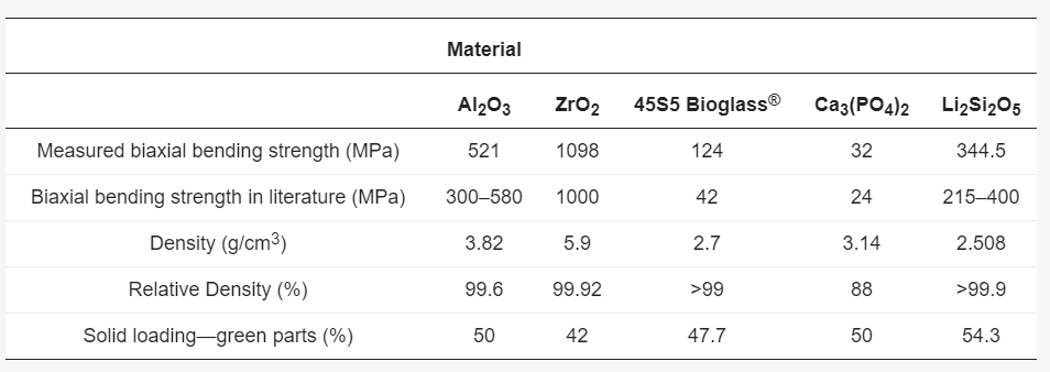
Material properties of different ceramics achieved with lithography-based ceramic manufacturing (LCM)
Several different scanning methods were used to evaluate part accuracy, including:
• Optical scanners
• Tactical scanners
• Micro-CT

The main components of the slurry.
The slurry base is made up of the following:
- Monomer compositions and solvents
- Photo absorber
- Photo initiator
- Ceramic filler with solid load of more than 50 vol %
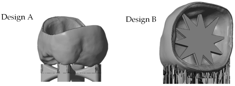
stl file of the molar crown with different support designs (from left: ‘cross’-support Design A, ‘star’-support Design B).
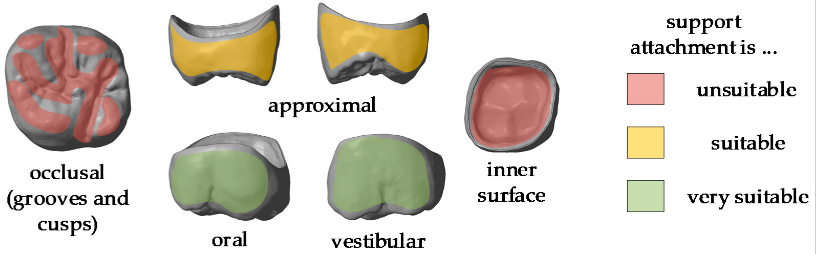
.stl-file of a molar crown with marking the different areas of support attachment.
Support structures are a critical part of the structures, enabling printing with overhanging areas, as well as preventing warping. The researchers printed a sample molar crown for the study, creating a cross-support for the crown’s occlusal side. Another star structure offered added support on the oral side, while the cusps, distal and mesial surfaces, and the gap between the crown and the core’s inner surface were to be avoided as areas for support placement.
To digitize samples, the research team used a variety of scanners, and 3Shape software. The star shape was adjusted in terms of the light absorber and printing parameters, with the potential for adjusting resolution further regarding wet layer thickness in the material vat.
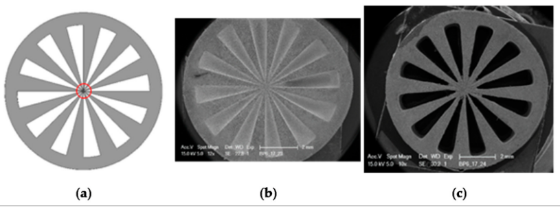
Siemens star, diameter 9 mm: (a) stl file used for the study; (b,c) SEM images of green bodies.
The scientists fabricated one crown and the star support structure sample for comparison, noting that different techniques produced varying results.
“There were challenges in reproducing the real surface due to reflections and translucencies of the ceramic, which resulted in the appearance of a so called ‘orange skin’ effect using the 3Shape D810 infrared scanner,” explained the researchers.

Images of crown A obtained with different scan methods.
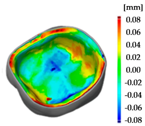
LCM-processed crowns were compared with the initial .stl file, as it was rescaled with the x, y, and z factors. The researchers noted that the best results were attained via micro-CT scans, so they used them for following analysis also. Analysis showed that the crown with the star support warped during sintering, not providing enough support. Further improvements resulted in much higher precision, no warping, and with a color scale that could be reduced to minus 80 µm to plus 80 µm.
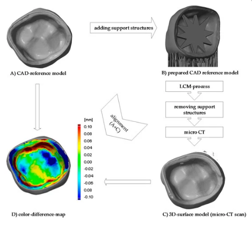
Procedure of evaluating the precision of the 3D-printed crown (a) CAD-reference model, (b) prepared CAD reference model, (c) micro-CT scan of 3D-printed and sintered molar crown, ‘star’ support was removed after sintering (d) comparison of CAD-reference model and 3D-surface-model using GOM Inspect.
“The improvement of the dimensional accuracy can be seen by using statistical information to compare all tests support types,” stated the researchers. “This was achieved by laying all analyzed crowns on top of the same original file and a surface comparison was done. After, the deviation scale was adjusted to highlight only relevant measurements and that all the colors in the false color diagram were represented. Finally, the maximal and minimal deviation of the color false scale is used for measuring scale in figure 9 [below].
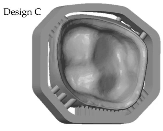
Comparison of 3D surface model (sintered molar crown with ‘milling’ support, design C) and stl file using GOM Inspect.
“To show the reproducibility of the process two LCM processed crowns were compared as illustrated in the pseudo color image in Figure 10 [below]. The maximum deviation amounts to 30 µm, which shows a high reproducibility of the whole process chain. These tolerances are also sufficiently low for allowing clinical use of such crowns. The maximum tolerance accepted for clinical use has been discussed during the years and is defined between 50 to 120 µm.”
What do you think of this news? Let us know your thoughts! Join the discussion of this and other 3D printing topics at 3DPrintBoard.com.
[Source / Images: ‘Stereolithographic Additive Manufacturing of High Precision Glass Ceramic Parts’]
The post Austria: SLA 3D Printing of High Precision Glass Ceramic Parts appeared first on 3DPrint.com | The Voice of 3D Printing / Additive Manufacturing.