How is 3D Printing Innovating Medical Research in 2020?

3D printing technologies are pushing the boundaries of what was once considered only possible in science fiction novels. The advances being made by engineers from around the world are contributing to a plethora of innovations that are having a major impact on conventional medical practice. Medical researchers have been able to develop solutions in the form of patient-specific prostheses and pre-operative models, tailored, corrective insoles and orthotics, new medical devices and instruments, and 3D bioprinting and tissue engineering. In this article, we will provide a brief review of some of the latest 3D printing technologies and methods that are inspiring medical research.

Pre-operative
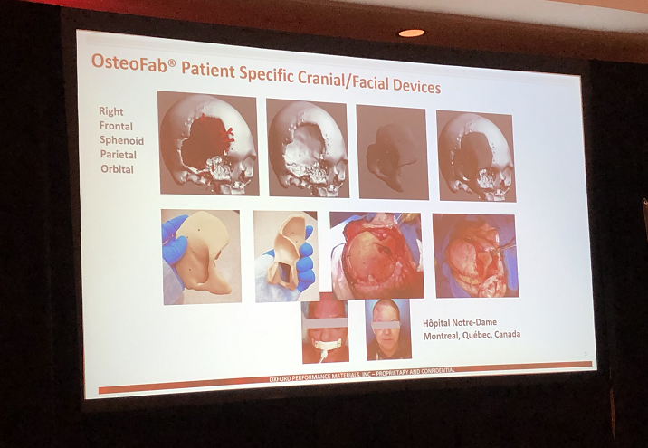
planning, prostheses, and implants

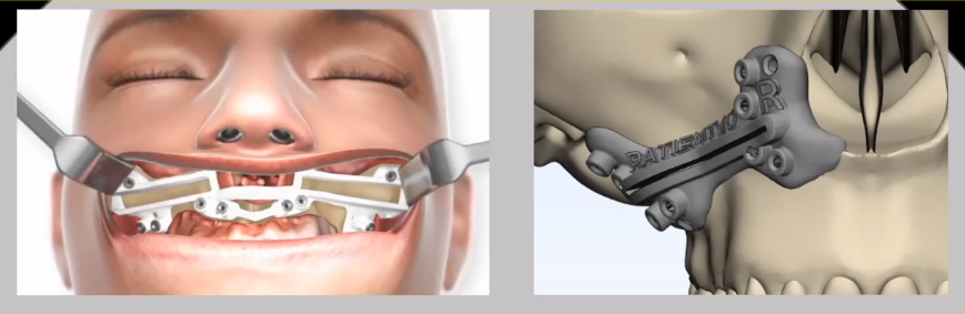
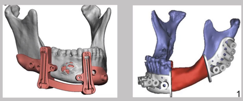
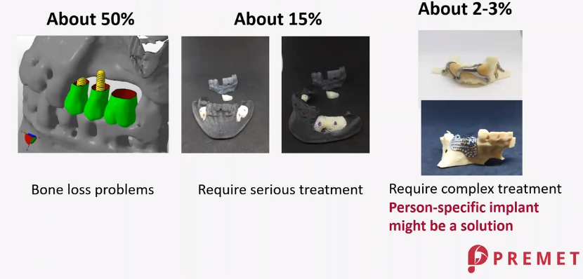
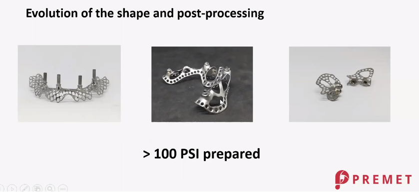
The rapid prototyping capability of 3D printing is offering the medical community a fast and cost-effective way of delivering life-altering medical interventions and solutions to patients. For individuals that require a prosthesis or implants such as a bionic hand or leg bone, 3D printing is providing a functional and affordable way to generate patient-tailored parts. The technology offers complete design freedom and rapid turn-around times. 

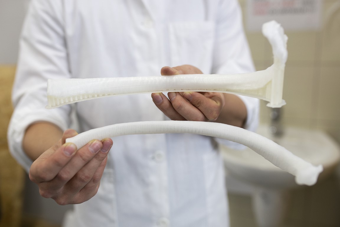
Using high-resolution images, 3D printing is able to generate accurate models of human anatomy. Image data can be exported as a common medical file format, DICOM (digital imaging and communication in medicine), which can then be converted into a stereolithography format (STL) file. From this file, a 3D virtual model can be created. For orthopedic surgery, implants can be made from these models to replace fractured bones. Further, virtual or physical models can be used by surgeons in pre-operative planning and for teaching patients, alleviating their stress and anxiety by explaining what a procedure would entail.

Biological tissue
generation

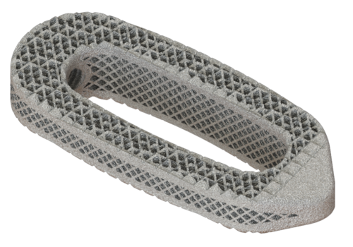
In early June of this year, scientists from the University of Colorado (UC) Denver and the University of Science and Technology in China were the first to use new material to 3D print structures that could mimic cartilage. Cartilage replacement has been a notoriously difficult hurdle to cross for scientists and healthcare professionals until now. UC Denver’s mechanical engineer, professor Chris Yakacki, led the team of researchers in using a 3D printing process called digital light processing (DLP) to create a liquid crystal resin-like substance. When exposed to UV-light the researchers observed that the substance cured and formed new bonds in several thin photopolymer layers. The final cured form constituted a strong, yet soft, and compliant elastomer. when printed as a latticed, honeycomb structure, that’s when Yakacki and his team saw that it began to resemble cartilage. Their research findings were published in the journal Advanced Materials.

In addition to utilizing this breakthrough material for cartilage replacement, Yakacki also believes there is potential for liquid crystal elastomer (LCE) to be used in the creation of a spinal cage prototype. The design of complex structures like LCE’s and the use of bioinks to help produce artificial live tissue will provide the medical research community with unique scaffolds with which to generate different components of the human body.

Bioinks

One particular area gaining interest by researchers and clinicians is
the design of patient-specific bone grafts. Associate professor at the Department of Biomedical
Engineering at Texas A&M University, Dr. Akhilesh Gaharwar, believes that developing
replacement bone tissues may be an exciting prospect in the generation of
treatments to help people with dental infections, arthritis, craniofacial
defects, and bone fractures. This is where bioinks enter the scene. In a recent
publication, Dr.
Gaharwar outlines the creation of a structurally stable, biodegradable, and
highly printable bioink. Garharwar’s nanoengineered ionic covalent entanglement
(NICE) bioinks involve two reinforcement techniques known as nonreinforcement
and ionic-covalent network. The use of these two techniques results in much
more stable tissue structures.

Following bioprinting, the NICE
networks form crosslinks with encapsulated stem cells to create stronger
scaffolds. Within the period of three months, the cells start to produce
cartilage-like extracellular matrix which calcifies to form mineralized bone.
The team used next-generation RNA-sequencing technology to establish the role
of nanosilicates (a component of the bioink) in inducing the formation of bone
tissue. Dr. Gaharwar and his team successfully demonstrated the ability of NICE
bioink to create patient-specific implantable 3D frameworks for the repair of craniofacial defects.

Orthoses

Medical research centered around the custom design of orthotics still
bears the stigma of a high price tag and inaccessibility which can be an
irritable deterrent for healthcare providers trying to do the best for their
patients and a disheartening prospect for patients respectively. The revelatory
story of Matej
and his son Nik, shows how powerful a tool 3D printing can be in advancing
medical and engineering research, efficient medical practice, and optimizing
patient care.

One of the latest uses for 3D printing in the world of orthotics was the design of a cervical collar using a novel workflow for a patient with a neurological disability with no alternative means of therapy. Dr. Luke Hale and Associate Professor Dr. Deepak Kalaskar from UCL’s Institute of Musculoskeletal Sciences (IOMS) led the research which was published in Scientific Reports. The research team scanned the head and neck of the patient with a handheld scanner to generate a 3D scan mesh. This framework was then imported into Houdini software (SideFX software, version 16.5). The geometry projected onto the 3D scan conforms with it completely to create a comfortable orthosis.

Using the scan, the design of the orthosis was optimized to incorporate modifications including a porous pattern to improve ventilation. This also reduces the cost and weight of the final orthosis. Four prototypes of the cervical collar were made to accommodate patient feedback and achieve the most comfortable design. The research validated the use of using 3D printing and scanning alongside a tailored workflow for clinically beneficial outcomes while allowing for iteration, modification, and improvement of the design.

These are only some of the latest medical research advancements coming
to fruition with the revolutionary technology of 3D printing. 4D printing and
the use of novel bioinks for organ tissue generation are some more fascinating
research prospects to look forward to in 2020. 

Are you a veteran of medical 3D printing looking for a bespoke manufacturing service, or, are you new to the scene and would like expert guidance? Find out how Shapeways can help with your medical 3D printing needs.

learn more

The post How is 3D Printing Innovating Medical Research in 2020? appeared first on Shapeways Blog.

3DHEALS 2020 Virtual Medical Summit: Comprehensive Look at Craniomaxillofacial 3D Printing

There were a multitude of sessions and sub-sessions to follow at 3DHEALS 2020 (running from June 5-6, 2020), with over 70 speakers and four workshops, covering many topics on complex design and patient-specific treatment. Here at 3DPrint.com, we have covered many stories on craniomaxillofacial surgeries, including exploring patient-specific applications, research into simulation, and the use of advanced 3D printed visualization and surgical planning guides.

In attending the virtual summit, however, we were able to hear from Dr. Devid Zille (Director of Applied Innovations at OsteoMed), Dr. Rui Coelho (CEO at BoneEasy), Zsolt Pasztor, PhD (Managing Director at PREMET), and Dr. Albert Woo (Associate Professor of Surgery at Brown University)—professionals and innovators who put the wheels into motion for fabricating complex, 3D medical designs for intricate surgeries. As an interesting side note, Dr. Zille actually used to work in the automotive realm as a car designer and, yes, he actually designed wheels (see below).

Now, as an oral and maxillofacial surgeon, Zille states that he sees parallels between virtual surgical planning (VSP) and the spokes of the wheel that he designed below:

Zille also discusses the numerous evolutionary phases in 3D printing with training models, patient-specific models, cutting guides, and patient-specific implants.

Older style on left; newer 3D-printed guide on the right, patented by OsteoMed, that extends into the nasal cavity for better fit.

Mandibular reconstruction cutting guide

Initially, the surgeon also stated that surgical planning with conventional methods can be extremely time-consuming and is not always predictable. Models allow for better diagnostics and treatment, and excellent training for everyone from medical students to surgeons.

Surgeries now can be simulated ahead of time, offering much better visual presentation and a foundation for nearly “anything to be developed over the mesh.” Zille points out that that includes cutting tools, but also patient-specific implants.

Dr. Rui Coelho went on to explain what is involved in biological design, as it requires engineering and architecture, as well as a surgeon.

“We should design a process where the experience of all of the designs could benefit new designs,” explains Coelho in his presentation. “Our design should answer to every anatomical detail. Not only to the bone but also the surrounding structures.”

Engineers must have time to examine the face of the patient, decide on materials for proper attachment (such as metal, PEEK, ceramics), create a prototype, and ultimately, improve the quality of life for the recipient.

Zsolt Pasztor, PhD, introduced himself from Hungary, explaining that Budapest is the capital of “dental tourism.” In using dental implants, placement is critical; however, 50 percent of patients do not have sufficient room for these medical devices, meaning that more ‘complex solutions’ like customized, patient-specific implants often offer success.

Bone loss is a challenge for treatment

Development of implants includes:

  • Design
  • Measurements
  • Simulation
  • Consultation
  • Continuous improvement

Sample implants

Fit for the patient, mechanical properties, and osseointegration are most important.

Measurements

As a surgeon, Dr. Albert Woo explains that procedures involving the eye sockets are usually the most delicate. Those are the cases that concern him the most, due to the extreme precision required in measuring. Insertion of implants in such surgeries requires state-of-the-art materials and technique.

Woo has also inserted implants in other severe cases; for instance, one patient was missing half of his nose due to cancer. With digital imaging, Woo was able to use a scan from the other side of his face to create an appropriate prosthetic. His team has also used 3D printing to sidestep the need for making molds. With 3D imaging, they are able to create the orthotic and go from there.

Dr. Woo concluded his presentation by explaining that he and his team are also using 3D technology for computational flow dynamics, creating new designs for treating spinal tumors.

Giant orbital fracture

Mirroring of normal activity

Although originally set for ‘the heart of San Francisco’ as a venue, this year’s 3DHEALS Global Healthcare 3D printing conference became a virtual—and inspiring—event. Focusing on the continued impacts to the field of medicine, rather than cancel the annual event due to the COVID-19 restrictions, founder and CEO, Dr. Jenny Chen, committed to an online format, and along with seeing every speaker conform to the changes, she was even able to add 25 percent more in programming.

What do you think of this news? Let us know your thoughts! Join the discussion of this and other 3D printing topics at 3DPrintBoard.com.

[Source / Images: 3DHeals 2020 – ‘3D Printing in CMF’]

The post 3DHEALS 2020 Virtual Medical Summit: Comprehensive Look at Craniomaxillofacial 3D Printing appeared first on 3DPrint.com | The Voice of 3D Printing / Additive Manufacturing.

AMS 2020: Panels on 3D Printing in Implants and Orthopedics, Regulation in Additive Medical Devices

There was much to enjoy and learn at the recent Additive Manufacturing Strategies event, held in Boston and co-hosted by 3DPrint.com and SmarTech Analysis, especially with the addition of a new metals track. There was the ever popular Startup Competition, time to network with colleagues, interesting keynotes, an exhibition hall, industry forecasts by SmarTech, and many different panels and presentations.

On the first day, I sat in on a panel on the event’s medical track about the use of 3D printing in implants and orthopedics. Martin Neff, the Head of Plastic Freeforming for German machine manufacturing company Arburg, spoke first, and provided attendees with a detailed explanation of its patented ARBURG Plastic Freeforming (APF), the process principle behind it, and what the Freeformer can offer.

“Our process can use standard resin, is already on the market and cleared by the FDA for medical devices, and it’s similar to injection molding,” he explained.

Moving on, Neff said the biggest thing to keep in mind for this application of 3D printed implants and orthopedics is how to achieve repeatability, traceability, and position. Additionally, he mentioned that the selection and freedom of materials in this sector is also a “very important area.”

Carissa Kennison, the Director of Marketing for New Jersey-based Additive Orthopaedics, explained that the company, which was founded in 2016, designs, markets, and manufactures medical implants.

“We’re inspired by the outcomes these 3D printed implants are having in our patients’ lives,” she said. “The patient testimonials are truly inspiring – often, patients go to multiple surgeons and are told to get a fusion, which limits joint motion, or an amputation. So we’re giving them an alternative solution, and challenging the solutions of standard of care. It’s pretty exciting to be a part of that.”

Jean-Jacques Fouchet on Skype

While he was unable to be at AMS 2020 in person, Jean-Jacques Fouchet, the VP Business Development and co-founder of 3D printing company Z3DLAB – Parc Technologique, was able to join the panel via Skype, and explained to attendees that “Z3DLAB is an expert in materials science,” and that it has developed a 3D printed implant for the dental field. The company’s mission is to deliver a new generation of advanced, titanium-based material for the AM market.

“We do two titanium materials, one based on Ti-64 that’s enhanced and one based in Ti-CP,” he explained. “Our 3D printed implant has an interior porous structure.”

Fouchet went on to say that Z3DLAB had completed a study with EnvA, LNE, and BAM, and that after just two months of implementation, “we got high-resolution scanner results that showed 84% bone inside the implant. Not bone tissue, but bone.”

The last panelist was Andrus Maandi, Sr. Product Development Engineer for Oxford Performance Materials (OPM). He explained that OPM was originally founded as a materials science company, working exclusively with PEKK (polyetherketoneketone), and began adopting 3D printing all the way back in 2008.

Discussing some of the company’s orthopedic applications, Maandi brought up OPM’s OsteoFab 3D printing process, which involves laser sintering with its high-performance OXPEKK material.

“We’ll get a CT scan, and in-house can deliver implants within 24-48 hours to healthcare facilities,” he stated.

OPM started with CMF and spinal implants, and its latest 3D printed device is a suture anchor, which will have its first case performed this month.

“One of the main benefits is the impact we can have on patient care and improving their lives,” Maandi explained. “We see the additive manufacturing industry moving, at least in the orthopedic market, and slowly going down the body…moving down to long bone defects and ankle reconstruction.”

The floor was then opened for questions, and someone asked if OPM had a roundabout price for its 3D printed implants; as we all know, custom medical devices can be pricey. But Maandi responded that it is “overall cheaper than something you could machine.”

John Hornick, the Chairman for the Medical and Dental track at AMS 2020, asked a question next, telling the panelists that two of his friends had recently received knee replacements. One friend had a 3D printed implant, while the other had a conventionally manufactured one, “because his doctor didn’t know anything” about AM technology. Hornick wanted to know how the panelists got the word out about what their companies could offer.

Kennison said that it really depends on the application – surgeons are more likely to engage in word of mouth, and do their own marketing and PR, for some of the more complex cases that use 3D printing.

“It can be challenging to market some of these cases,” she said. “You can’t promote custom devices, so there are some restrictions here.”

Maandi acknowledged that it can be tough, because many of the people they deal with in the healthcare field just aren’t aware of all of the available 3D printed options.

Later that same day, I sat in on a panel called “Regulation of Additive Manufacturing of Medical Devices and Its Impact on Products Liability,’” which I had not originally planned on attending; however, after sitting at the same lunch table as panelist Sean Burke, a partner with the Duane Morris law firm in Washington D.C., I was intrigued.

Panelist Bob Zollo, the President of Avante Technology, was unable to make it, so Burke had the floor all to himself. Acknowledging with good humor that he was the only thing standing in the way of happy hour, he moved through his topic efficiently.

According to his bio, “Mr. Burke’s practice focuses on representation of manufacturers of medical devices in products liability cases across the country, including in consolidated multi-plaintiff matters in both federal court and state courts in California, Illinois, and Tennessee.” In terms of defense experience, he has worked with many things, including surgical instruments and fusion plates, and recently became interested in the use of AM, advising and consulting his clients on best practices in the early product development stages in order to help them lower their risk of liability exposure.

But, as Burke told the room, “Basically, at the end of the day, there are always risks.”

He explained that while many people look at it as more of a barrier, FDA regulation and compliance is “really the best shield that medical device companies have.”

“You’re on a bit of an island if you don’t have the same regulations.”

Burke explained that the FDA is trying to “play catch-up” in determining how exactly to regulate this kind of technology. The agency has issued guidance on design, testing, and manufacturing controls for AM, but this doesn’t mean that it’s offering a solution.

From a products liability standpoint, if a company has standards to fall back on when telling a jury about the testing that’s been completed on a 3D printed medical device, the chances are more likely that the jury will be able to understand.

“But when there aren’t standards or testing, but the FDA wants to look at it, that’s a recipe for exposure,” Burke said.

As an example, Stryker’s 3D printed tritanium spinal cage was recalled last year for updates; after conducting a Google search, Burke found four different attorneys who were looking to take these cases to court…bad news for the AM industry.

Burke moved on to current trends about litigation involving 3D printed medical devices. While there haven’t been too many class action suits because the cause for each patient’s failure is usually different, the number of cases is rising.

He listed some of the factors that drive litigation, including the media, company field actions, FDA safety communications and labeling changes, pending litigation, and scientific and medical literature. Burke also provided an explanation on the different types of product liability claims – strict liability, negligence, and fraud/misrepresentation.

In terms of manufacturing defect claims, evidence must be presented that shows there has been a “deviation from the original design.” This can be difficult to validate, but there are many variables involved with AM, such as powder use. To protect against Failure to Warn claims, medical device companies that use 3D printing need to broadcast if there are any developments, and provide up to date information as well.

Burke gave attendees some pretty solid advice at the end:

“I know you all consider this, but think about why you’re 3D printing, and make a concise statement about why you’re doing it, and not just because you’re trying to “keep up with the Joneses” in terms of cool new technology.”

Then it was time for some questions. One attendee said that patient-specific products are not always treated as customized, and wanted to know why this makes a difference in terms of regulation. Burke explained that if a 3D printed patient-specific device is deemed a custom product, then it does not have to go through the same regulatory channels.

Joris Peels, 3DPrint.com’s Editor-in-Chief, was chairing this panel, and asked about the use of 3D printing in courtroom settings, and if regulatory bodies consider it. Burke said that his firm’s experts are definitely on board with this, especially in terms of patient anatomy models. He mentioned a big case centered around hip dysplasia, and how a 3D printed exhibit – I’m guessing it was an acetabular cup – was used to show how things were not fitting correctly in the patient’s body.

Another attendee asked Burke for best practices that startups not yet looking to liabilities could use.

“I think it depends on what they want to do,” he answered. “I work with startups and large companies, but there are some things to do up front to advise them.”

He suggested setting up a meeting with the FDA to hear their thoughts and concerns, and document the meeting.

“It’s an exhibit – the FDA knew we were doing this and that test, and still cleared it,” Burke explained.

Stay tuned to 3DPrint.com as we continue to bring you the news from our third annual AMS Summit.

Discuss this and other 3D printing topics at 3DPrintBoard.com or share your thoughts below.

[Photos: Sarah Saunders]

The post AMS 2020: Panels on 3D Printing in Implants and Orthopedics, Regulation in Additive Medical Devices appeared first on 3DPrint.com | The Voice of 3D Printing / Additive Manufacturing.

Scott Dunham: SmarTech Industry Forecasts for Metal and Medical/Dental 3D Printing

The 2020 Additive Manufacturing Strategies (AMS) event ended earlier this week in Boston. The summit was focused on the business of 3D printing in medical, dental, and metals, so it makes sense that Scott Dunham, the Vice President of Research at SmarTech Analysis, was on hand to give everyone a sense of where we are in these industries, just like at last year’s AMS. SmarTech provides the additive manufacturing industry with industry analysis reports and consulting services, and Dunham began with the company’s metal additive quarterly advisory services. These reports are compiled using data from 10-12 consecutive quarters.

Dunham noted that the messaging and adoption rates have changed for metal AM, and that while we’re all still “working towards the same goals,” we are “drilling down to specific solutions and challenges.”

“Metal additive manufacturing is in a strange place right now,” he said. “From 2016 to 2018, there was lots of hype, lots of investments and growth and attention paid, and the growth was aggressive and accelerated. But now, the past couple of years, we’re in this period where people are saying, ‘What’s happening? We though this technology was supposed to revolutionize things.’ Growth rates don’t always line up with perceptions.”

He got into some of the specific factors that are going into the challenges the metal AM market is facing. There’s a large disparity between metal AM hardware and metal powder sales, which Dunham said tells us that metal 3D printers are viewed much differently than the machine tool systems to which people compare them.

“Right now, the machines are not viewed or utilized in the same way that other popular manufacturing tools are, so people are still looking at this as a longer-term opportunity that still needs development work and may not necessarily always be the right tool for high-volume serial production,” he explained. ” Users now understand they can’t just drop it on the shop floor like a CNC machine. This in some ways is a barrier to growth. There are still plenty of investments being made, though, but maybe we don’t expect those days to last forever now. We may be ending the phase of early adopters and innovators who want to make these investments.”

In the years 2014-2016, the sale of metal machines was averaging just below 30%, then climbed up closer to even, but are now dropping again a bit. According to SmarTech, non-metal 3D printers are still generating most of the hardware sales, but Dunham said we should see more of a 50/50 split into the mid 2020s.

SmarTech has a theory that this leapfrog effect is due to the current two-tiered market scenario. The advanced market focuses on serial applications and high-volume production, while the legacy market consists of applications that have around for a long time, maybe resembling a factory floor, such as injection molding and tool inserts, jigs and fixtures, prototyping or limited series, medical and dental models, and one-off high volume components. Dunham said these markets are both important, but that they each have a “different set of considerations.”

He pointed out that this advanced market will soon grow to over $4 billion worth of AM hardware sold.

“We consider this side of things a little bit further ahead of polymer machine sales,” Dunham explained. “That’s why there’s so much focus on metals.”

So, where is all this growth in the metal AM market coming from? Dunham said that hardware sales is a “good indicator of the pulse of the industry,” and that SmarTech is seeing a lot of growth on what Dunham called “the fringes,” like some of the new companies coming up over the last few years, as well as the legacy manufacturing companies adopting the technology for the first time. He referred to the newer companies, such as Desktop Metal, HP, Markforged, Trumpf, and VELO3D, as “challengers,” while the legacy companies were called “incumbents.”

Next, he talked about metal 3D printing service bureaus, which see a global market of a little over $2 million.

“It’s a pretty big opportunity on the metals side, but not as big as we think it should be, or as big as polymer service bureaus,” Dunham said. “But the footprint of metal additive manufacturing in the healthcare industry is very important, and will continue to be so.”

Dunham pulled up a slide about powder bed fusion technology, noting that because the dental industry was so mature in terms of AM adoption, it actually skews the production data in the top two graphs

Bound metal processes, like binder jetting, are currently used often for tooling, and SmarTech forecasts that applications for this technology in prototyping and end-use components will rise. Dunham said that powder-based DED 3D printing is currently “heavily skewed” towards end-use components, in addition to prototyping, and that the “vision of this will likely not change much in the future.

Moving on to the market value of metal parts produced with 3D printing, Dunham said that this number is “hard to assign,” but that investments by end users are likely just south of $5 billion. However, there are lots of high-value parts to consider, which contributes to that number.

“By 2025, we expect that all metal 3D printed parts will exceed 20 billion,” he stated.

In terms of project applications for metal AM, healthcare leads the pack, with crown and bridge substructures and hip implant components at the top of the list. If you remove medical applications from the equation, we’re looking at using the technology to repair high-value turbine blades and aircraft parts, valves and pumps in the oil & gas & energy sector, and more medium-sized industrial components.

“If you’re a supplier in the industry, these are what will succeed,” Dunham said. “The incentive here is to invest in different approaches to metal additive manufacturing.”

Dunham summed everything up by saying that while metal AM is still demonstrating value, entry barriers, such as financial reasons, are also high, which does deter growth somewhat, and that a multidisciplinary approach to it is necessary for growth to continue.

Then I followed Dunham out and into the next room for the SmarTech medical 3D printing forecast, which was wisely titled “Healthcare – the Backbone of Additive Manufacturing.”

“Within the healthcare segment, there are many ways that AM has been and will continue to be leveraged,” he stated. “There are some very industrialized serial, serious manufacturing applications in healthcare, so emphasis is put on the customization of these devices.”

He noted AMS 2020 has a theme of looking at business cases, which is why it’s so heavily focused on dental and orthopedic 3D printing applications.

“We don’t think these are more impactful or important, but these are areas that we’re seeing more challenges and work here,” Dunham explained.

Excluding software numbers, the healthcare portion of the AM market – combining medical and dental applications – is a little over $3 billion dollars; truly, “the backbone of the industry.” These revenue numbers have gone down a bit, because there’s a lot of attention being paid to industrial markets, but Dunham said that SmarTech forecasts a stabilization, stating that healthcare will “continue to be important to overall industry structure for at least the next several years, and into decades.”

As has been previously mentioned, in comparison to other industries, dental is “fairly mature overall in its adoption of additive manufacturing.” If you’re looking at metal AM used in healthcare, you get into the orthopedic sector, which means you’re looking at implants.

“The longer that we can gather clinical evidence for these implants the better,” Dunham said, noting that this will help ‘build confidence’ with metal 3D printing in the medical field.

Some OEMs are bringing AM in-house, so that they can better control the process to try and ensure a good outcome. A lot of factors go into making medical implants, and if something goes wrong, “clinical efficacy is damaged.”

As of yet, there isn’t a huge push by OEMs for non-metal 3D printed implants, but SmarTech believes this is coming later, for materials like ceramics, and especially for craniomaxillofacial (CMF) implants.

There are plenty of business use cases for metal orthopedic 3D printed implants, and while the hip is still in the lead, about a third of 3D printed implants made now are are spinal. But Dunham said that hip implants won’t dominate the production numbers forever, as the 2025 forecast shows more diversification coming.

Moving to the dental side of things, companies are seeing a lot of success with high speed vat photopolymerization technologies, which Dunham said was expected. But what they didn’t count on was the aligner segment looking to get into powder bed fusion.

“No one process has everything locked down, and we can all benefit from more competition to push the technology forward,” he said.

Dunham said we should expect that 3D printing will ultimately follow the “trend of machines in dentist offices.”

“We expect a pretty healthy growth in investment by dental offices and clinics, though dental labs are still where it’s at from a hardware perspective.”

Dunham pulled up a slide that showed numbers from 2018, and forecast out to 2027, that show specifically what’s going to keep driving the sale of materials and hardware for dental applications. Looking at things like direct aligners and aligner tools, models, surgical guides, and denture bases and trays, it’s clear that he’s correct when he said that there is a lot of “diversification going on out there.”

Stay tuned to 3DPrint.com as we continue to bring you the news from our third annual AMS Summit.

Discuss this and other 3D printing topics at 3DPrintBoard.com or share your thoughts below.

[Photos: Sarah Saunders]

The post Scott Dunham: SmarTech Industry Forecasts for Metal and Medical/Dental 3D Printing appeared first on 3DPrint.com | The Voice of 3D Printing / Additive Manufacturing.

Overview: Powder Bed Fusion for 3D Printing Optimized Biomedical Implants

L.E.Murr, a researcher from the University of Texas at El Paso, presents an overview of medical devices for 3D printing, outlined in the recently published ‘Metallurgy principles applied to powder bed fusion 3D printing/additive manufacturing of personalized and optimized metal and alloy biomedical implants: an overview.’

Powder bed fusion, using both SLM and EBM technologies, has continued to rise in popularity around the world, including in hospitals where commercial systems are used to create patient-specific medical devices.

EBM system (Fig. 1c) for Ti-6Al-4 V product fabrication. (a) EBM schematic showing electron gun at (1) operated at 60 kV potential, focused electron beam (2) scanned by CAD to selectively melt raked (r) powder layer gravity fed from cassettes (3) to 3D print product (4). (b) Ti-6Al-4 V powder in EBM cassette as shown in (a). (c) Optical micrograph showing Ti-6Al-4 V EBM solid, as-built product α-phase, lenticular grain microstructure.

“The main difference between EBM and SLM fabricated metal or alloy products involves the lower SLM powder bed temperatures and more rapid cooling or cooling rate,” explains Murr. “This produces internal strain in some SLM, products which often requires hot isostatic processing (HIPing) to relieve this intrinsic strain, and product warping or other distortions. SLM product surfaces are also often smoother, and for some applications this can be an important feature.”

While challenges continue in 3D printing, additive manufacturing processes, and materials selection, researchers around the world continue to innovate with implants, whether for the brain, teeth, spine, or more, heightening the quality of life for many and in some cases, saving lives altogether. Murr points out that many attempts have been made to produce porous materials that are functional and effective. Medical device manufacturers have been working steadily to create new ways for producing implants with suitable porosity.

EBM-fabricated Ti-6Al-4 V rhombic dodecahedron mesh cranial insert in patient specific, CAD-generated, polymer skull model. Form Murr

“As of 2019, there were hundreds of so-called point-of-care 3D printing centers or hubs in hospitals world-wide; some in orthopedic units and other serving as service units for a variety of surgical departments, including developing 3D printed surgical planning models,” states Murr.

Today, Ti-6Al-4 V powder is used by most 3D printing point-of-care centers and service laboratories responsible for making implants for:

  • Maxillofacial reconstructions
  • Skull plates
  • Spinal inserts
  • Total hip and knee implants

Relative elastic modulus (E/Es) versus relative density (ρ/ρs) plots for EBM-fabricated Ti-6Al-4 V mesh and foam and Co-Cr-Mo alloy mesh and foam samples. Adapted from Murr [9]. Note arrows correspond to E/Es = 0.02 and ρ/ρs = 0.18 along the fitted line whose slope corresponds to the exponent 2 in Eq. (11).

“While considerable research on the development of a wide range of Ti-alloy systems has been conducted over the past 2 decades, including alloys such as Ti-24Nb-4Zr- 7.9 Sn, having a Young’s modulus less than half that for Ti-6Al-4 V, there is little incentive, either biomedical or economical, to adopt these alloys for implant fabrication,” concluded the researchers.

“For Ti-6Al-4 V, the principal microstructures, consisting of various α-phase dimensions or αʹ-(martensite) phase dimensions, allow the hardness (Vickers) to be adjusted over a range of roughly 3.5–4.5 GPa, as well as the corresponding manipulation of strength and ductility. While this can assure a high degree of biomechanical compatibility with bone for porous, open-cellular implant designs, optimal bone ingrowth not only assures effective fastening of the implant but also renders the implant a bone scaffold, or ideal bone replacement.”

What do you think of this news? Let us know your thoughts! Join the discussion of this and other 3D printing topics at 3DPrintBoard.com.

EBM-fabricated Ti-6Al-4 V porous mesh pelvic girdle custom manufactured for patient specific polymer T-CAD model (a). (b) Shows the inverted Ti-alloy pelvic insert with arrow indicating attachment for acetabular prostheses for connecting the right leg. From Murr [42]. Courtesy of S.J. Li, Institute of Metal Research, Shenyang, China.

[Source / Images: ‘Metallurgy principles applied to powder bed fusion 3D printing/additive manufacturing of personalized and optimized metal and alloy biomedical implants: an overview’]

The post Overview: Powder Bed Fusion for 3D Printing Optimized Biomedical Implants appeared first on 3DPrint.com | The Voice of 3D Printing / Additive Manufacturing.

University of Nottingham: 3D Printed PG/PLA Composites for Repairing Fractures

In ‘Mechanical properties and in vitro degradation behavior of additively manufactured phosphate glass particles/fibers reinforced polyactide,’ authors Lizhe He, Jiahui Zhong, Chenkai Zhu, and Xiaoling Liu explore a new level of material for 3D printing with phosphate glass/polylactide (PG/PLA) composites for use in medical applications such as fabrication of customized bone fixation plates for repairing fractures.

While bone regeneration is an area of great interest in 3D printing and additive manufacturing, so is the more common element of healing breaks, as researchers continue to look for better ways to improve the process—often accompanied by a range of bone fixation plates, screws, pins, and rods. Materials are key, along with integrity in design. Implants must be biocompatible, but the process is seamless when they are biodegradable too, thus eliminating the need for surgery.

The materials were tested for suitable mechanical properties as well as in vitro degradation behavior after creating models designed with PTC Creo Parametric, which were then imported into Simplify3D and the PG/PLA composites were 3D printed on an Ultimaker 2+. With the ability to fabricate complex geometries, the researchers could also control the level of porosity for bioprinting and tissue engineering purposes.

“Comparisons were made with PLA, and PLA reinforced with different loadings of PG particles (PGPs) as well as composites with reinforcements of different geometries [PGPs or milled phosphate glass fibers (PGFs)].”

The aim was to evaluate the AM composites as fracture fixation plates. A three-point bending test was performed, along with in vitro degradation for examining the strength and hydroscopy of the composites. There was a pH value check, along with dynamic mechanical analysis, and fiber length and laser particle size analysis. Both microscopy and statistical analysis were performed also.

Initial flexural properties of the FDM fabricated PLA, PGP/PLA, and PGF/PLA composites. Error bars represent standard deviation. Significance was marked with: * (p < 0.05, n = 5), ** (p < 0.01, n = 5) in black (strength) and red (modulus).

In continuing to compare with PLA specimens, the authors noted the following:

  • Improved flexural modulus
  • Reduced flexural strength
  • Reduced strain at break
  • Intensified effects with increased PGP loading

Typical stress–strain curves of the three‐point bending test of the FDM fabricated PLA, PGP/PLA, and PGF/PLA composites.

“Embrittlement and strength reduction are associated with of stress concentration and low interfacial strength. It is likely here that the stress concentration effect was augmented by the incorporation of particulate with sharp corners. With increased filler loading, stress concentration sites also increased and led to more pronounced strength reduction and the same effect on strain at failure,” noted the authors.

Here, the average fiber length was 54 μm, and median and mode of fiber length were even lower. In comparison to authentic cortical bones, the PGF 10 composite was noted by the researchers to be ‘a close approximation,’ although flexural modulus was found to be considerably lower.

“Stiffness matching is recognized as the ‘gold standard’ for bone fixation implants, as fixation implants with such mechanical properties are strong and stiff enough for the load‐bearing activities without leading to ‘stress shielding.’ As such, it is probably necessary to consider the use of higher/longer fiber loading for this type of application,” stated the researchers.

Continuous PGF/PLA composites are more ‘suitable,’ according to the authors, in regard to load-bearing fixation—a feature connected with continuous fibers leading to stiffness. The flexural modulus of these materials, however, was reduced by ~80% after 28 days of degradation. The PGF 10 composites lost ~30% of initial flexural modulus after a degradation period of 56 days. The rapid flexural modulus could have been a result of the fiber ends being exposed in degradation media.

“Based on the consideration of both the initial mechanical properties and the facility to produce composites with desired geometries straightforwardly, the additive manufacturing of PG/PLA composites exhibits good potential in the making of patient‐specific fixation implants for bone that has low demand for load‐bearing, for example, zygoma, ankle, and maxilla,” concluded the researchers.

“These bones have been previously reported to be successfully restored using PLA‐based biodegradable fixation devices. Compared to PLA alone, it was demonstrated that the incorporation of PGF enhanced the flexural modulus of implants. It is also anticipated that the degradation of PGF releases magnesium, calcium, and phosphate to upregulate bone regrowth. Moreover, the FDM process allows fixation implants with customized geometries to be built directly and may remove the need for contouring of implants for anatomic fit during the operation.”

A serious interest in 3D printing today translates into a serious interest in materials—and most likely composites too, as they are able to add significant strength and improved properties to prototypes and parts, including that of polymers, bioprinting applications, and metals like titanium. What do you think of this news? Let us know your thoughts! Join the discussion of this and other 3D printing topics at 3DPrintBoard.com.

SEM images of polished/pristine fractured surfaces of virgin PLA (a,b); PGP 10 (c,d); PGP 20 (e,f); and PGF 10 (g,h) composites

SEM images of pristine fractured surfaces of PLA (a,b); PGP 10 (c,d); PGP 20 (e,f); and PGF 10 (g,h) degraded at 37 °C in PBS for 28/56 days.

SEM images showing the fusion of PGPs (a) and PGFs (b) into excrescences, captured on Day 56.

[Source / Images: ‘Mechanical properties and in vitro degradation behavior of additively manufactured phosphate glass particles/fibers reinforced polyactide‘]

The post University of Nottingham: 3D Printed PG/PLA Composites for Repairing Fractures appeared first on 3DPrint.com | The Voice of 3D Printing / Additive Manufacturing.

Researchers Evaluate 3D Printed Mandibular Grafts for Effectiveness as Implants

Researchers outline findings from their recent study in ‘Analysis of biomechanical behavior of 3D printed mandibular graft with porous scaffold structure designed by topological optimization.’ Their main point is that in past years there have been glaring improvements needed for implants such as those created for the lower area of the jaw known as the mandible, due to hazards like patient rejection and infection.

While the mandible allows for successful mastication, it also lends solidity to the facial structure along the jawline. When an issue such as a tumor or infection arises and requires portions of it to be cut out, this can interfere with chewing as well as the patient’s appearance, which often has a close emotional connection to their identity. As implants are needed to help with jaw movement and aesthetics, complications often occur. This is common with anything artificial placed into the human body. Infection can easily occur, or the body may reject the implant, and especially if it is ill-fitting or ill-designed.

“The limitations of autografts are donor site morbidity, lack of bone volume for a large defect, and possible nerve damage. While allografts are limited by anatomical variations, genetic differences, and possible disease transmission. It is important to restore a critical-sized mandibular defect to its original size and shape to achieve desirable facial aesthetics and functional outcome for subsequent prosthetic reconstruction,” state the researchers.

One of the greatest benefits in 3D design and 3D printing, especially as it relates to the medical field, is the potential for creating patient-specific medical treatment and devices. The researchers point to current successes with such procedures, using implants created via FDM 3D printing and PLA as the material. Bioprinting with live cells generally takes success to another level as use of the patient’s own cells reduces the chances of rejection even further. Here, they used cone beam computed tomography (CBCT) to create a mandible—with one part acting as a control group and other blocks as experimental groups. Samples were printed with PLA, at 0, 45, and 90 degrees.

3D design of the mandible was created using Mimics software. The porous scaffold structures were comprised of defined mesh configurations, and an intricately connected microporous network. 3D printing of multiple sample grafts was completed on a CEL Robox 3D printer, with a 210 × 150 × 100 mm build space. The samples were then submitted to testing as the researchers examined how anisotropy affected the grafts, and their mechanical properties.


Schematic of mandibular grafts with printing angle 0° (left) and 90° (right)

A three-point-bending jig was used to test the 3D printed beams:

“The results show good repeatability of samples in the same group. The yield stress, failure stress, failure strain, ultimate strength and flexural modulus were calculated for each sample to better understand the mechanical performance of the printed material,” say the researchers. “The yield stress was calculated using the method of 0.2% offset; failure stress corresponds to the stress at failure strain, ultimate strength obtained from the maximum stress value. Flexural modulus was along the linear portion of the stress-strain curve. All these five properties were evaluated for each printed graft.”

Failure mode of three beam specimens with printing angle 00, 450, 900 (from top to bottom)

Direction of 3D printing was shown to have definite effect on mechanical properties at a 0-degree angle, with strains and cracking appearing as the load increased. The researchers obtained similar results with beams printed at 45-degree angles. This was not the case at 90 degrees though.

“After reaching the peak point, the crack propagates suddenly penetrated through the whole cross-section and the beam fractured into two parts,” state the researchers. “This is different from the flexural curves for the specimens printed at 0-degree or 45-degree angles, which show slight plastic failure before their rupture occurred.”

Overall testing showed that the 3D printed grafts offer required strength, stiffness, and porosity for such applications discussed by the researchers in their paper.

 “Although the results of this study are based on PLA material, the proposed methodologies are also applicable to other promising 3D printing materials such as Polyetheretherketone (PEEK),” concluded the researchers. “3D printing technology and topological optimization are useful tools in fabrication and designing bone analogs for mandibular reconstruction.”

Find out more about materials being used today to 3D print implants and other devices. The key to these innovations is that they are patient-specific, offering a new level of care, with orthopedic implants, dental implants, and even devices made specifically for veterinary care. Discuss this article and other 3D printing topics at 3DPrintBoard.com.

Graft with topological optimized-pore scaffold structure

[Source / Images: ‘Analysis of biomechanical behavior of 3D printed mandibular graft with porous scaffold structure designed by topological optimization’]

TU Delft Researchers Discuss Microstructural Optimization for 3D Printing Trabecular Bone

Trabecular bone, also known as spongy or cancellous bone, is one of two types of bone found in the human body. It is found at the end of long bones, in the pelvic bones, ribs, skull and vertebrae. Trabecular bone is one of many microstructures with spatially varying properties found in nature. In a paper entitled “Compatibility in microstructural optimization for additive manufacturing,” a group of researchers points out that these microstructures can now be created by additive manufacturing. One challenge in the computational design of such materials is ensuring compatibility between adjacent microstructures. The researchers’ work aims to find the optimal connectivity between topology optimized microstructures.

“Given the fact that the optimality of connectivity can be evaluated by the resulting physical properties of the assemblies, we propose to consider the assembly of adjacent cells together with the optimization of individual cells,” the researchers explain. “In particular, our method simultaneously optimizes the physical properties of the individual cells as well as those of neighbouring pairs, to ensure material connectivity and smoothly varying physical properties. This idea is substantiated on the design of graded microstructures with maximized bulk moduli under varying volume fractions. The graded microstructures are employed in designing an implant, which is fabricated by additive manufacturing.”


When designing orthopedic implants, the researchers point out, it “may be desirable to have a continuous transition from denser microstructures in the central region to highly porous microstructures at the bone-implant interface.” This functional gradation promotes bony ingrowth at the bone-implant interface, they continue, while maintaining structural integrity and increasing mechanical properties in areas where bony ingrowth is not relevant.

The researchers presented a method of ensuring mechanical compatibility among topology optimized microstructures.

“Our results show that the bulk moduli of individual cells reach the theoretical bounds predicted by the Hashin–Shtrikman model, meaning that the optimization of compatibility does not compromise the performance of individual cells,” they state. “Furthermore, the bulk moduli of neighbouring pairs also agree well with the Hashin–Shtrikman bounds.”

The method was extended to allow maximum length scale and isotropy in microstructures. The researchers demonstrated the effectiveness of their proposed method in a number of designs, including functionally graded materials and multiscale structures. They also showed that the optimized microstructures can be fabricated by additive manufacturing technology. This has implications for a number of applications, including orthopedic implants, which 3D printing can optimize for better growth of new bone.

“As future work, we are particularly interested in the following aspects,” the researchers conclude. “Firstly, this method is directly applicable to 3D design problems. To alleviate the computational burden in 3D, the GPU-based topology optimization framework can be used. Secondly, while we have applied the compound formulation for maximizing bulk modulus, its applicability to other physical problems such as conductivity is left to be demonstrated.”

Authors of the paper include Eric Garner, Helena M.A. Kulken, Charlie C.L. Wang, Amir A. Zadpoor and Jun Wu.

Discuss this and other 3D printing topics at 3DPrintBoard.com or share your thoughts below. 


Aries-L Patient Specific 3D Printed Spinal Device Implanted for the First Time

On Tuesday, January 8th, Dr. Samuel Joseph, Jr. and Dr. Andrew Moulton of Joseph Spine became the first in the world to implant the 3D printed titanium Aries-L interbody fusion device from Osseus Fusion Systems. The device features a proprietary multi-axis mesh and optimized micro-surface topology that are designed to facilitate fusion, as well as a lattice structure that helps increase the implant’s porosity to 80 percent. This allows for excellent in situ radiovisibility, especially compared to other titanium implants. The Aries-L is designed to aid in faster recovery thanks to its anatomic profile, anti-migrational teeth and streamlined insertion.

“As a surgeon, it’s very exciting to participate in the device development process and see your ideas brought to life so quickly,” said Dr. Joseph, Founder of Joseph Spine. “Today we were able to successfully implement this cutting edge, patient-specific spinal implant that addresses the complex anatomy of the spine and the delicate nature of it surrounding structures.

“Through 3D printing we are able to manufacture spinal implants that are customized to the patient. Meaning the size of their disc, the size of their vertebra, how high  the vertebra may be, as well as the curvature they may have in their back we are trying to create. With this cutting edge technology we can offer our patients implants such as the Aries L Interbody Fusion Device which can be created at a certain height, a certain length, as well as a certain angle, which offers our patients better, customized care, which results in better outcomes for the patients.

“Working with Osseus and utilizing their technology for spinal implants provides us with a great opportunity for superior patient care. Osseus understands that the future of advanced spine care is in the technology of custom implants as well as creating partnerships with doctors to create customized implants that offer:

  • better solutions for our patients with spinal problems,
  • in a way that is more effective,
  • that is less traumatic to the patient’s body,
  • with a minimally invasive technique, which also becomes,
  • more cost effective as it may reduce
  • the amount of biologics necessary to allow for bones to heal

This, in turn allows better outcomes for our patients.”

No details were provided about the patient who received the implant, but it can be expected that he or she will benefit from faster recovery time as well as reduced risk of complications. The Aries-L is only one of Osseus’ novel healthcare technologies, which also include the Black Diamond Pedicle Screw System, a fixation system technology; the White Pearl Preferred Angle Anterior Cervical Plate, a cervical spine fixation device; and the Red Ruby Anchored Cervical Interbody, a revisable cervical interbody system. Osseus was founded to develop solutions for minimally invasive spinal surgery using advanced technology like additive manufacturing.

Discuss this and other 3D printing topics at 3DPrintBoard.com or share your thoughts below. 


Patient Receives a 3D Printed Rib for the First Time in Bulgaria

Ivaylo Josifov (R) with one of his surgeons

35-year-old Ivaylo Josifov was an active, healthy individual who thought he only had a case of tonsillitis when he went to the doctor recently. A chest X-ray, however, showed that he had a growth in the area of his fifth right rib. It was a congenital disease that could lead to the weakening of the chest and issues like difficulty breathing. Because of the risk that the growth could spread, the best option would be to remove the rib entirely and replace it with an implant. Josifov’s doctors decided to try something that had been done only a few times in the entire world before, and never in Bulgaria – to use 3D printing technology to create a new rib for their patient.

The doctors chose 3D printing because it could guarantee a perfect replica of the original rib shape, both in thickness and curvature. They started by scanning the original bone, then sending the scan to Bulgarian service bureau 3dbgprint. The service bureau prepared the file for 3D printing, adjusting it to be sure that it fit into the chest wall. 3dbgprint then 3D printed the implant using a 3DGence 3D printer.

“Our 3D printers ensure high 3D dimensional accuracy which was crucial in this particular medical procedure,” said Filip Turzyński, Quality Development Manager at 3DGence.  “Individually designed rib model allowed for very accurate implementation of the new element in place of the removed bone. Replacing the missing rib with a 3D printed segment with the same shape, curve, width, and thickness was possible with the use of 3DGence 3D printer.”

The new rib was 3D printed using an FDA-approved flexible, durable polyamide.

“When we had the first model ready, we started working on its sturdiness,” said Georgi Tolev of 3dbgprint. “Each consecutive model was analyzed and improved until we attained the perfect 3D print of the rib.”

Before the rib was implanted, 3 mm holes were drilled in it to facilitate broaching and proliferation of connective tissue. The implant was thoroughly sterilized, using ethylene oxide as well as gamma radiation and autoclave at 140ºC. The surgery took place at Tokuda Hospital, and the patient is back to excellent health.

Due to the success of the procedure, doctors are already planning new projects using 3D printed implants. Next, they plan to create an implant of three ribs attached to a sternum.

“This is a new era in thoracic wall reconstruction for patients with tumors that require bone-cartilage structures to be removed,” said Professor Minchev, Head of the Cardiothoracic Surgery Department at Tokuda Hospital. “The material used has proven tissue compatibility and the accuracy of reproduction allows for large chest wall resections and their single substitution with individually designed implants.”

Without 3D printing, doctors would have had a much more difficult time creating an implant that fit perfectly and matched the patient’s original rib. An ill-fitting implant could have led to pain and complications, possibly requiring additional surgery in the future. With 3D printing, however, it was possible to perfectly recreate the original rib, allowing the patient to recover quickly and go about his normal life.

Discuss this and other 3D printing topics at 3DPrintBoard.com or share your thoughts below.