AMS 2020: 3D Printing Metals II Keynote by Craig Sungail, Global Advanced Metals

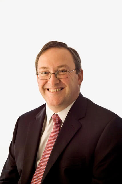
The final keynote presentation at our recent Additive Manufacturing Strategies, held in Boston and co-hosted by SmarTech Analysis, was given by Craig Sungail, the Vice President of Global Research and Development for Global Advanced Metals, which just so happened to be one of the event sponsors. Sungail was part of our new 3D printing metals track, and presented a very interesting talk about tantalum, “the other gray metal.”

“We’ve used other metals for years, like cobalt chrome and stainless steel, to make implants,” Sungail said. “In 80% of the cases, for most people, it’s successful. But 20% of the time, patients aren’t happy with the results.”

He went on to say that there is a 10% revision rate each year for surgical implants 3D printed out of these other materials, for reasons such as infection, fracture, and becoming dislodged. That’s why he said that we should all “consider tantalum as an alternative.”

“This metal has a long history. We’ve been reviewing the literature for the past 25 years. The authors vary – physicians, universities, etc. But there is a broad, diverse group of people investigating this metal for medical devices.”

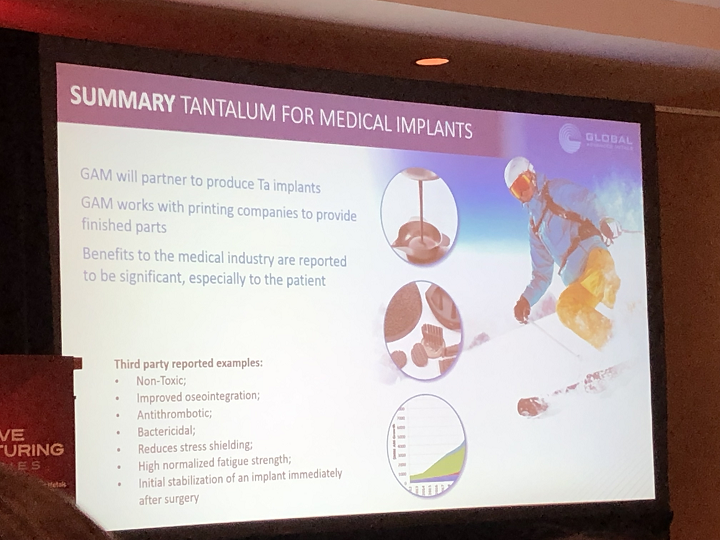
Sungail explained that these journals have determined that tantalum (Ta) is not toxic, which “can’t be said for some of the other metals out there today.” Additionally, the research shows that when using tantalum for implants, the osseointegration (bone ingrowth) of the implant into existing bone is pretty good, and perhaps even better than implants made with straight titanium or the Ti-64 alloy.

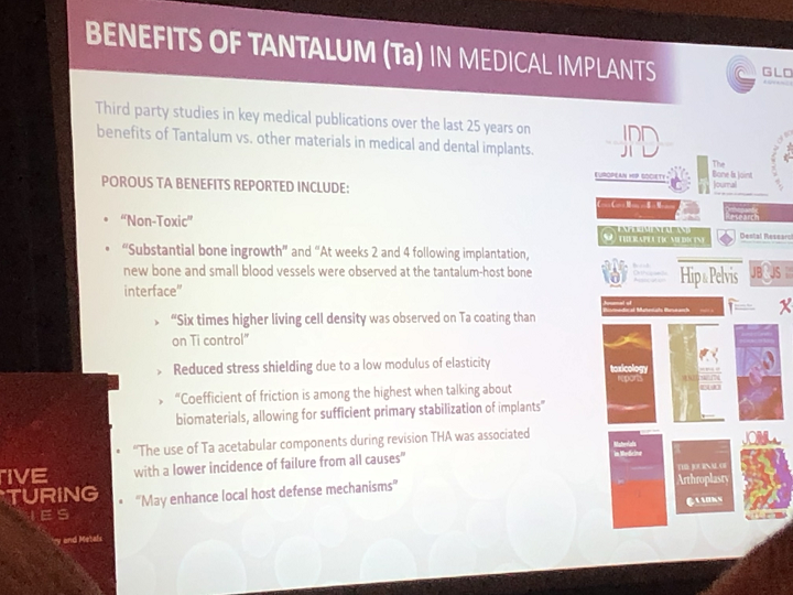
He pulled up a slide listing some of the other benefits of using tantalum to fabricate medical implants, including the fact that it could enhance local host defense mechanisms, and that it may even have some antibacterial properties.

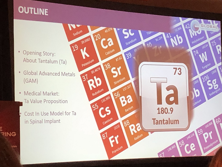
Sungail offered a brief history about tantalum, which is a transition metal/element. He explained how the material got its name, bringing up a slide about Greek mythology, which I had not been expecting and was very interesting. Tantalus, the son of Zeus and a nymph, stole ambrosia and nectar from his father, and the punishment definitely fit the crime in this case – he was forced to stand in a pool of water that was tantalizingly close to a fruit tree.

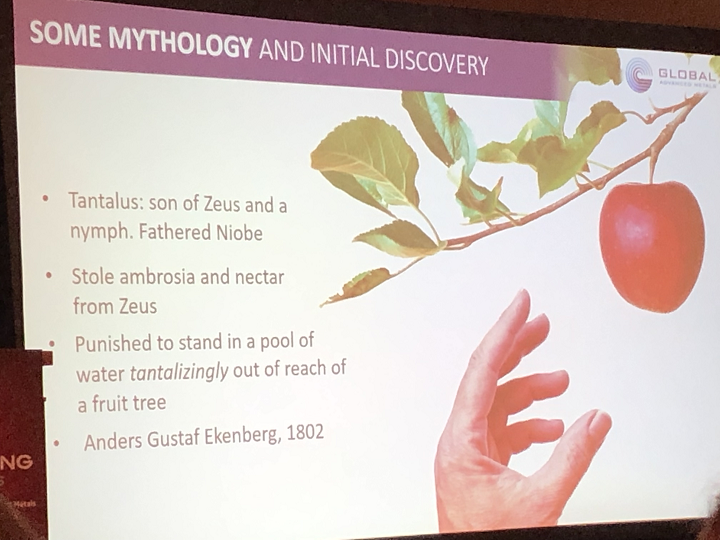
“The water would fade away, and the fruit was just out of reach,” Sungail went on.

Then, in 1802, Swedish analytical chemist Anders Gustaf Ekeberg became the first person to discover tantalum when he successfully separated it from nyobium. Ekeberg was tantalized for a long time attempting to achieve what many others had not, and once he’d succeeded, he was given the honor of naming both of the new elements.

“I’m confident that every one of you has been touched by tantalum in some way,” Sungail said. “It’s highly conductive, with a high melting point, chemical and corrosion-resistant, dense, hard, ductile, and biocompatible. We have to use biocompatible carefully, but I’m using it with the FDA definition – it’s been implanted in some way into the body, and studies concluded that the implant was biocompatible.”

Sungail said that the most common application for tantalum is in the capacitor sector, such as when it’s used for cell phones. It does have a 40-year history in medical devices, and it can be mixed with materials in order to make super elements, which can be used in turbines for jet engines and energy generation.

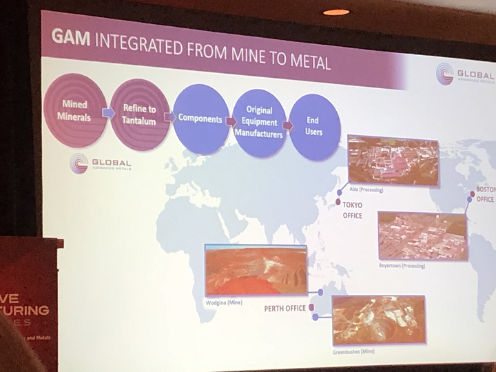
He explained that the company is “truly global,” with locations in the US and Japan. GAM also has a controlling interest in the largest reserve of tantalum in the world, which is in Australia. I’m skipping ahead a little, but I thought this was a good question – at the end of the presentation, an attendee asked Sungail about the potential environmental impact of mining tantalum. He explained that GAM does what he referred to as a “bag and tag” when they receive ore from a conflict country.

“We ensure the money isn’t going to terrorists, we do it ethically. If it wasn’t mined ethically, we wouldn’t have sales,” he stated.

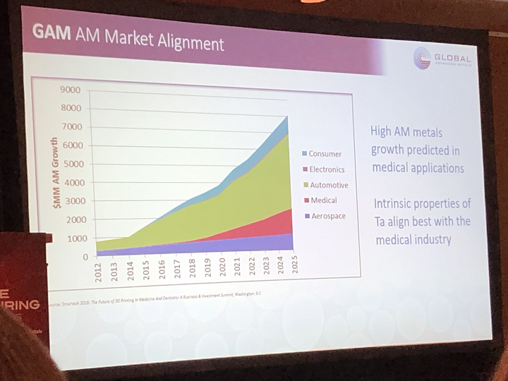
Back to where we were, Sungail said that two years ago, the company was taking a look at the various AM markets, wondering which would be the best to participate in with its tantalum. Just like the above graph shows, GAM determined that its “value proposition was best in medical, and not automotive.”

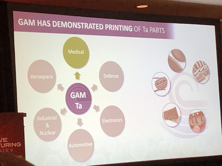
“We realized we’d have to bridge the chasm between early adopters and later innovators. We’d have to teach the industry about tantalum and that it can be printed,” he said.

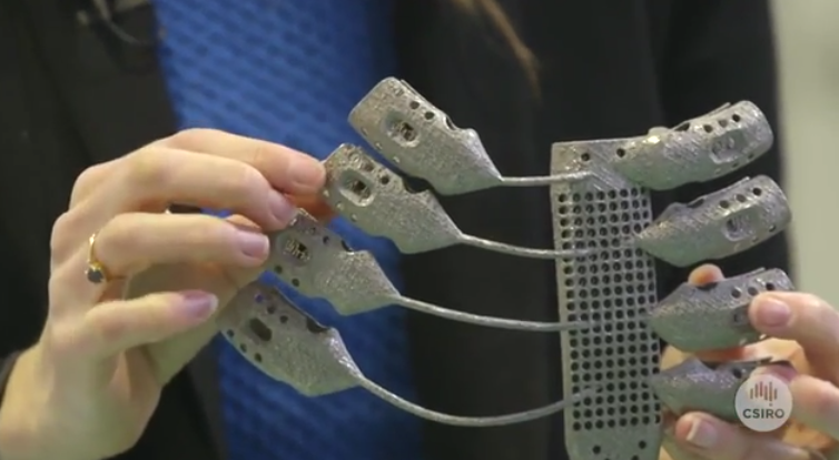
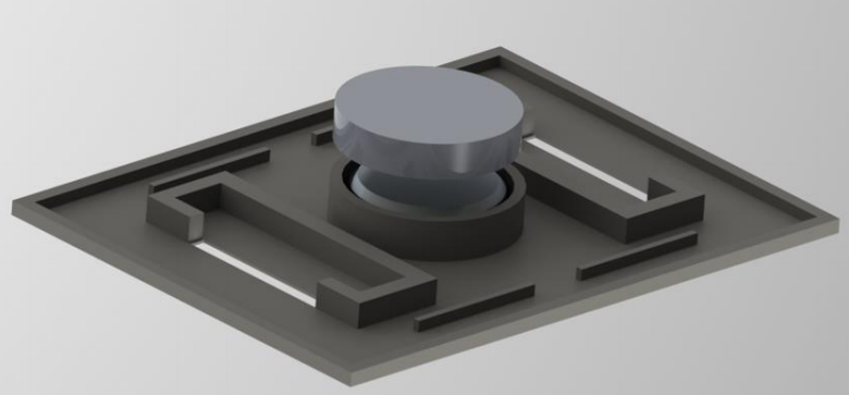
So the company got to work, using 200W and 300W lasers to 3D print medical devices like spinal implants and baseplates out of its tantalum; these fully dense parts are now in testing.

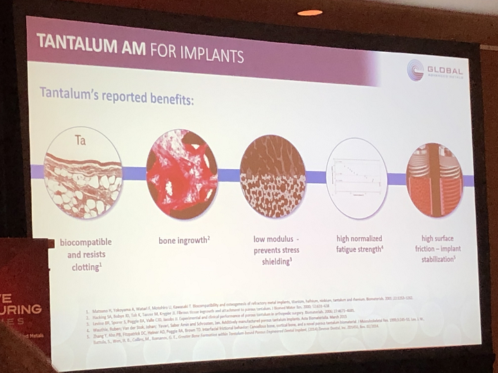
Sungail listed several reasons why tantalum is a good material to use in 3D printed medical devices – it resists blood clotting, so it can be used to fabricate stents, and its high surface friction, proven through several research studies done on animals, is good for implant stabilization.

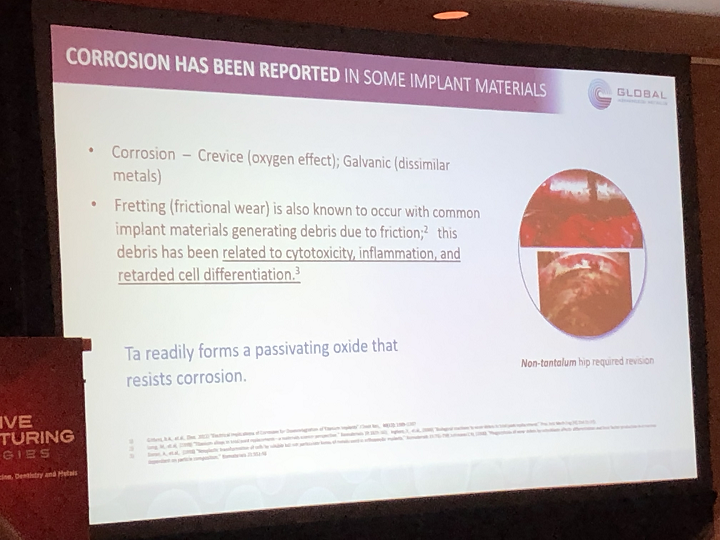
Tantalum also has no problem with corrosion, which has been reported as being an issue with other implant materials. Sungail had a slide that showed a picture of a non-tantalum 3D printed hip implant, which required revision post-surgery due to corrosion; researchers determined that it was caused due to crevice (the oxygen effect) and galvanic (dissimilar metals). He explained that debris due to friction can lead to even more issues with implants, such as inflammation in the tissue around the joint, which can cause severe pain, and that cobalt chrome and Ti-64 implants can even lead to toxic effects, like bone degradation, if absorbed into the body.

“Tantalum doesn’t corrode in a normal body,” Sungail said. “Its only attacker is hydrofloric acid, and threading should also not occur with tantalum.”

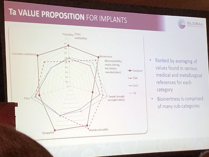
Looking at the graph above, you can see that the material’s printability comes down to several factors, of which bioinertness combines several; Sungail explained that “these are generic combinations of various features for easy reading.”

“It’s significantly more printable than some other metals we use for medical devices,” he continued. “Tensile and elongation properties unfortunately aren’t well reported, so we turned to engineering handbooks for this informnation, and modulus can be tuned with this material. There are four to five papers out now from researchers who printed tantalum and made it 70-80% porous, because this is the sweet spot for osseointegration. They noticed that the elastic modulus exactly matched bone in this range.”

Sungail said that he’s been at many conferences where people have concurred that tantalum is a great material, but don’t know how to justify using it since it’s more expensive than Ti-64.

“That’s the wrong question,” he said. “Ask the cost to the patient.”

While looking for a well-documented surgical study, GAM found an example with a 3D printed transforaminal lumbar interbody fusion (TLIF) implant, which is shown in the slide below with the cost benefit example analysis.

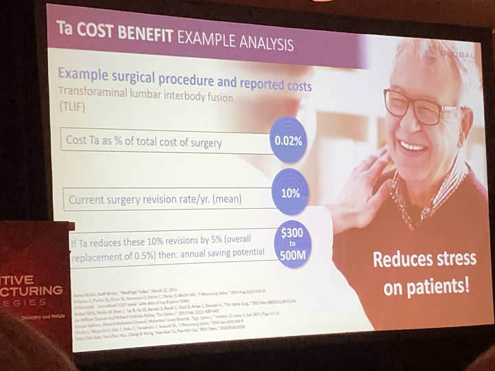
“We looked at the whole process, buying the raw material and printing and cleaning it and sterilizing it, packaging, surgery, to the point where the patient walks out,” Sungail explained. “Tantalum’s contribution to this implant on the slide is .02%. I think that’s nearly negligible. Tantalum will allow the patient to walk out much quicker and recover much quicker.”

3D printing isn’t even the most expensive part of the whole process – it’s the surgery itself. If annual implant surgery revisions can be prevented by even 5% from switching to tantalum, the medical industry will save $300-500 million a year.

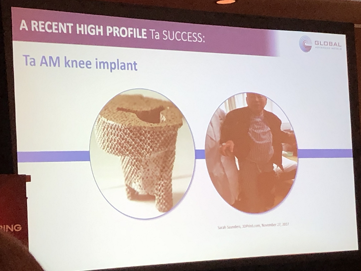
Another example Sungail shared was a 3D printed knee implant made out of tantalum. The surgery took place in China back in 2017, and the patient was actually able to stand up two hours post-op…that’s a pretty impressive feat.

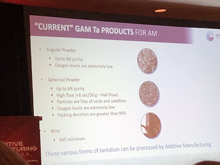
Wrapping things up, he pulled up a slide showing GAM’s “current” tantalum products for 3D printing. In its angular powder form, the material works for cold spray technology and DED printing, while spherical powder can be used with laser AM technologies. He said that the company is also working on tantalum tungsten, and is “always looking for partners,” especially since GAM doesn’t have its own 3D printing system yet and relies on its partnerships to print tantalum for them. However, Sungail said they are considering a 3D printer purchase…perhaps this is an announcement we’ll see in the near future?

Stay tuned to 3DPrint.com as we continue to bring you the news from AMS 2020.

Discuss this and other 3D printing topics at 3DPrintBoard.com or share your thoughts below.

[Photos: Sarah Saunders]

The post AMS 2020: 3D Printing Metals II Keynote by Craig Sungail, Global Advanced Metals appeared first on 3DPrint.com | The Voice of 3D Printing / Additive Manufacturing.

Copper3D Antimicrobial Filament Device Attempts To Reduce HIV Transmission From Breastfeeding

3D printing startup Copper3D, based in Chile and the US, uses nano-copper additives, and adds antimicrobial properties to polymers like PLA and TPU to create antibacterial 3D printed objects. Last year, Copper3D partnered with NASA to study microbial risks in outer space, but now the startup is working on an important project that’s a little closer to home.

According to UNICEF, the number of children and adolescents living with HIV in 2017 reached 3 million, with 430,000 newly infected people and 130,000 deaths from AIDS-related causes. UNAIDS reports that in 2018, 26,000 new HIV infections among children up to the age of 14 resulted from withdrawal of treatment during pregnancy, and breastfeeding. But even with this knowledge, the World Health Organization reports that 37.9 million people around the world were living with HIV at the end of 2018, 8.1 million of which didn’t even know they had the disease to begin with.

Companies and scientists around the globe are working to use technology to help control dangerous bacteria and viruses with high replication rates, like HIV. Copper3D has created a 3D printed device, with its copper nanotechnology, that can effectively inactivate the HIV virus under the right conditions on certain objects- a project that the startup’s Director of Innovation Daniel Martínez tells us is “the result of more than one year of research in antimicrobial polymers and the role on inactivating high replication rate viruses like HIV.”

Dr. Claudia Soto, Copper3D’s Medical Director, said, “Understanding the global problem behind the HIV statistics and analyzing the role that our antimicrobial materials could have in containing the transmission of HIV virus led us think that we could develop some kind of device that acts like an interface between mother and child to prevent the spread of this virus through breastfeeding, which is one of the main routes of infection.

“The initial idea is based on some of the few available studies that establish that copper based additives and filters can inactivate HIV virus in a solution of breastmilk, acting specifically against the protease (essential for viral replication) where copper ions non-specifically degrade the virus phospholipidic plasmatic membrane and denaturalize its nucleic acids; nevertheless, several issues such as toxicity levels, milk nutritional degradation, time for virus inactivation, or the optimal size/form of these filters remain unsolved.”

3D concept of the Viral Inactivator (patent pending)

Copper3D, led by co-founders Martínez, Dr. Soto, and CEO Andrés Acuña, began work on a project with, as the startup stated in a release sent to 3DPrint.com, “two lines of research.” Last year, they submitted a patent application for the project, called Viral Inactivation System for a Breastmilk Shield to Prevent Mother-to-Child Transmission of HIV. First, the viral inactivation effectiveness of its PLACTIVE material was tested with samples of HIV-infected breast milk, and then the team designed an object that optimizes the “viral inactivation of HIV” in the milk, acting as a mother-to-child interface during breastfeeding.

“Our purpose as a company has always been related to make a global impact through innovation in materials and nanotechnology. This line of research of active/antimicrobial medical devices and applications that opens with these studies, fills us with pride as a company. We believe that we are marking a before and after in the industry and we take this honor with a great sense of responsibility,” stated Acuña. “We will continue on the path of applied innovation, always thinking of playing an important role in the most urgent global healthcare challenges, where our antimicrobial materials, intelligent 3D designs, rigorous processes of technical validations and laboratory certifications, can generate a new category of antimicrobial/active devices that can avoid infections at a global scale and save millions of lives.”

Virology Laboratory at Hospital Clínico Universidad de Chile

The startup commissioned a proof-of-concept laboratory study at the Hospital Clínico Universidad de Chile’s Virology Laboratory to validate PLACTIVE’s potential HIV viral inactivation capacity. The study used a split-sample protocol to test and treat 20 sub-samples of HIV-1 (subtype B, cultivated from infectious clone NL4-3, with CXCR4 co- receptor).

The sub-samples were randomized into different groups: A, B, and Control. Samples for A and B were placed in either a green or blue 3D printed box, with and without the nano-copper additive; for a proper blind study, the researchers did not know which was which. The samples were exposed to the medical device for 15, 60, 120, and 900 seconds, and then cultured with HIV-1 Jukat reporter cells LTR-luciferase Cells (1G5); Copper3D performed culture measures on the samples 24, 48, 72, and 96 hours post-treatment.

“The preliminary results showed a reduction of viral replication up to of 58.6% by simply exposition of the samples to the 3D printed boxes containing copper nanoparticles. Fifteen (15) seconds of exposition were enough to achieve such a reduction. These data allow us to infer that by increasing the contact surface by a factor of 10X, we could obtain much higher inactivation rates, very close to 100% (log3) and according to our calculations, most probably in less than 5 seconds,” explained Martínez. “These results are coherent with the hypothesized reduction times proposed by Borkow, et. al. To the best of our knowledge, this is the first essay aiming to study the inactivation of HIV virus by using this new kind of polymers with antimicrobial copper nanotechnology in 3D printed objects.”

3D model of the Viral Inactivator (patent pending)

These results are pretty promising, which bolstered the team as they moved on to the second part of the study – designing a device, with a surface of contact expanded 10X, for HIV-contaminated milk, that’s embedded in nano-copper for use during breastfeeding.

“Like any innovation project, this is a constantly evolving process. We have learned a lot along the way, and we will continue designing, iterating, testing, validating and learning about antimicrobial materials and devices in the future. The preliminary results obtained in the first phase of our investigation with viral inactivation on active/antimicrobial nanocomposites materials gives us a great drive to continue in that line of research,” said Martínez. “We hope in the coming months to conclude the second phase of this study. For these purposes we develop a new antimicrobial flexible TPU based material (MDflex), with the same nanocopper additive as PLACTIVE, to test with new iterations of the design of this viral inactivation device with expanded surfaces of contact that we believe will be much more effective. These new insights will allow the development of a whole new range of active medical devices and applications, with incredible capabilities to interact with the environment, eliminating dangerous bacteria and viruses and protecting patients and users around the globe. This second and final phase of the study will be concluded in Q2 of 2020.”

Copper3D’s concept for its Viral Inactivator is to study how the antimicrobial capacity of its nano-copper materials impacts HIV inactivation, and how different shapes and designs for the 3D printed device can increase the surface of contact with breast milk, while using the nano-copper to enhance effectiveness. The device was made with various layers and “rugosities” in order to imitate what has been observed in the human gastrointestinal tract.

Collaborators at the University of Nebraska at Omaha’s Department of Biomechanics will perform mechanical characterization testing of Copper3D’s prototype.

“Copper3D has once again disrupted the field of medical devices by creating this revolutionary device that can have a tremendous impact in reducing mother-to-child transmission of HIV,” said Jorge Zuniga PhD, Associate Professor of Biomechanics with the university. “Our laboratory is fortuned to partner with Copper3D, in such an impactful project.”

Concept of applications with the Viral Inactivator

Discuss this and other 3D printing topics at 3DPrintBoard.com or share your thoughts below.

The post Copper3D Antimicrobial Filament Device Attempts To Reduce HIV Transmission From Breastfeeding appeared first on 3DPrint.com | The Voice of 3D Printing / Additive Manufacturing.

Costa Rica: Researchers Design 3D Printed Medical Device for Suturing Extremities

Our skin protects us from invading microorganisms and foreign substances, eliminates harmful toxins, helps to regulate our core body temperature, and is in charge of receiving both tactile and thermal stimulation. But, it’s fragile and easily damaged, which can lead to open wounds that get infected. Michelle Orozco-Brenes, José A. Jiménez-Chavarría, and Dagoberto Arias-Aguilar, researchers out of Costa Rica, published a paper, titled “Design of a medical device for superficial suturing upper and lower extremities,” about their work creating a medical suturing device.

“This work presents the design for a class 2 medical device that meets the basic requirements of the current and known suturing methods in Costa Rica,” the abstract states. “The design process was achieved in three main stages, (i)Research on similar technologies; e.g. The operation principles of a sewing machine, materials used; (ii) The study of types of skin traumas; (iii) General approach toward the suturing device, including device functionality, integration with the human body and manufacturing process. The device model was designed and fabricated using 3D printing technology, this allowed the team to analyze ergonomics, the assembly of the parts and the equipment’s motion. The printed prototype made it possible for potential users to provide feedback on the design and suggestions for improvement.”

Figure 1. SolidWorks design of the medical device to be printed.

Suturing means to connect blood vessels with a specific material, such as thread, when tissue is torn in a way that halts natural healing. You can find many suturing devices on the market around the world, but Costa Rican hospitals don’t typically use them, as they are complex and costly. So the team set out to design a class 2 FDA electronic medical device that could both reduce tissue damage and uniformly, and quickly, suture a wound so an “aesthetically acceptable” scar is left behind.

“The idea for a medical device to suture arose for three main reasons,” the researchers wrote. “First, physicians were noticing poorly sutured wounds that would result in large scars. These in some cases required further procedures like plastic surgery. Also, time consumption, making the search for a device that would make the method faster a necessity. Finally, sutures stitched by hand are sometimes left too loose or too tight, causing bleeding from the wound.”

Table 2. Schematic representation of the function of the suturing medical device.

Device specifications were functionality, cost, durability, modularity, and reliability. They used SOLIDWORKS software to create the design for their model, which required three specific functions:

  • Stabilize the skin
  • Rotate the needle on its axis to join tissue sections
  • Initiate and finish with the least possible amount of user interference

“The final design was oriented to have the area and volume of the shell as similar as possible for the needle to rotate 360° without any problem,” the researchers explained.

In order to test out several functionality features, they 3D printed a prototype first, using Polyjet technology to fabricate the piston and and an FDM printer for most of the other parts. Due to its high strength and toughness, corrosion and fatigue resistance, and low friction coefficient, they used the AISI 316L alloy for the prototype.

The suturing device has seven main components. The shell encases the device, while two guides allow the movement of the guide pin, which is used to tie a double knot. Rollers provide the rotational movement that allows for the suturing, while a piston gives the rollers their movement. The final parts are a ½ circle needle with tapered tip, and nylon thread, which has good elasticity for skin retention and closure.

Figure 2. Final design for the suturing medical device.

To use the device, the needle is first threaded in its initial position at the top of the shell, and then set in the rollers. The piston lowers the shell, and the needle is rotated 270° to pinch the tissue for suturing. The knot is initiated when the rollers, guided by the holder, turn 45° to the right, and the pin is set in place over the guide. The needle makes a 360° turn on its axis, and the guides turn over the shell and let go of the guide pin, “letting it fall due to gravity over the guides” beneath it to finish the first knot. The first few steps are repeated, and after the final full turn, the user tenses the thread through the top hole, until it’s kept that way through the guide pin. The lower guides will release, and the guide pin is removed, completing the double knot.

“After the prototype was assembled and design functions checked, the final step required a survey,” the team wrote. “The study contained questions about the medical device presented via prototype and they were asked to elaborate on their answers regarding their opinion as health professionals.”

Table 3. Survey on trained medical physicians.

The 3D printed prototype device was presented to Dr. Stephanie Gómez Najéra, Dr. Pamela Villareal Valverde, and Dr. Tatiana Piedra Chacón. The numbers listed in the survey results are the average between these three Costa Rican physicians, and the scale, based on the Likert scale, goes from 1-5, with 1 being strongly disagree and 5 being strongly agree.

“The comments reference that the usefulness depends on the context of where it would be applied, for example a jail or emergency room,” the researchers wrote of the doctors’ opinions on their device.

“One main drawback is that the device may not be suitable for all types of wounds. Other concerns raised by the physicians were related to the price and size of the device.”

Based on observations from the survey, the researchers modified the final prototype to “improve its ergonomic factor” by adding a holder at the top of the shell for more stability and easier manipulation.

Next steps include standardizing parts of the prototype so that some pieces can be purchased in the market, and optimizing the mechanisms, like the servomotor, sensors, and motors, that generate the device’s movements.

Discuss this story and other 3D printing topics at 3DPrintBoard.com or share your thoughts in the Facebook comments below. 

The post Costa Rica: Researchers Design 3D Printed Medical Device for Suturing Extremities appeared first on 3DPrint.com | The Voice of 3D Printing / Additive Manufacturing.

What Does the Future Hold for Additive Manufacturing for Medical Devices in 2020?

Personalised medical devices are often cited to create the greatest opportunity in additive manufacturing (AM). There are a number of notable advantages that AM can bring to enhance the development of medical technologies for the benefit of patients. However, in this highly regulated sector, there remains a number of challenges, some that are understandably sector-specific and others that impact the broader manufacturing community.

So how can medical device manufacturers capitalize on this innovative technology and manufacture more personalised and complex medical devices, faster, more efficiently, and more cost-effectively?

Paul Unwin, Chairman of the Additive Manufacturing UK Strategy Steering Group

Ahead of the Additive Manufacturing for Medical Devices Conference we asked Paul Unwin, Chairman of the Additive Manufacturing UK Strategy Steering Group to share his insights and top predictions for 2020 on the main benefits and challenges of using additive manufacturing to produce the most innovative medical devices for commercialisation.

Read the full article here to receive:

  • 6 top predictions to help medical devices manufacturers to leverage the benefits of additive manufacturing
  • A full analysis on the key benefits and challenges associated with adapting additive manufacturing in highly regulated industries.
  • Solutions to close the gap between research and commercialization

The post What Does the Future Hold for Additive Manufacturing for Medical Devices in 2020? appeared first on 3DPrint.com | The Voice of 3D Printing / Additive Manufacturing.

UK Heart Patient Undergoes Rare Surgery for 3D Printed Titanium Sternum

A Fleetwood, Lancashire woman in the UK is enjoying better health today, able to perform daily tasks at home, not flinching when she coughs or sneezes—but best of all, she is now able to hug her one-year-old granddaughter. All this progress is due to a 3D printed implant fitted and inserted by surgeon, Dr. Ehab Bishay, at University Hospitals Birmingham NHS Foundation Trust.

The 52-year-old patient, Linda Edwards, had been suffering for years with angina, but even after surgery, she suffered further complications as her breastbone became extremely fragile.

After her chest plate collapsed twice post-surgery, it was obvious another solution was needed—but without anything to attach another metal plate to, her previous doctors were running out of options; however, Linda had watched a documentary featuring Dr. Bishay’s work, and she made contact with him after finding him on social media.

Although she was told she would need to ‘apply’ to have the surgery both for funding and to be cleared for the operation, she steadfastly did so and waited two years to have her 3D printed sternum implanted by Dr. Bishay—making her case the third in Britain (and fifth internationally) to undergo such a procedure.

Scan shows Ms Edwards’ ‘collapsed’ sternum before she underwent the operation

“I woke up from the operation feeling terrible and, at one point, I thought I had died, but I am feeling better every day,” she said, also mentioning that the doctors told her to take it easy and even joked with her about not falling over because she had so much money’s worth of metal in her body to protect now.

“I still feel numb because I am on a lot of drugs, but the main thing is my ribcage doesn’t keep shifting about,” explained Linda. “It feels incredible I have had an operation as advanced as this. I feel like I’ve got my life back.”

“It’s priceless. I can hold my granddaughter and that’s the best feeling in the world.”


Dr. Bishay and his team were able to open Linda’s chest again while being careful to avoid any trauma to the previous bypass area or her heart.

“It’s fantastic to see Mrs. Edwards is doing extraordinarily well given the complexity of the procedure she has undergone,” said Dr. Bishay. “My team and I removed Mrs. Edward’s original breastbone and inserted the custom-built prosthesis.”

“The plastic surgery team, led by Mr. Haitham Khalil, harvested several muscle flaps to cover all the extensive components of the prosthesis, a fundamental step in this operation,” continued Dr. Bishay. “Fortunately, complications such as those experienced by Mrs. Edward’s following previous heart surgery are rare but are notoriously difficult to manage.”

While 3D printing is an amazing technology spawning countless, fascinating inventions, we would still be going a bit far to say such processes have changed the world; they have, however, changed the lives of many patients already, worldwide—with some receiving chest implants and titanium 3D printed sternums, and even composite sternums and rib cages. What do you think of this news? Let us know your thoughts! Join the discussion of this and other 3D printing topics at 3DPrintBoard.com.


Linda Edwards, and her granddaughter, Sienna

[Source / Images: Daily Mail]

The post UK Heart Patient Undergoes Rare Surgery for 3D Printed Titanium Sternum appeared first on 3DPrint.com | The Voice of 3D Printing / Additive Manufacturing.

GE Additive Customer Uses DMLM 3D Printing to Manufacture Blades for Medical Cutting Device

endoCupcut

As the population continues to age, the number of necessary hip replacements rise, which means we’re seeing more 3D printed hip implants and hip cups. Implanting a hip cup is fairly straightforward these days, but removing one, for reasons ranging from abrasion and infection to loosening, is another story. Surgeons typically have to use a hammer and chisel for this, which can damage tissue and bone and make it hard to reinsert a new implant.

Germany medical device company Endocon, a GE Additive customer, is using additive manufacturing to make it easier for surgeons to remove hip replacement cups. The company isn’t 3D printing the cups, but instead created a new device, called an acetabular cut cutter, with 3D printed blades. This product has improved not only the surgical experience for the patient and physician, but the cost savings and product reliability as well.

“We’ve also been able to reduce the cost per blade by around forty to forty-five percent. That means cost savings for us and in turn for our customers,” said Klaus Notarbartolo, the General Manager at Endocon. “When you combine that with a reduction in product development time, higher efficiency and lower rejection rates, then the business case for additive really becomes attractive.”

Typically, traditional casting is used to manufacture cutting blades, but for an end product that comes in a variety of shapes and sizes, it could take up to three and a half months to produce a single batch of blades. Casted blades can also have a rejection rate of about 30% due to issues like non-repeatable quality, corrosion, and consistent hardness.

The company called on GE Additive’s Concept Laser Mlab Cusing 100R, which uses direct metal laser melting (DMLM) technology, to 3D print the blades for its endoCupcut in 17-4 PH stainless steel. This reusable device allows surgeons to quickly loosen and extract cementless hip cups without damaging the surrounding bone, as its blades allow for more precise cutting along the edge of the acetabular cup. Additionally, it can be combined with up to 15 different 3D printed stainless steel blades in sizes ranging from 44 mm to 72 mm, and makes it possible to implant the same size cup that was originally there.

The 3D printed blades for the endoCupcut, which had only minimal changes from the original model, can be available in just three weeks, including post-processing. The device now has a rejection rate of less than 3%, can achieve consistent outcomes, and the 3D printed blades show excellent corrosion resistance. Rather than cracking after 600 N, the blades show a plastic deformation after applying 1,8 kN, and their hardness level has improved to 42+-2 HRC, compared to 32 HRC.

“Endocon’s ability to solve multiple challenges using additive is impressive example of how it can have a positive impact for smaller companies targeting the orthopedic industry,” said Stephan Zeidler, Business Development Manager Medical for GE Additive. “What started with the need for a reduced time-to-market in terms of product development and flexible production of various shapes and sizes has resulted in a smart, innovative medical product that enhances patient outcomes.

“Moving the entire production process from casting to additive manufacturing was a logical step and that shift continues to provide inspiration for future projects.”

Metal 3D printing specialist and service bureau Weber-KP manages the entire process, including data preparation, build platform orientation, 3D printing, surface finishing, hardening, and bead blasting, for Endocon. The company has even improved the manufacturing process of the blades in order to, as GE Additive put it, “maximize the best possible outcome” and can fit between two and six blades on the Mlab Cusing 100R’s build platform, depending on orientation and size.

Using DMLM technology to 3D print the blades has improved their mechanical properties, and also ensures high density and accuracy. By using stronger, harder, and more reliable blades on the endoCupcut, the device performs better for the surgeon in the operating room, and also makes things safer for the patient by lowering the risk of breakage and splinters being embedded in their tissue. Using this device, surgery time has been decreased from 30 minutes to just three, and its precise cutting method preserves the highest possible amount of bone substance, which “supports an accelerated healing process for the patient.”

Other benefits of fabricating the endoCupcut blades with DMLM 3D printing include:

  • High-fitting accuracy of blades through modular system of ball-shaped heads
  • Perfect fitting of ball-shaped heads in a 38-60 mm width
  • Reusable for multiple operations
  • Wear-resistant and easy to sterilize

Lowering surgical risk saves hospitals money and time, and the world is definitely taking notice of Endocon’s innovative work. The endoCupcut is already being used by several medical professionals around Germany, and the company itself is a finalist in the TCT Awards next week.

Discuss this story and other 3D printing topics at 3DPrintBoard.com or share your thoughts below. 

[Images provided by GE Additive]

Pakistani Researchers Create 3D Printed Drug Delivery Device


Modern-day medicine has advanced enormously in methods for pain management in the last decades, and thankfully so for patients who are in dire need. If you have ever been seriously injured or debilitated by an illness hat was accompanied by severe physical discomfort, then you are aware how helpful a fast drug delivery system can be. And as 3D printing has lent the power of innovation to so many corners of the medical field, it may also begin to play a larger role in dressings and pain relief mechanisms.

Munam Arshad recently authored ‘Controlled Drug Delivery System for Wound Healing and other Biomedical Applications,’ outlining how useful such techniques can be in serving (and healing) the patient better, as well as creating more efficiency for medical professionals. With a 3D printed device, the drugs are delivered via a vibrating mechanism that moves the medications through small ‘slits.’

“The device is made up of a biocompatible polymer which is 3D printed and one complete unit which houses the drug reservoir and the haptic motor,” states Arshad in his paper. “Different dosage regimes can be constructed for different drugs; the concentration can be modified by changing the amount of time the device is turned on. The static and dynamic studies perform in the research provide the guidelines for constructing different dosage regimes.”

The dressing of wounds is a process that has been necessary since the beginning of medicine itself, meant to eliminate both infection and pain, and promote healing. The goal for any human is to see them heal as quickly as possible too, and Arshad points out that scientists today seek smarter ways to dress wounds while promoting better elimination of bacteria and offering relief from what can sometimes be enormous physical discomfort. Conventionally, wound dressing has meant the patient was either in the hospital being cared for continually or having to travel back and forth to a doctor’s office.

Researchers like Arshad see the need for more progressive devices and techniques and are looking toward both FDM and SLA 3D printing to advance these goals—and with the accompaniment of smart electronics for monitoring. Materials science comes into play here heavily as well as dressings must be compatible with the human body, and affordably so too.

“The synthetic polymer helps to reduce scab formation in the wound also managing to elevate the movement of the growth cells into the wound. Use of synthetic polymer also manages to obtain a better rate of epithelial cell organization,” states Arshad. “Thus, we can say that a functional wound dressing should be able to wrap around the given site of the wound firmly and at the same time enable the better healing environment to the wound, it should also help to reduce the amount of pain making it easier for the patient to transition back to their normal routine.”

The researchers settled on PMC-744 as the material of choice due to its biocompatibility and flexibility—both requirements for the device. After that, the research team began working on the vibrating mechanism for delivering the drugs, with several different iterations in place before they settled on the final 3D printed model featuring one system with both a haptic motor and drug reservoir with drug release area. The model was created in SOLIDWORKS and then 3D printed in PLA.

The 3D printed device should prove to be helpful in two different areas, at least:

“The first one is the use of the device on the living beings such as rats and ultimately for humans which is the desired goal, however the prototype is still far off from how the final device would be like, but in the future it can be used to replace conventional methods of drug delivery and bandages for better wound healing solutions,” states Arshad. “The second application involves the use of the drug delivery system in the laboratories to study the anti-microbial activities of the drugs for better study of wound healing.”

“The device can be programmed to deliver specified amount of drug at specified intervals in a day or even for greater time intervals. This can be used as an automated drug dispensation system for the clinicians without the requirement of frequent intervention to manually deliver drugs.”

Initial PMC 744 model

Extensive testing was performed on the device, but there is still plenty of room for improvement, according to the research team. Read more about this drug delivery device here. What do you think of this news? Let us know your thoughts; join the discussion of this and other 3D printing topics at 3DPrintBoard.com.

[Source / Images: ‘Controlled Drug Delivery System for Wound Healing and other Biomedical Applications’]

Father Takes Up 3D Printing, and Founds New Company, to Create Son’s Custom Orthosis

Some of the most heartwarming aspects of the 3D printing industry involve the people who do everything they can to develop and provide affordable 3D printed prosthetics to people who need them the most. Just in time for Father’s Day, Formlabs has shared a beautiful story about a dad who worked tirelessly to help his young son walk on his own…and ended up helping others along the way.

Cerebral palsy (CP) causes more than 17 million people around the world to have limited control of their own bodies. Seven years ago, Nik, the son of Matej and Mateja Vlašič, was born one month early, and due to difficulties during childbirth, suffered brain damage that led to the diagnosis of CP, and an inability to stand or walk on his own.

To help CP patients walk, many doctors will prescribe standard orthoses meant to correct spine and limb disorders. Patients can purchase pre-made orthotics, and some can even be slightly modified to better fit the patient, but it’s not easy to use one device to help with several symptoms, and they can even lead to skin irritation and pain.

Custom orthoses, CNC machined based off of a plaster or foam box impression, generally fit better, but the cost can be astronomical, even with insurance, and delivery can take weeks. On top of that, children outgrow them quickly.

Matej, who has an engineering background, said, Based on my knowledge, I knew that a piece of plastic could not cost so much money.”

Matej has worked hard all of Nik’s life to help him move on his own, even using ski boots to stabilize his ankles when he got older.


When you’re looking at your child, you instinctively know what to do in order to help him. When Nik was unable to turn on his side, I decided to build a ramp so that he could easily flip on his belly. When he found out that this was fun, he was trying to do it all by himself,” Matej said.

He instantly felt confident, and you could see it in his eyes that he loved it and that he wanted to progress. This is what kept us going.”

Unfortunately, Nik’s short Achilles tendon and low muscle tone kept him on his toes.

He was afraid of walking because his feet were in a really bad position,” said Petra Timošenko, Nik’s physiotherapist. “If he had tried to walk longer like that, he would have injured the bones and the joints.”

Matej knew he had to find a better way to help his son.

The lack of comfort and high price combined with all the cons were enough that I decided to do something about it. I didn’t have the solution at that time, but I wanted to find a better way to design it,” Matej said. “I was just trying to help my son the best possible way.

I didn’t know how orthoses are produced currently, so I was able to look outside of the box.”

He had heard of 3D printing, and after conducting some research, determined that the technology was accurate enough to create a properly-fitted orthosis. One of the benefits of 3D printing, especially in the healthcare field, is its ability to design customized products at a more affordable cost, and Matej was confident he could create a custom, 3D printed orthosis that would give Nik the correction and support he needed.

After a few attempts, Matej successfully digitized Nik’s feet, learned 3D modeling, and spent the next six months researching and experimenting, and eventually developed an innovative workflow, which starts with placing the patient’s feet, in the corrected, standing position, on a vacuum bag.

An iPad-mounted structure scanner scans the footprints from the bag, while the feet are also 3D scanned from above, and the data is merged and cleaned up into an accurate representation. The custom orthosis is designed right on the 3D scanned foot in CAD software, and then 3D printed in high resolution on a Form 2 3D printer with Durable Resin.

The first 3D printed prototype reached almost to Nik’s knee and kept him from walking freely, so Matej got to work on the second iteration, creating a prototype that fit inside a regular shoe. Finally, a successful prototype was created.

“In two or three days he was walking, and we were not needed to take care of him so that he doesn’t fall anymore,” Matej said. “The change was immediate, it was unbelievable.”

Nik’s orthosis is barely visible.

Just how braces align teeth, the 3D printed orthosis keeps Nik’s foot in the corrected position. It’s best to use orthoses at a young age, as children’s bodies can adapt while they grow. Physiotherapy also helps to strengthen ligaments and muscles.

When he’d been using the orthosis for two or three months, for the first time, I saw Nik smiling,” said Timošenko. “After four or five months, he started to become faster and faster. His steps became longer, and his walking more smooth. He actually started to dance.

Now I can do much more sophisticated exercise with him. We can run on a treadmill, we can jump, because I know that his feet are in the right position and I can’t cause any deformation to his bones or joints, that might, on the long term, require an operation to correct. If he didn’t have this orthosis, his feet would be in danger.”

Matej created four versions of Nik’s 3D printed orthosis.

The first version gave him confidence and stabilized him. The second version improved his overall walking smoothness,” Matej explained. “Then the third helped him get better posture, and that’s when he really started to enjoy the walking and started to play around. The fourth orthosis corrected his right foot that was off the center of his body, so now he’s able to stand with his feet together in a straightened, upright position.”

After looking at the workflow, and measuring Nik’s feet with and without his 3D printed orthoses, certified orthotist and prosthetist Dejan Tašner knew that Matej had created a novel solution. He is able to make an affordable custom orthosis in less than 24 hours, and the devices are also comfortable.

3D printing allows us to create orthotics with different thicknesses in different areas. We can apply a more thick area where it’s needed and minimal thickness to the areas where correction is not required,” Matej explained. “This is not possible with current solutions.

Orthoses don’t need to hurt, only without pain can the children accept them.”

Matej and his wife decided to certify the workflow, which is now patent-pending, so the process and components will meet standard requirements for medical devices and allow for clinical trials. Matej quit his job to focus on 3D printed, patient-specific 3D printed children’s orthotics full-time and, together with Mateja, Tašner, and Timošenko, formed a new company called aNImaKe.

At the moment, we are testing with several patients with different pathologies from age three to 11,” Tašner said. “We already see improvements in terms of biomechanics, which is the main goal. But also, crucially, a positive change in sentiment that the parents see in the daily life of their children because they need to feel comfortable to use the orthosis often enough to improve their walking.”

aNImaKe hopes to expand the technique to other parts of the body, such as a hand brace that helps young CP patients spread their fingers.

We want to enlighten others in the medical industry about the tools that are available today to provide better options to the children,” Matej said. “Orthotics should be built for a person, and should treat only the symptoms, not be standardized solutions that put them in boxes.”

Discuss this and other 3D printing topics at 3DPrintBoard.com or share your thoughts below. 

[Source/Images: Formlabs]