FDM 3D Printing & PLA: Fabrication of a Splint for Mallet Finger

Researchers from Australia and the UK are finding ways to improve treatments for injuries to the hand, detailing their results in the recently published ‘Patient-specific 3D-printed Splint for Mallet Finger Injury.’ The goal is to offer better outcomes for patients who may have experienced damage to the tendon responsible for straightening the joint found at the end of the finger (or thumb).

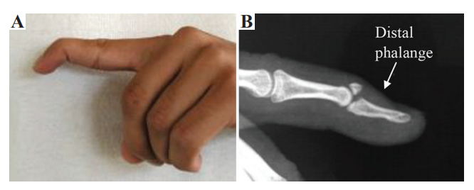
Mallet finger injuries most commonly occur due to trauma after being struck by an object that bends the finger or thumb tip painfully.

(A) Mallet finger fracture (Image courtesy from Sachin J Shah, MD, online), (B) anatomy of finger

While there are many injuries and illnesses far more seriously affecting lives today, losing the use of a finger or hand can be debilitating as we must use them for so many tasks—primarily at work. Many types of splints may be uncomfortable for the patient to wear, but are also labor-intensive and expensive to make, resulting in the use of excess material during production too.

3D printing and additive manufacturing processes are beginning to make an impact in manufacturing of orthotics, offering benefits like greater affordability, speed in production, and best of all—total customization. Innovation around the globe has resulted in new software and improved workflow for creating medical devices like orthotics, the production of more comfortable foot/ankle orthotics, myoletric orthotics, and more.

“Mallet finger is often left untreated by patients unless severe restriction in extensor ability is present, or there is lingering pain. This injury, in the case, that there are functional shortfalls, can impede the whole hand in everyday fine motor skill tasks,” explained the authors. “Furthermore, this deformity can develop additional medical conditions in the finger and hand as overcompensation can create hyperextension of proximal interphalangeal joint, a swan neck deformity.”

“Effective use of AM may lead to a reduction in size and weight of the splint making it more comfortable for the user.”

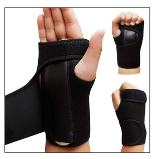
Today, the most common splints are the prefabricated stack splint, the dorsal aluminum splint, and the customized thermoplastic splint. Generally they are worn for six to eight weeks, but with the first two choices there may be ongoing comfort issues as the splints may not fit well causing the skin to become irritated, the patient may experience added pain, and the splint itself may break due to inferior materials or manufacturing. Many patients are also unhappy with the size and style of their splints.

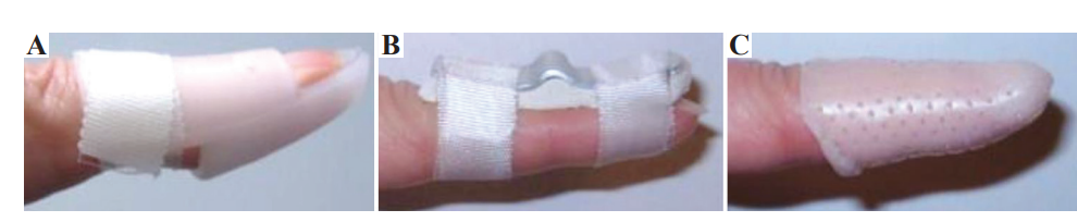
(A) Stack, (B) dorsal aluminum, and (C) personalized thermoplastic splints

“… customized splints, fitted to the exact dimensions of a patient’s finger, have the ability to provide successful treatment in more cases than the other two splint types,” stated the authors.

FDM 3D printing was chosen for the course of this study as the researchers made samples, noting that they would be taking advantage of the ability to fabricate complex geometries not previously possible through conventional techniques in manufacturing. More importantly is the ability to offer patient-specific treatment like never before—especially with medical devices like prosthetics and orthotics. In this study, the issue of comfort could be addressed with comprehensive customization, splints made quickly, and revised simply by changing the 3D design and printing a new one in the case of growth (these issues are common with children who may outgrow an orthotic made with conventional methods before it even arrives to them!), damage, or other requirements.

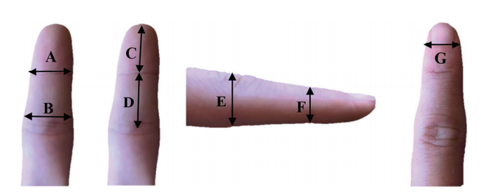
The seven measurements required to create a personalized finger splint computer-aided drawing model.

The research team used Autodesk Inventor for entering the measurements for their sample, including a built-in constraint:

“When the user clenches their fist, without this material removed, the skin of their middle phalanx finger can push into the back of the splint dislodging its correct position. The stack splint is designed with an open ventilation section above the fingernail to allow some airflow to reduce sweat when being worn and to allow limited access for washing.”

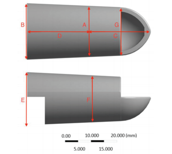
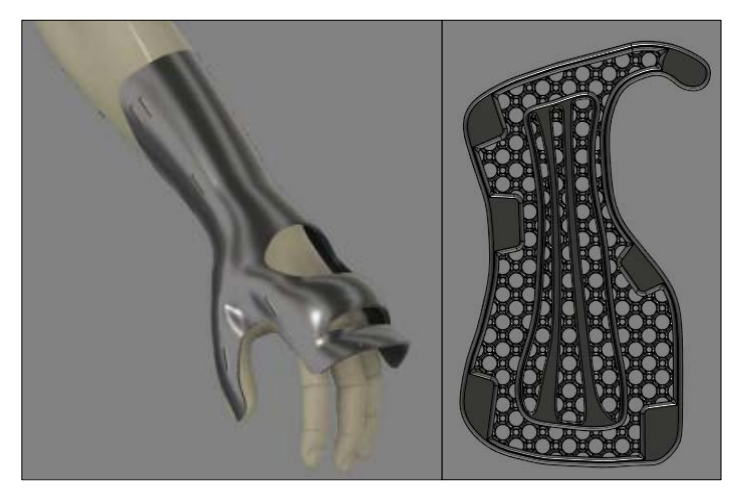
Geometry of a sample 100% mass design according to a patient’s finger.

Addressing the issue of wasted material and recycling of products later, the researchers point out that patient-specific devices like the splints they designed cannot be re-used by another individual later due to the level of customization, so they must be disposed of; however, with the use of PLA as their 3D printing material, a discarded splint can be ‘composted’ within six to eight weeks.

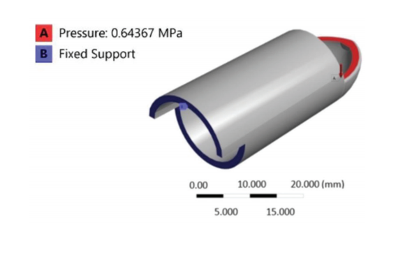
Schematic view of boundary conditions on a sample splint design.

One of the main objectives in the study was to develop a highly functional orthotic with topology optimization, eliminating as much material as possible while still maintaining the splint’s required volume fraction and stiffness.

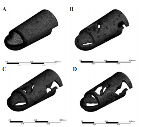
Domains discretization with tetrahedral elements for (A) 100%, (B) 79.49%, (C) 71.13%, and (D) 62.51% mass

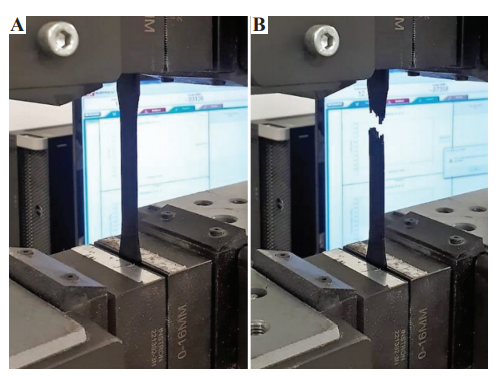
A three-dimensional-printed dog-bone poly-lactic-acid sample (A) before, and (B) after tensile test.

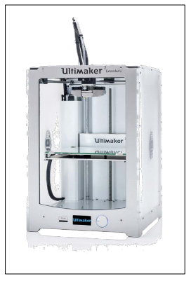
An Ultimaker2 Extended+ 3D printer was used for fabrication, while tensile testing was performed on an Instron 300LX.

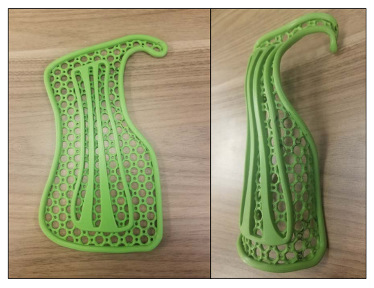
“In FDM topology optimized and original design, finger splints were fabricated, original (100% mass), 62.51% mass, 71.13% mass, and 79.49% mass, by the same 3D printer and processing parameters used for the dog-bone specimens,” stated the researchers.

Noting that deflection results did not correlate exactly with heat dissipation, a trade-off analysis was required in choosing the best splint since the initial splint operating at 100 percent mass offered the best mechanical performance, but the 71.13 percent mass splint performed optimally in heat dissipation.

From left to right 62.51%, 71.13%, 79.49%, and 100% mass splints

“This splint is simpler to print than lower percentage mass splints that can require more printing support structures. It was found that inevitably reducing the amount of material in a load-bearing finger splint would increase the deflection of it. However, when the distribution of that material is chosen to optimize the stiffness in that situation, the deflection value was low enough to justify its use,” concluded the researchers.

“The results of this project would pave the way for the medical industry to utilize superior advanced manufacturing and minimum materials that have been shape optimized to better serve their purpose while improving patient comfort.”

What do you think of this news? Let us know your thoughts! Join the discussion of this and other 3D printing topics at 3DPrintBoard.com.

[Source / Images: ‘Patient-specific 3D-printed Splint for Mallet Finger Injury’]

The post FDM 3D Printing & PLA: Fabrication of a Splint for Mallet Finger appeared first on 3DPrint.com | The Voice of 3D Printing / Additive Manufacturing.

3D Printed Occlusal Splint Provides Relief in Just a Few Weeks

In the recently published ‘Digital manufacturing of occlusal splint: from intraoral scanning to 3D printing,’ researchers explored the use of a new digital workflow for creating intraoral occlusal splints. These devices are used by dentists to treat temporomandibular disorders as they force muscles to relax, protect the teeth, and more.

As 3D technology continues to progress, so do manufacturing processes within dentistry, featuring 3D scanning, CAD technology, and 3D printers for fabrication of dental prostheses and a range of other devices.

For this case, the researchers chose to study a 44-year-old man presenting with issues in his jaw muscles. As doctors examined his masticatory system, they were unable to find any health conditions, except for pain emanating from his anterior temporalis muscles and external pterygoid muscles. It was suspected that he may have been in the habit of clenching his jaw, thus leading to the pain. The doctors decided to fit him with an intraoral occlusal splint to stop the pain and begin ‘repositioning’ the mandible.

“With the accuracy and efficiency of the digital workflow, the splint would be fabricated with digital intraoral scanners and a 3D printer,” stated the researchers.

The team used a scanner to photograph all the teeth, saving an .stl file read for 3D printing. The first scans, however, related both the lower and upper virtual models. The initial setting allowed parameters to include:

  • 40 °TMJ eminence angle
  • 10° Bennet angle
  • 40° incisal guide angle

“The bite plane was designed to be retained by the mandibular teeth, according to patient’s preference, and the maxillary teeth had a single contact with the appliance,” explained the researchers.

Splint designed on the digital casts.

A 3D ProJet MJP 3600 Dental by 3D Systems was used to 3D print the intraoral splint, with VisiJet® M3 Stoneplast acrylic resin, made for dental practices. Once inserted and evaluated, the doctors found that the intraoral occlusal splint was in need of little adjustment. The patient was to wear the splint while sleeping or while at home during the day. Pain subsided after three weeks; however, the patient was instructed to keep using the splint for another six months.

“The presented workflow allowed for a noticeable reduction of the complexity and of the total time of the laboratory procedures. Although in this case the authors sent the impression scan to the dental laboratory, the possibility of producing the same appliance in the dental clinic should be considered as 3D printers are becoming more popular within the dental office. However, chairside production could be time consuming for the dentist and the authors prefer at this time to delegate the design and the production to the dental technician more familiar with CAD software and 3D printers,” concluded the researchers.

“After centric relation was recorded with bimanual manipulation, the technique included intraoral scans of the maxillary and mandibular arches, digital registration of vertical relationship, computer-aided design of the intraoral occlusal splint and then manufactured with a multi-jet 3D printer. The presented technique allows for time efficient laboratory manufacturing, which could also be performed chair-side in the dental office. The delivered splint is accurate and precise and could be reproduced anytime if needed.”

3D printing has made huge impacts in the dental world, and has improved the quality of life for many patients from dental implants to orthodontics to new manufacturing systems. What do you think of this news? Let us know your thoughts; join the discussion of this and other 3D printing topics at 3DPrintBoard.com.

Splint produced with a multi-jet 3D printer and verified on the prototyped casts.

Intraoral pictures of the splint immediately after delivery

[Source / Images: ‘Digital manufacturing of occlusal splint: from intraoral scanning to 3D printing’]

The post 3D Printed Occlusal Splint Provides Relief in Just a Few Weeks appeared first on 3DPrint.com | The Voice of 3D Printing / Additive Manufacturing.

3D Printed Splints & Braces: Just As Effective & Comfortable, Cheaper & Faster to Make

University of Nebraska researcher James Pierce has delved further into the potential for 3D printed assistive devices in ‘Efficacy of Assistive Devices Produced with Additive Manufacturing.’ He begins by stating that it is surprising there have not been more previous ventures into the use of devices like 3D printed splints and casts, despite the obvious need—and an enormous amount of injuries each year resulting in sprains and breaks.

Traditional wrist splint used for
functional comparison.

The goal is to eliminate older, conventional methods that may promote healing but take more time in production, are expensive, and often result in uncomfortable, poorly-fitted devices. If you have endured wearing a splint or a cast or watched someone else try to deal with wearing one, then chances are you are nodding your head right now and getting pretty interested in hearing what 3D printing can do differently for orthopedic patients.

Pierce explores how all the most intrinsic benefits of 3D printing could be used in creating customized medical devices, beginning with affordability—after all, healthcare is expensive, and cost drives many of our choices, not to mention controlling insurance coverage too. In terms of design, the researcher was hoping to find a way to fabricate parametric assistive devices that could be made quickly. This is important in nearly any consumer scenario, but especially when something is being created to fit a patient who may be in pain, whether acute or chronic.

Even more interesting about this research is the potential Pierce sees for applications in space:

“Knowledge gained from this study will validate novel assistive devices which could be used in the treatment of musculoskeletal injury for astronauts’ both during spaceflight and after return to Earth,” said Pierce. “These novel solutions will require less expert intervention and less on-site modifications for fitting.”

Pierce states that it has already been noted in previous studies that 3D printing for these types of devices is significantly faster, but still there have been no parametric devices of this specific kind actually produced using AM methods, which should create a way for patients to recover more quickly, avoid further injury, and allow for better use of effort and materials. Initial data regarding a 26-year-old male wearing a wrist orthosis showed results ‘not significantly different’ from a conventionally made device.

A) 3D model of a parametrically-defined hand exoskeleton design, scaled to fit a participant in CAD before part production. B) 3D model of a wrist orthosis, which is printed flat and thermoformed to the
contours of the users’ upperlimb.

The patient did say that the 3D printed model was more comfortable, and Pierce attributes this to the breathability of the design. The 3D printed version was also preferred due to aesthetics, featuring a more streamlined style, bright colors, and better design overall.

An Ultimaker 3D printer was used in production of the orthoses.

One of the most exciting aspects of 3D printing is that it allows users to challenge conventional methods and promotes positive change for consumers as well as a wide range of medical patients today who are benefiting from the use of 3D printed medical models, a variety of different devices, and a vast number of forays into bioprinting too.

For patients healing from sprains and fractures, the usual visit to the doctor for X-rays and a cast made of plaster may soon be a completely different experience. Find out more about additive manufacturing for parametric devices here, and check back for a full analysis of the study in April 2019.

What do you think of this news? Let us know your thoughts; join the discussion of this and other 3D printing topics at 3DPrintBoard.com.

[Source / Images: Efficacy of Assistive Devices Produced with Additive Manufacturing]

A) Unformed wrist splint; B) A 3D printed wrist splint, thermoformed to fit the user.