Rice Researchers 3D Print with Lasers and Sugar to Build Complex Vascular Networks

A team of researchers from Rice University has uncovered a promising strategy to generate vascular networks, one of the most daunting structures in the human body. Using powdered sugar and selective laser sintering, the researchers were able to build large structures from complex, branching, and intricate sugar networks that dissolve to create pathways for blood in lab-grown tissue.

This is the team’s latest effort to build complex vascular networks for engineered tissues to show that they could keep densely packed cells alive for two weeks. The findings of their study—published in the Nature Biomedical Engineering journal—prove that developing new technologies and materials to mimic and recapitulate the complex hierarchical networks of vessels gets them closer to providing oxygen and nutrients to a sufficient number of cells to get a meaningful long-term therapeutic function.

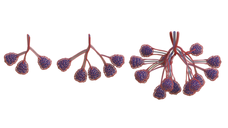
“One of the biggest hurdles to engineering clinically relevant tissues is packing a large tissue structure with hundreds of millions of living cells,” said study lead author Ian Kinstlinger, a bioengineering graduate student at Rice’s Brown School of Engineering. “Delivering enough oxygen and nutrients to all the cells across that large volume of tissue becomes a monumental challenge. Nature solved this problem through the evolution of complex vascular networks, which weave through our tissues and organs in patterns reminiscent of tree limbs. The vessels simultaneously become smaller in thickness but greater in number as they branch away from a central trunk, allowing oxygen and nutrients to be efficiently delivered to cells throughout the body.”

Overcoming the complications of 3D printing vascularization has remained a critical challenge in tissue engineering for decades, as only a handful of 3D printing processes have come close to mimic the in vivo conditions needed to generate blood vessels. Without them, the future of bioprinted organs and tissues for transplantation will remain elusive. Many organs have uniquely intricate vessels, like the kidney, which is highly vascularized and normally receives a fifth of the cardiac output, or the liver, in charge of receiving over 30% of the blood flow from the heart. By far, kidney transplantation is the most common type of organ transplantation worldwide, followed by transplants of the liver, making it crucial for regenerative medicine experts to tackle vascularization.

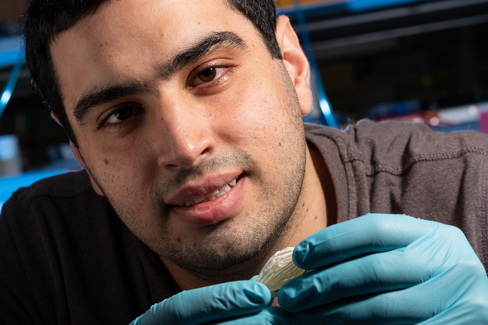
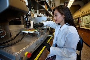
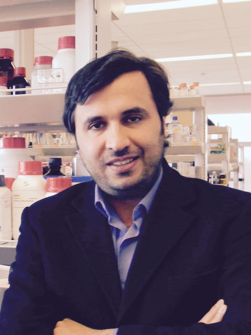
Ian Kinstlinger with a blood vessel template he 3D printed from powdered sugar (Credit: Jeff Fitlow/Rice University)

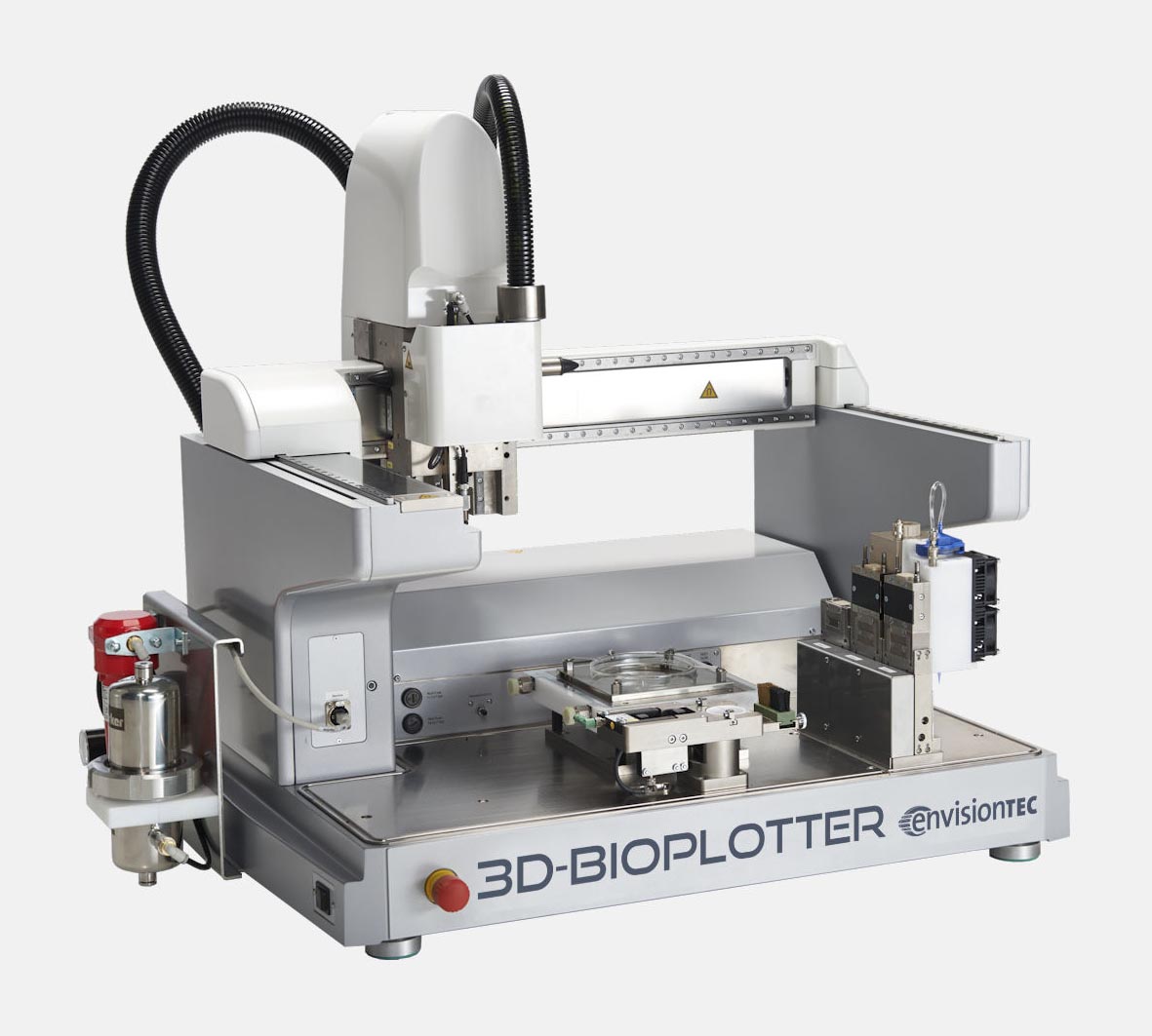
In the last few years, extrusion-based 3D printing techniques have been developed for vascular tissue engineering, however, the authors of this study considered that the method presented certain challenges, which led them to use a customized open-source, modified laser cutter to 3D print the sugar templates in the lab of study co-author Jordan Miller, an assistant professor of bioengineering at Rice.

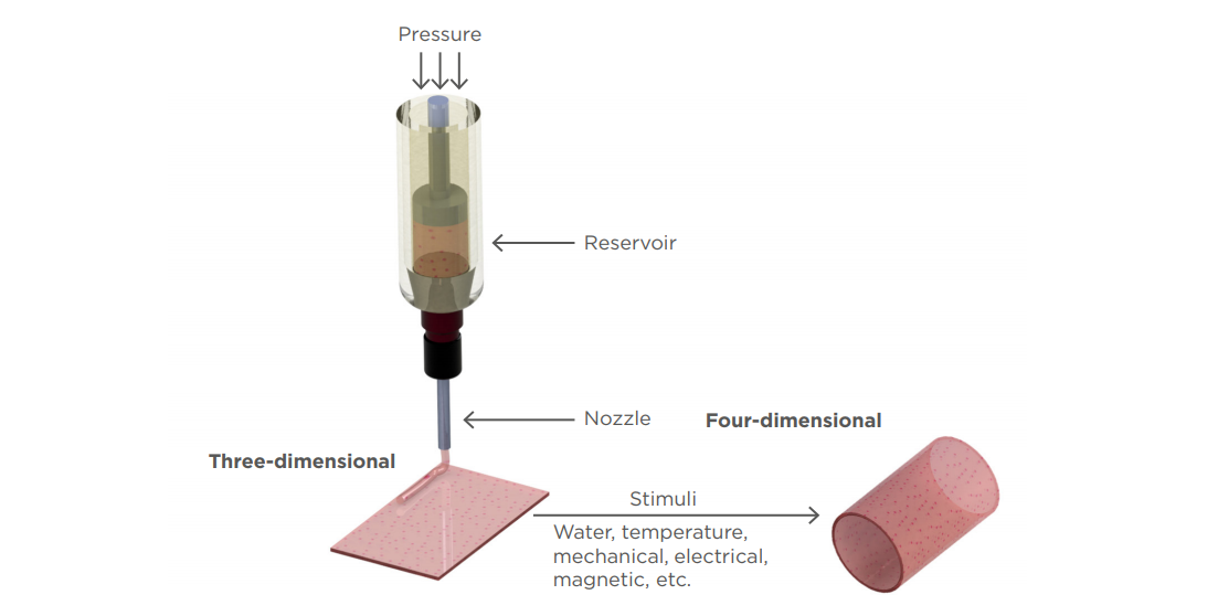
Miller began work on the laser-sintering approach shortly after joining Rice in 2013. The 3D printing process fuses minute grains of powder into solid 3D objects, making possible some complex and detailed structures. In contrast to more common extrusion 3D printing, where melted strands of material are deposited through a nozzle, laser sintering works by gently melting and fusing small regions in a packed bed of dry powder. According to Miller, “both extrusion and laser sintering build 3D shapes one 2D layer at a time, but the laser method enables the generation of structures that would otherwise be prone to collapse if extruded.”

“There are certain architectures—such as overhanging structures, branched networks and multivascular networks—which you really can’t do well with extrusion printing,” said Miller, who demonstrated the concept of sugar templating with a 3D extrusion printer during his postdoctoral studies at the University of Pennsylvania. “Selective laser sintering gives us far more control in all three dimensions, allowing us to easily access complex topologies while still preserving the utility of the sugar material.”

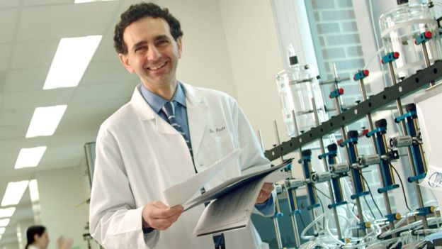
Assistant professor of bioengineering at Rice University, Jordan Miller (Credit: Jeff Fitlow/Rice University)

Generating new 3D printing processes and biomaterials for vascularization is among the top priorities for the researchers at Miller’s Bioengineering Lab at Rice. The lab has a rich history of using sugar to construct vascular network templates. Miller has described in the past how sugar is biocompatible with the human body, structurally strong, and overall, a great material that could be 3D printed in the shape of blood vessel networks. His original inspiration for the project was an intricate dessert, even going as far as suggesting that “the 3D printing process we developed here is like making a very precise creme brulee.”

To make tissues, Kinstlinger chose a special blend of sugars to print the templates and then filled the volume around the printed sugar network with a mixture of cells in a liquid gel. Within minutes, the gel became semisolid and the sugar dissolved and flushed away to leave an open passageway for nutrients and oxygen. Clearly, sugar was a great choice for the team, providing an opportunity to create blood vessel templates because it is durable when dry, and it rapidly dissolves in water without damaging nearby cells.

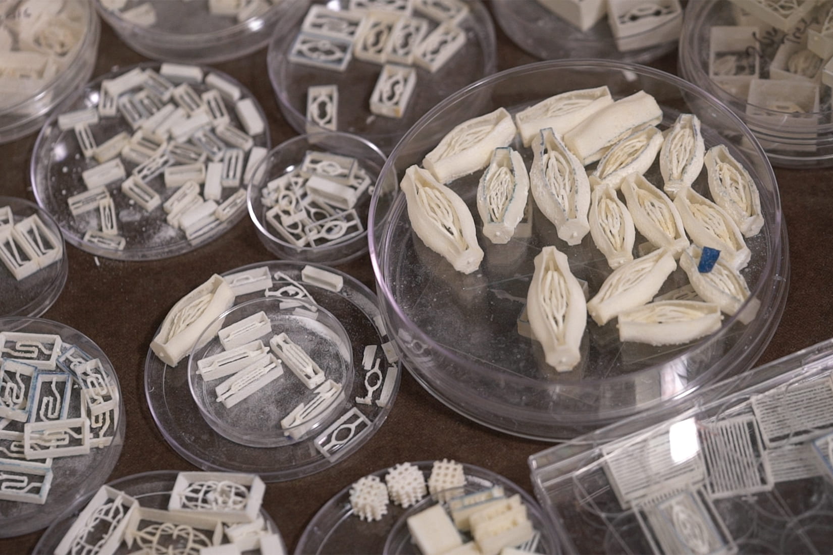
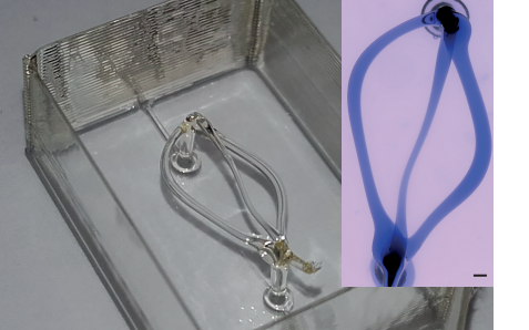
A sample of blood vessel templates that Rice University bioengineers 3D printed using a special blend of powdered sugars. (Credit: B. Martin/Rice University)

In order to create the treelike vascular architectures in the study, the researchers developed a computational algorithm in collaboration with Nervous System, a design studio that uses computer simulation to make unique art, jewelry, and housewares that are inspired by patterns found in nature. After creating tissues patterned with these computationally generated vascular architectures, the team demonstrated the seeding of endothelial cells inside the channels and focused on studying the survival and function of cells grown in the surrounding tissue, which included rodent liver cells called hepatocytes.

The hepatocyte experiments were conducted in collaboration with the University of Washington (UW)’s bioengineer and study co-author Kelly Stevens, whose research group specializes in studying these delicate cells, which are notoriously difficult to maintain outside the body.

“This method could be used with a much wider range of material cocktails than many other bioprinting technologies. This makes it incredibly versatile,” explained Stevens, an assistant professor of bioengineering in the UW College of Engineering, assistant professor of pathology in the UW School of Medicine and an investigator at the UW Medicine Institute for Stem Cell and Regenerative Medicine.

The results from the study allowed the team to continue their work towards creating translationally relevant engineered tissue. Using sugar as a special ingredient and selective laser sintering techniques could help advance the field towards mimicking the function of vascular networks in the body, to finally deliver enough oxygen and nutrients to all the cells across a large volume of tissue.

Miller considered that along with the team they were able to prove that “perfusion through 3D vascular networks allows us to sustain these large liverlike tissues. While there are still long-standing challenges associated with maintaining hepatocyte function, the ability to both generate large volumes of tissue and sustain the cells in those volumes for sufficient time to assess their function is an exciting step forward.”

The post Rice Researchers 3D Print with Lasers and Sugar to Build Complex Vascular Networks appeared first on 3DPrint.com | The Voice of 3D Printing / Additive Manufacturing.

Envisiontec 3D-Bioplotter: New Bioprinting Capabilities

The 3D-Bioplotter from EnvisionTEC has been at the heart of over 333 scientific papers, from 3D printing human tissue to 3D printing an ovary for mouse implantation. It is one of the most commonly used (and earliest) 3D bioprinters in the industry for tissue engineering and biofabrication research.

Now, the global provider of professional-grade 3D printing solutions is expanding the bioprinting capabilities of their star product just in time for the European Society for Biomaterials (ESB) Annual Conference in Dresden, Germany. Launched in 2000, the 3D-Bioplotter is probably one of the most seasoned bioprinters in the market, and now it’s getting two new print head options that will help advance biomaterial research.

Researcher using EnvisionTEC 3D-Bioplotter

The first is an upgrade of the Photo-Curing head, now allowing up to five wavelengths or combinations thereof during one project. The second is an Ink-Jet Low-Temperature head designed to dispense materials through a non-contact process.

One of the several solidification processes available to 3D-Bioplotter customers is photo-curing. According to EnvisionTEC, while the wavelength of 365 nanometers (nm) remains the most commonly required by photoinitiators used by academic and industrial users, this wavelength has a negative impact on cell survivability during prolonged or repeated use, especially in the research field of bioprinting.

Therefore, the company sought out a way to solve this need to shift towards photoinitiators that react to the visible light range, to which cells can be exposed to with minimal biological effect.

To avoid the constant manual exchange of light sources when using different materials, as well as to allow combinations of photoinitiators, EnvisionTEC came up with an upgrade for their machine, a multi-wavelength pen upgrade which now provides five wavelengths into one single source pen. Through the 3D-Bioplotter software, individual wavelengths, or combinations thereof, are user-selectable and can be assigned to individual parts.

Current customers with existing photo-curing heads can have their existing heads upgraded to allow the use of the new source pens. The wavelengths included are 365, 385, 395, 405 and 455 nm.

The firm’s second highlight, the Ink-Jet Low-Temperature head, is aimed at dispensing low viscosity hydrogels as coatings while 3D printing parts or for hybrid scaffold fabrication. The key is that the built-in microdispensing valve can be controlled through the software to dispense individual, unconnected dots of material, or to connect them into lines of dispensed droplets.

With a 100 micron aperture, this head is restricted to low viscosity materials, such as hydrogels. Additionally, all components in contact with the dispensing material can be autoclaved, allowing for cell-suspensions to be dispensed as well. And depending on the choice of materials, the Ink-Jet head can be used to create fast dot printing projects, to dispense materials in specific positions on the platform (organ-on-a-chip projects), to fill pores in hybrid scaffolds, or to dispense coatings onto simultaneously printed 3D scaffolds.

EnvisionTEC states that the whole cartridge mount is heatable, from a room temperature range of up to 70 degrees centigrades, in order to keep the materials in the cartridge at their proper processing temperatures. Also, this head was designed to use 10 ml cartridges but also fits 3 ml disposable cartridges, with a dispensing duration of between 0.4 ms to 100 ms, and a frequency range of 1-100 Hz.

The 3D-Bioplotter

The 3D-Bioplotter family of printers consists of three models: the Starter series, the Developer series and the Manufacturer series, each with increasing capabilities. The original 3D-Bioplotter is now in its fourth generation, and more than 15 years of hardware and software development have gone into it.

One of the frequent users of the 3D-Bioplotter, Teja Guda, Assistant Professor of Biomedical Engineering at the University of Texas at San Antonio, said recently that “what’s so unique about the printer is that it is capable of printing living cells within the material as you print it.”

The modular 3D printer is easy to use while being capable of advanced research at the same time. The Bioplotter series prints with open-source biomaterials, using air or mechanical pressure to extrude them through a variety of syringes. Both new heads are currently on display in Dresden, at the EnvisionTEC booth, where attendees can see them in action and learn more about the research these new additions will make possible.

[Images: EnvisionTEC]

The post Envisiontec 3D-Bioplotter: New Bioprinting Capabilities appeared first on 3DPrint.com | The Voice of 3D Printing / Additive Manufacturing.

Nervous System Works with Rice University Researchers 3D Printing Vascular Networks

Nervous System has been heavily engaged in experimenting with 3D and 4D printing of textiles in the past years, and all their research is paying off now as they find themselves engaged in the realm of tissue engineering. The Somerville, MA company is known for their generative design process, combining both programming and art within most of their serious projects, drawing bioengineers from Rice University to turn to them for added expertise.

Assistant professor Jordan Miller invited the Nervous System team to join his researchers on an incredible journey to fabricate examples of possible vascular networks via bioprinting—harnessing their knowledge of software and materials to find a way to create soft hydrogels. Kind of not a phantom but more a path towards ideas that can lead to concepts that may let us build true vascularized structures at one point. As Miller explains, in their research they were able to create large tissue blocks easily, but as so many scientists engaged in bioprinting today have discovered before them, it is extremely challenging to keep cells alive. Viability becomes the goal, and as that becomes more comprehensively mastered overall in bioprinting, it may finally unlock the door to true fabrication of organs that can be transplanted into the human body.

Open-source technology, mainly centered around 3D printing has offered huge opportunity for the bioengineers from Rice University to make progress in their work—and that was what drew them to Nervous System in the first place. Jordan became ‘captivated’ with the structures they were creating, specifically in their Growing Objects series, which was featured as an exhibit at the Simons Center for Geometry and Physics in Stonybrook, NY in August and September of 2014. In speaking with Nervous System, his proposal involved what they describe as an ‘epic task,’ to create simulated synthetic tissue and human organs.

Rendering showing lung-mimicking structures generated within different volumes

“The idea of taking our generative systems which are inspired by nature and using them to actually make living things was a dream come true,” states the Nervous System team in their case study.

Elsewhere the research did,

“…show that natural and synthetic food dyes can be used as photoabsorbers that enable stereolithographic production of hydrogels containing intricate and functional vascular architectures. Using this approach, they demonstrate functional vascular topologies for studies of fluid mixers, valves, intervascular transport, nutrient delivery, and host engraftment.”

As Miller and his expanding team continued to work on developing the necessary tools for bioengineering, part of their research resulted in a new 3D printing workflow called SLATE (stereolithography apparatus for tissue engineering). Their proprietary hardware can bioprint cells encased in soft gels that act just like vascular networks. Nervous System accompanied them (going back as far as 2016) in this bioprinting evolution by designing the materials for the networks—but with their background in programming, the contribution went far beyond designed materials and included customized software for creating ‘entangled vessel networks.’ These networks can be connected to both inlets and outlets for oxygen and blood flow, as they use specific algorithms to ‘grow’ the branching airways.

“Air is pumped into the network and it pools at the bulbous air sacs which crown each tip of the network,” states Nervous System in their case study. “These sacs are rhythmically inflated and deflated by breathing action, so called tidal ventilation because the air flow in human lungs is reminiscent of the flows of the ocean tides.

“Next we grow dual networks of blood vessels that entwine around the airway. One to bring deoxygenated blood in, the other to carry oxygen-loaded blood away. The two networks join at the tips of the airway in a fine mesh of blood vessels which ensheathes the bulbous air sacs. These vessels are only 300 microns wide!”

This project, bringing together scientists and art designers, was featured in the American Association for the Advancement of Science (AAAS) in ‘Multivascular networks and functional intravascular topologies within biocompatible hydrogels,’ authored by Bagrat Grigoryan, Samantha J. Paulsen, Daniel C. Corbett, Daniel W. Sazer, Chelsea L. Fortin, and Alexander J. Zaita.

The recently published article goes into great detail about SLATE 3D printing, indicating that this hardware is capable of rapid bioprinting, and offering possible sustainability to human cells—along with maintaining functionality of stem cells and necessary differentiation.

The project was created by Jordan Miller at Rice University and Kelly Stevens at the University of Washington, and included 13 additional collaborators from Rice, University of Washington, Duke University, and Rowan University.

Nervous System is undeniably one of the most fascinating companies producing 3D printed innovations today. Their versatility has led them to create everything from 4D textiles and 3D printed stretched fabrics to their famed Kinematics Petal Dress. With their latest project delving into 3D printed tissue, the stakes become higher—and their impact on the world much greater. Find out more here.

What do you think of this news? Let us know your thoughts! Join the discussion of this and other 3D printing topics at 3DPrintBoard.com.

The Miller Lab fabricated and tested the architectures we generated showing that they can withstand more than 10,000 ventilation cycles while being perfused with human red blood cells. Study of the printed gels shows that the architecture we designed promotes red blood cells mixing and bidirectional flow which is hypothesized to occur in the human lung.

[Source / Images: Nervous System]

McGill University Researchers: Can We Use PLA for Desktop Bioprinting?

Bioprinting has proven to be useful for bone regeneration, as researchers learn to create more stable structures that mimic human tissue. In ‘Three-Dimensional Printed Polylactic Acid Scaffolds Promote Bonelike Matrix Deposition in Vitro,’ authors Rayan Fairag, Derek H. Rosenzweig, Jose L. Ramirez-Garcialuna, Michael H. Weber, and Lisbet Haglund explore the uses of desktop bioprinting with PLA.

Even in conventional medicine today, surgeons find difficulty in repairing bones that have undergone trauma, whether due to an accident, tumor, or other serious issue. Grafting can still be challenging to complete, and then problematic later in terms of pain, infection, and the need for multiple procedures. Materials such as calcium phosphate bone cement (a synthetic graft) have become more popular for repairing bone defects, but there are also limitations due to lack of mechanical strength. While poly-cements have been used also, they can cause stress around the ‘target area,’ and lead to secondary fracture, which defeats the purpose of healing altogether.

Here, the researchers have investigated the use of tissue engineering for bone repair in growing cells, scaffolds, and using numerous bioactive factors. 3D printing has been successful in fabricating scaffolds using different polymers like PLA.

“The ideal material for scaffold development should fulfill specific criteria,” state the researchers. “The material must be biocompatible and must be capable of being generated with an interconnected network to mimic the natural tissue architecture.”

Cell sustainability is the greatest challenge, along with creating stable structures. The researchers sought to create scaffolds that would allow for complete cell sustainability, along with the best environment for encouraging tissue to form. They must also allow for the following:

  • Fabrication in different, complex shapes
  • Resistance to inflammation and toxicity
  • Strong mechanical properties
  • Appropriate porosity
  • Affordability

In previous studies, the researchers were aware that PLA 3D printed from the desktop was suitable for both chondrocyte and nucleus pulposus tissue engineering applications. Here, they tested PLA scaffolds with pore sizes of 500, 750, and 1000 μm, fabricating accurate structures with good porosity; in fact, all scaffolds reflected pores in line with the initial designs, leaving the authors to conclude that this ‘suggested accuracy’ with desktop 3D printer—in this case, the Flashforge Creator Pro.

Pore size results were as follows:

  • Small pore scaffolds – 585.61 μm ± 26.40
  • Medium pore scaffolds – 769.94 μm ± 12.98
  • Large pore scaffolds – 1028.85 μm ± 57.54, p < 0.0001

“The scaffold fabrication and replication process manifests high accuracy and precision as evidenced by μCT analysis, which proves the value of low-cost printing in tissue engineering applications,” stated the researchers.

The authors reported the following for mechanical properties:

“Significant differences in stiffness were observed between the three sizes (p < 0.05, p < 0.0001) in which Young’s modulus for the small pore size was 206.7 MPa ± 0.17 SD, medium size scaffold was 137.5 MPa ± 6.98 SD, and 116.4 MPa ± 5.97 SD for the large size PLA scaffold.”

Mechanical properties of 3D-printed scaffolds. (A) Young’s modulus representing 5−10% compressive stress/strain curves of printed PLA scaffolds. For each set, (n = 3), error bars represent ±SD and (* = P value < 0.05), (# = P value < 0.0001). (B) Stress/strain curves of 500, 750, and 1000 μm showing the amount of deformation, elastic (proportionality) limit, and plastic region. For each set, (n = 3).

“The failure point of each scaffold was determined from the stress/strain curves in which the small-size failure point was around 21.63 MPa, around 11.86 MPa for the medium size, and around 8.53 MPa for the large-pore scaffold. Our results demonstrated an overall higher compressive modulus with smaller pores because of the addition of bulk material (smallest pore size has the highest amount of material and is the stiffest).”

The use of PLA was successful, indicating both accuracy and reproducibility, and the scaffolds presented properties like native bone. The authors stated that the data reflected structures stable enough for an environment recruiting host stem cells and repairing bone.

Morphological characterization of 3D printed scaffolds. (A) Representative images of the 3D models with dimensions and printing process. (B) Quantification of scaffold weight, (n = 6), error bars represent ±SD (** = P value < 0.005), (# = P value < 0.0001), with a representative image of printed scaffolds (Canon EOS 350d Camera). (C) Pore size was calculated by scanning electron microscopy, and porosity was determined by μ-CT. For each set, (n = 3), error bars represent ±SD and (* = P value < 0.05), (** = P value < 0.005), (# = P value < 0.0001).

“In vivo studies will be necessary to determine potential adverse effects, bone repair, and scaffold resorption rates,” stated the researchers. “It comes without surprise that 3D printing has been strongly adopted by orthopedic surgery clinical practice, medical education, patient education, and orthopedic-related basic science.

“Whereas 3D printing has been used for some time to generate patient models of defects for presurgical planning, there is a growing shift in using this technology in actual bone or tissue repair. One major focus in orthopedic and reconstructive surgery is to use 3D printed constructs for filling bone defects, substituting current standard therapies as an innovative approach for bone repair. Several studies have shown applicability and clinical relevance of using different types of 3D-printed polymers as a graft substitute.”

From 3D printing in hospitals to bioprinting in outer space and bringing forth materials which may eventually yield fabricated human organs, researchers are driven to create what used to be considered impossible, with a wide range of innovations already in use around the world. Find out more about desktop bioprinting here. What do you think of this news? Let us know your thoughts! Join the discussion of this and other 3D printing topics at 3DPrintBoard.com.

SEM of acellular and cell-seeded scaffolds. Representative SEM images of acellular, osteoblasts, MSC−OST seeded scaffolds at 80×,450×,1500×, and 22 000× magnifications and scale bars represent 1 mm, 200, 50, and 5 μm with the rectangular marker indicating the region of the scan (n = 3).

[Source / Images: ‘3D-Printed Polylactic Acid (PLA) Scaffolds Promote Bone-like Matrix Deposition In-vitro’]

Eindhoven University of Technology: Researchers 3D Print Microvascular Structures with Carbohydrate Glass

As we go about our busy lives each day, it is easy to forget what a miracle the human body is—and when you are feeling tip-top, you can thank the well-oiled machine in the form of the human body that keeps you breathing, talking, and walking. While you are probably familiar with the vascular system (in relation to your circulatory system), you may not also realize that the microvascular system is a vital player in your body, formed of tiny vessels that are, not surprisingly, responsible for microcirculation.

The microvasculature is made up of arterioles, capillaries, metarterioles, and more—and the vascular system overall has been connected with 3D printing numerous times over the past few years from bioprinted vascular scaffolds to 3D printed models for microvascular surgery to viable 3D printed tissue.

Today, researchers are still challenged to find ways to imitate and re-create the microvasculatur system with fabrication in 3D printing trending toward using devices such as an “organ-on-chip.” A team of scientists at Eindhoven University of Technology has been exploring this route further, as they explain in ‘3D printing of round microfluidic channels to mimic the microvasculature,’ presented last year in Montreaux at the Nano Bio Tech Poster Sessions.

Obviously, many parts of the human body are complex and hard to mimic; for example, consider that we still are not able to 3D print human organs. We may be getting closer, but it will be the holy grail of bioprinting when it happens. Just trying to make something like microvascular ‘components’ is a substantial undertaking, and the researchers explain this because of the difficulty in translating the cross sections, smaller diameters, and network architectures that are intricate.

Free-standing structure of carbohydrate glass inside a printed casting frame mimicking the vascular architecture. Insert: Perfused network with dye solution after dissolving carbohydrate glass cast in PDMS. Scale bar 500 µm.

3D printing with carbohydrate glass is one viable option that has been suggested by researchers, but the Eindhoven scientists want to use multiple types of materials in fabrication, along with making the parts smaller:

“Our main focus was to reduce the diameter to a size closer to the microvasculature, namely in the 10-500 μm range and be able to engineer hierarchical 3-dimensional branching networks that can change diameter along the vessel.”

The team set up a 3D printer with a heated barrel connected to a Nordson EFD performus III pressure control system. Standard nozzles were applied with a .4 mm diameter, and the researchers were able to adapt the diameter limits through limiting or speeding up the movement. In using self-supporting carbohydrate glass as the material of choice, there is greater latitude in printing complex geometries. As in so many 3D printing research projects, temperature is a significant consideration—and is often an obstacle when it cannot be manipulated properly, resulting in deformation of parts.

Carbohydrate fibers strung from droplets horizontally across a
printed frame with a speed of 600 mm/min. Insert: Microscopic image of 3 fibers, top and bottom strung left to right and middle right to left at 600 mm/min. Fibre diameter ~100 µm

The researchers state that a great portion of their work in 3D printing microvasculature will be centered around controlling the thermal elements in fabrication.

“This will offer even greater freedom in network design, and it will give the possibility to exactly control reflow of fibers to form a single in-plane junction,” conclude the researchers. “In the end, the printed models will be used to investigate the flow of blood and particles inside the blood through a microvascular network, leading to a better understanding of perfusion and particle distribution/interaction in the microvasculature.

What do you think of this news? Let us know your thoughts! Join the discussion of this and other 3D printing topics at 3DPrintBoard.com.

Boxplot of diameter for different movement speeds of the stage. Increasing speed reduces variance and average diameter of the fiber insert: The high variance of the diameter at 300 mm/min can be explained by the strong tapered shape of the fibers, resulting in a different diameter at the start and end of the fiber. This is probably caused by solidification of the droplet at the nozzle tip leaving less material to string from.

Portuguese Researchers Review Potential of 4D Bioprinting in Regenerative Medicine

3D printing has been progressing rapid-speed over the past few years, with numerous forays to the next level into 4D printing, whether in serious world-changing endeavors or those that are creative and sometimes even whimsical, such as fashion. Now, researchers see that 4D advancements in bioprinting may trickle down to doctor’s offices too. Authors Pedro Morouço and João Gil of Biofabrication RDi Group, Centre for Rapid and Sustainable Product Development at Polytechnic Institute of Leiria (Portugal) expand on their findings in Four-Dimensional Bioprinting for Regenerative Medicine: Mechanisms to Induce Shape Variation and Potential Applications.

Regenerative medicine is a central focus in bioprinting as researchers around the world try to overcome the challenges of sustaining living tissue in the lab. The end goal, the holy grail of bioprinting, will be to eventually fabricate human organs in the lab—or perhaps even in clinical practice—meaning the elimination of donor lists, organ rejection, and failing quality of life for a multitude of patients globally.

“Regenerative engineering has come to be considered an inevitability and a promising approach for the regeneration of tissues or organs by culturing patient cells into biological substitutes (scaffolds) and subsequent implantation into the patient for the regeneration of new tissue,” state the researchers, pointing out that scaffolds can be made through both conventional and newer techniques—both offering a list of pros and cons.

The team leans toward using 3D printing and venturing into the 4D however, pointing out the potential for greater strides in medicine and tissue regeneration, with so much more control over pore size, shape, and interconnectivity. With 4D printing, researchers can move beyond the restrictions of implants unable to transform according to their biological environment.

Illustrative representation of the transformation from three-dimensional to four-dimensional bioprinting,
which can be triggered by different types of stimuli.

“The potential of 3D printing enhanced by a fourth dimension makes it possible to contribute significantly to the bioprinting of engineered tissues, such as the liver and heart, which will represent a major breakthrough in the area of regenerative medicine,” state the researchers.

Examples of current applications of four-dimensional bioprinting.

Bioinks are commonly used in fabricating viable cells, but they must meet certain criteria for success and researchers must navigate fragility to temperature as well as peripheral chemicals, stress, and issues like UV light exposure. Significant progress has been made with ‘smart materials’ also, namely with bioprinting and some level of shape-morphing. And while creating patient-specific organs may seem to be right around the corner, the research team points out that there is still much progress to be made in creating human tissue structures in the lab.

“The interdisciplinary combination of life sciences with engineering is demonstrating noteworthy advances for healthcare,” conclude the researchers. “Although 3D bioprinting has opened minds to biofabrication, the absence of response to planned stimuli should be considered. However, it does provide an appropriate tool to create hybrid, versatile, and functional tissue constructs; thus, coupling biofabrication with stimuli-responsive materials, novel maturation processes, and validation procedures will bring us one step closer to successful regenerative medicine.”

What do you think of this news? Let us know your thoughts! Join the discussion of this and other 3D printing topics at 3DPrintBoard.com.

[Source / Images: Four-Dimensional Bioprinting for Regenerative Medicine: Mechanisms to Induce Shape Variation and Potential Applications]


Creating Vascular Structures Using Low Cost Desktop 3D Printers

In a thesis entitled “Engineering of vascular networks within biocompatible hydrogels using 3D printing technology,” a PhD student named Juan Liu discusses the need for new technologies in wound healing. While skin flaps and grafts are the “gold standard” in clinical treatment of large skin and subcutaneous tissue defects, there are many complications that can arise from these treatments, and there is also the issue of not having enough skin to be able to harvest to cover particularly large wounds.

3D printing, however, using stem cells, allows for an unlimited amount of tissue to be created to heal large wounds. Using cells from the patient reduces the risk of rejection, as well. In order to create and maintain live tissue, however, vascularization is required, meaning that blood vessels need to be established, which is the tricky part of tissue engineering. Liu hypothesizes that open source desktop 3D printing technology can be used to design and fabricate “customized bio-artificial multicellular tissues with embedded vessel-like supply channels and corresponding bio-reactors for long-term 3D tissue culture.”

Liu outlines the following objectives:

  • Use open source software to design 3D tissues with vascular patterns and fabricate them with desktop 3D printers
  • Analyze cell survival and function over time in 3D tissues in respect to vascular patterns
  • Generate 3D printable bioreactors that allow online monitoring of the process
  • Prevent shrinking of 3D tissues
  • Generate multilayer 3D tissues with different cell types

Liu goes on to demonstrate how 3D printing technology can be used to precisely form vascular structures within cell-laden hydrogels. He also creates “customized PLA and PDMS bioreactors for continuous perfusion and real-time operation.” The vascular structures formed within the cell-laded hydrogels, which were created by 3D printing, were able to increase the viability of the cells surrounding the vascular structures.

“In addition, the final system based on PDMS has been proven to sustain long-term 3D cell culture, which is the basic for 3D cell proliferation and tissue formation in vitro,” says Liu. “Among others this approach exhibits unique advantages to fabricate a hydrogel-based multilayer vascular device in a cost-effective and fast manner, which has a huge potential for viable 3D cell culture, complex tissue engineering, disease modeling as well as drug screening.”

This model, he continues, could also be used to mimic natural tissue architecture and create bigger multilayer hydrogel constructs with corresponding layers of functional cells to study cell morphology, differentiation, and potential function of engineered tissue constructs.

“Further optimization of the hydrogel concentration in each layer, cell density and perfusion parameters may enable the preparation of 3D vascular tissues under the conditions mimicking the natural environment with better functions and matrix compositions,” he adds. “In order to overcome the tendency of shrinking in soft hydrogels, combination with eletrospun fibers are promising. The system presented in this thesis allows fabrication of vascular networks in both soft and stiff cell laden hydrogels. It may serve as a novel platform for vascularized tissue engineering, that facilitates the generation of more functional, engineered vascular tissue. It may be useful for studies of wound coverage and tissue regeneration and eventually aid the treatment of wounds.”

Discuss this and other 3D printing topics at 3DPrintBoard.com or share your thoughts below.


Penn State Researchers 3D Print Porous Tissues

3D bioprinting still has a lot of issues that need to be worked out before we can see anything like a 3D printed organ transplant. One issue is figuring out how to grow blood vessels in printed or engineered tissue, but researchers at Penn State have found an alternative to that idea, creating tissues with micropores that allow nutrient and oxygen diffusion into the core.

Ibrahim T. Ozbolat

“One of the problems with fabrication of tissues is that we can’t make them large in size,” said Ibrahim T. Ozbolat, Associate Professor of Engineering Science and Mechanics. “Cells die if nutrients and oxygen can’t get inside.”

Creating tissue building blocks with micropores is an alternative to vascularization, or growing blood vessels inside the tissue, according to the researchers. They refer to the building blocks as “porous tissue strands.” They began with stem cells derived from human fat and mixed them with sodium alginate porogens. Sodium alginate, which is derived from seaweed, can be printed into tiny particles that leave holes, or pores, behind in the fabric of the tissue when dissolved. The researchers used the stem cells and sodium alginate to 3D print strands of undifferentiated tissue, which were then combined, by 3D printing them next to and on top of each other, to form patches of tissue.

The researchers then exposed the tissue to the chemical cocktail that causes stem cells to differentiate, allowing the cells to turn into bone or cartilage. The pores allow the fluid to flow to all of the stem cells. According to the researchers, the strands were able to maintain 25 percent porosity and 85 percent pore connectivity for at least three weeks.

“These patches can be implanted in bone or cartilage, depending on which cells they are,” said Ozbolat. “They can be used for osteoarthritis, patches for plastic surgery such as the cartilage in the nasal septum, knee restoration and other bone or cartilage defects.”

Cartilage tends to be easier to produce than bone because in the human body, cartilage does not have blood vessels running through it. Some bone is naturally porous, however, so porosity in engineered tissue means greater potential for repairing or replacing natural bone. Only tiny patches of tissue can currently be made, but they are still easier to fabricate than growing artificial tissue on scaffolding.

The research was documented in a paper entitled “Porous tissue strands: avascular building blocks for scalable tissue fabrication.” The work has a lot of potential for bone and cartilage regeneration, and the researchers are also considering applying their technique to muscle, fat and other tissues as well.

Authors of the paper include Yang Wu, Monika Hospodiuk, Weijie Peng, Hemanth Gudapati, Thomas Neuberger, Srinivas Koduru, Dino J. Ravnic and Ibrahim T. Ozbolat.

Discuss this and other 3D printing topics at 3DPrintBoard.com or share your thoughts below.


3D Bioprinting Makes Progress as Lab-Grown Bladder Helps Young Man to Lead a Normal Life

Luke Massella, aged 10 in 2001 with his mother and uncle

Luke Massella was born with spina bifida, a condition that left a gap in his spine. By the age of 10, he had undergone a dozen surgeries and, contrary to doctors’ expectations, was able to walk. But then his bladder malfunctioned, causing his kidneys to fail.

“I was kind of facing the possibility I might have to do dialysis for the rest of my life,” he says. “I wouldn’t be able to play sports, and have the normal kid life with my brother.”

Dr. Anthony Atala [Image: Wake Forest Institute for Regenerative Medicine]

Dr. Anthony Atala of Boston Children’s Hospital had a better idea. He took a small piece of Massella’s bladder and, over the course of two months, grew a new organ in the lab. The new bladder was transplanted into the patient in a 14-hour surgery and, according to Massella, he has been able to live a normal life since then. Now 27, Massella underwent 17 surgeries before he was 13, but has not had to have a single operation since then.

Dr. Atala is an expert in bioprinting, and he and his team have developed eight cell-based tissues that they have transplanted into patients. These include skin, urethras and cartilage grown in the lab. The organs are currently going through clinical trials for approval by the US Food and Drug Administration.

“You need to know how to make these organs by hand, then the bioprinter is really a scale-up tool,” said Dr. Atala, now Director of the Wake Forest Institute for Regenerative Medicine in North Carolina.

According to Dr. Atala, the easiest structures to 3D print are flat structures like skin, followed by tubular structures like blood vessels and urethras. Hollow, non-tubular structures like bladders are more difficult – Massella is one of 10 people who currently has a bladder grown from his own cells. The most difficult of all are solid organs like hearts, lungs and kidneys, which have “so many more cells per centimeter,” Dr. Atala said.

A 3D printed urethra in progress [Image: Wake Forest Institute for Regenerative Medicine]

For a patient like Massella, technology like that Dr. Atala is working with allows a safer transplant, with much less risk of rejection. Massella, who feared he would never be able to have a normal life, went on to become a wrestling coach and now runs events in the jewelry industry.

Massella’s case was an early example of what tissue engineering can accomplish, but advances in bioprinting are making such cases more common.

“A lot has happened in the last couple of years,” said Steven Morris, Chief Executive of bioprinting startup BIOLIFE4D.

BIOLIFE4D has become known as the startup that plans to be the first to 3D print a functioning human heart – and it is getting closer to its goal. Initially, the company will be printing smaller versions of hearts to be used for pharmaceutical testing purposes, but its ultimate goal is to be able to print organs that can be transplanted into patients. That is the dream of most bioprinting companies and research institutions, and while some still doubt that it can be done, the technology is getting closer every day.

Patients like Massella are evidence that functioning organs are possible to create in the lab, and one day – perhaps sooner than anyone expects – cases like his may become much more commonplace. Will we ever be able to eliminate organ donor waiting lists thanks to bioprinting? It’s not nearly as impossible as it once seemed. Generally most practicioners in the field see true 3D printing of organs as something that will happen some 20 to 25 years from now. Approvals, developing all of the tools and assessing long term risk will all take time. We’re still very far from this becoming a day to day procedure for many people. The signs are encouraging however that bioprinting will lead to improvements in patient’s lives.

Discuss this and other 3D printing topics at 3DPrintBoard.com or share your thoughts below. 


Exclusive: 3D Printed Cardiac Patch Shows Promise for Healing Damaged Hearts

Hexagonal scaffolds induce iPSC-CM contractile properties and maturation. iPSC-CM characterization before combination with scaffold, showing A) proliferating cells, B) sarcomeric structures, C) Connexin 43 (Cx43), and D) mitochondrial localization. iPSC-CMs localize to both E) hexagonal and F) rectangular fiber scaffolds, and confocal microscopy shows G,H) Cx43 expression, and increased sarcomere density, I,J) alignment, and K) length in hexagonal scaffolds compared to rectangular scaffolds. L) Beating rate at days 2, 7, 10, 11, and 14. M) Cardiac marker and maturation-related gene expression in hexagonal and rectangular scaffolds at day 7 and day 14.

After a person suffers a heart attack, they lose about half the cells in their heart, greatly weakening the organ and increasing the odds that further attacks will occur. Doctors have begun injecting cells into the heart to grow into muscle and help contractions, but 99% of those cells get washed away. But there are alternative approaches – like 3D printing. Scientists have developed 3D printed cardiac patches that can be used to repair hearts damaged by heart attacks, but only about five have been produced worldwide.

Injectability and in vivo placement of cardiac patch with hexagonal geometry. A) In vitro culture of cardiac patch consisting of iPSC-CMs in cardiac-like ECM on large hexagonal scaffolds. B) In vitro injectability, shape recovery, and C) macro image of cardiac patch after injection. D) Application and shape recovery of cardiac patch on beating porcine heart. E) Cell viability of in vitro F,G) noninjected and H,I) injected cardiac patches. J) Spontaneous beating rate of in vitro noninjected and injected cardiac patches 30 min and 2 d after injection.

In a new study entitled “Melt Electrowriting Allows Tailored Microstructural and Mechanical Design of Scaffolds to Advance Functional Human Myocardial Tissue Formation,” a group of researchers 3D printed a world-first stretchable microfiber scaffold with a hexagonal design. They then added specialized stem cells called iPS-Cardiomyocytes, which began to contract unstimulated on the scaffold. The work was then demonstrated on the actual hearts of pigs.

We spoke with the authors of the paper to learn more about the technology used by the scientists and its implications for the future.

What are the advantages of MEW relative to other technologies? 

“Melt Electrospin Writing (MEW) has distinct advantages over other 3D Printing technologies for tissue engineering applications. MEW utilises common thermoplastics used in biomedical engineering, including Polycaprolactone (PCL). The advantage of this approach is that we are able to create microfibres with diameters often in the 10 micron range, but nano-fibres also also achievable. To put this into perspective, a single strand of hair is about 50-100 microns. Printing at such resolutions allows us to mimic the native extracellular matrix (ECM) components, and allows cells to bind onto the small fibres that at similar to collagen fibrils. Additionally, MEW allows for the control over scaffold architecture, creating scaffolds with aligned fibres and controlled fibre diameters. This specific control over architecture, allows us to tailor the mechanical properties, direct cells growth and  control the movement of nutrients. Because we can modify the scaffolds properties so well, they are often used to reinforce hydrogels (as a backbone), which are typically much weaker and less tailorable.”

What is the significance of your paper?

Representation of the workflow and fabricated microfiber scaffolds. A) Schematic illustration of the in house-built MEW device used. B) Designed hexagonal microstructure. C) 3D fiber scaffold combined with iPSC-CMs and further application in vivo through minimally invasive delivery. D) Optical images of the fabricated scaffolds: detail of microstructure with hexagonal cells (with a side length of 400 µm) composed of multiple stacked aligned microfibers. Images acquired from top and lateral perspective.

“We developed patches with controlled hexagonal micro-fibre structures that had unique flexibility and shape-recovery properties, meaning that the patch can be highly deformed without sustaining damage to its structure or cells. Moreover, such novel paths allowed the maturation of contractile human iPSC-derived cardiomyocytes, which is a breakthrough in creating a functional patch that could match an adult heart. Finally, due to the patch’s flexibility, it can be compressed and pushed through a catheter for delivery in vivo with minimally invasive laparoscopic surgery.”

Why are collagen-based hydrogels so important?

“Collagen is the most abundant protein in the human myocardial tissue and for that reason an ideal biomaterial for use in myocardial tissue engineering.”

How close are we to using 3D printing in a clinical setting?

“There are already reports of 3D printed implants used in clinics, mostly metallic or ceramic based for bone repair. However, in what concerns to our heart patch’s that include biological derived components (new cell therapies, iPSC) we believe a few more years will be required. We need first to demonstrate the efficacy and safety of such approach in animal models, which we are currently planning. Also, important challenges like integration with surrounding tissue will need to be solved: for example, currently the patches can contract autonomously, but we don’t know if they will sync with the beating of the heart once implanted.”

What work will you be doing next?

“We want to concentrate our efforts in conducting more extensive animal studies to assess feasibility and show functional effects, such as improved cardiac function. We also intend to make the patches more complex, by integrating other cell types in the patch.”

Authors of the paper include Miguel Castilho, Alain van Mil, Malachy Maher, Corina H.G. Metz, Gernot Hochleitner, Jürgen Groll, Pieter A. Doevendans, Keita Ito, Joost P.G. Sluijter, and Jos Malda.

Discuss this and other 3D printing topics at 3DPrintBoard.com or share your thoughts below.

[Images: Castilho et al, “Melt Electrowriting Allows Tailored Microstructural and Mechanical Design of Scaffolds to Advance Functional Human Myocardial Tissue Formation,” Advanced Functional Materials, 2018. Copyright Wiley – VCH Verlag GmbH & Co. KGaA. Reproduced with permission.]