Cellink Teams with Lonza to Advance Bioprinting Cell Cultures

For over four years biotech company Cellink has been helping researchers pursue creative ways to promote organ regeneration, test novel drugs, develop vascularized bioconstructs, and more. Through collaborative partnerships and pioneering research, the firm has quickly become a market leader for cell-based applications. Along that road, they have partnered with several startups and multinationals to open up a broader range of possibilities in the field. Their latest partnership with Lonza—a leading suppliers to the pharmaceutical, biotech, and specialty ingredients markets—will provide researchers and scientists with even more options to enhance bioprinting of complex 3D human tissue constructs.

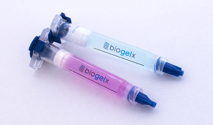
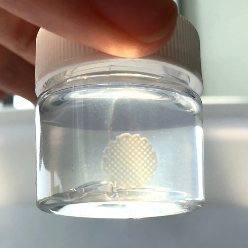
The companies have joined forces to offer a comprehensive 3D bioprinting solution designed to optimize and increase access to complete 3D cell culture workflows. The solution integrates Cellink’s 3D bioprinting instruments and pioneering commercial bioinks with Lonza’s broad selection of human-derived primary cells and supporting culture media.
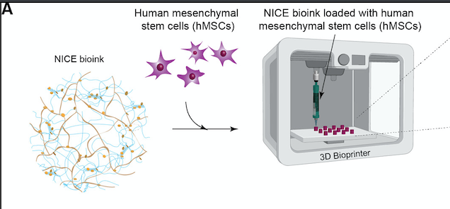
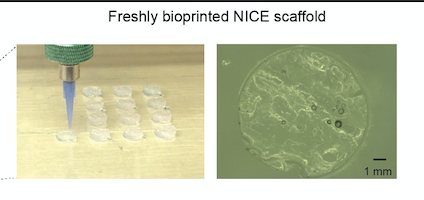
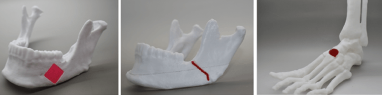
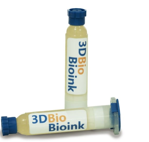
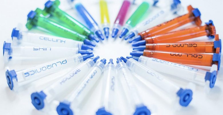
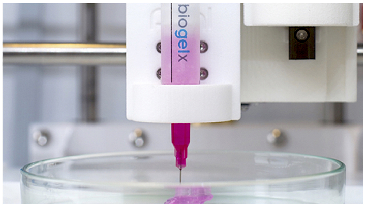
Credit: Lonza

“Everything we do at CELLINK, from live cell imaging to our innovative bioprinting systems and bioinks, is meant to support our customers with the products and services needed for them to more effectively and efficiently research solutions to some of the most important challenges of our time. Challenges such as cancer therapeutics, regenerative medicine and the testing and development of drugs, to name a few,” said Ginger Lohman, Biodispensing Product Manager at Cellink’s Gothenburg headquarters in Sweden. “When it came time to expand our portfolio into complete 3D cell culture workflows, we knew it was critical that we brought the right partner onboard. We’re confident that Lonza is that partner.”

According to Cellink, cell biologists will now be able to rely on a high-performing product portfolio to successfully execute some of the most demanding work on 3D bioprinting and boost their scientific research. The proposed solution under the partnership combines Cellink’s 3D bioprinting instruments and bioinks with Lonza’s primary cells and culture media, to meet the needs of cell biologists for enhanced bioprinting of complex 3D human tissue constructs.

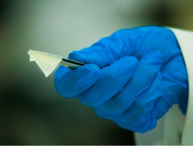
Since the origins of cell culture more than a century ago, cells have been cultured in two-dimensions; however, 3D cell culture has proven to be a better model for representing in vivo conditions, offering a more accurate and reliable means of predicting and analyzing cell behavior. In fact, it has proven to be a great enabler for scientists to handle cells in vitro while obtaining results that are closer to the in vivo environment without relying on animals, thereby avoiding many ethical issues.

With so many benefits, 3D cell cultures are being widely adopted in numerous laboratories—even more so as researchers look to create increasingly complex 3D constructs and find solutions to structural and material engineering challenges. For years, Swiss-based Lonza has been developing the building blocks needed to create 3D cell culture models, offering an extensive array of human-derived primary cells and culture media, ethically sourced and authenticated thorough quality control testing.

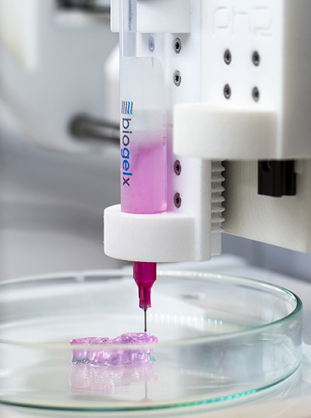
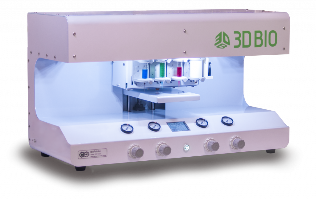
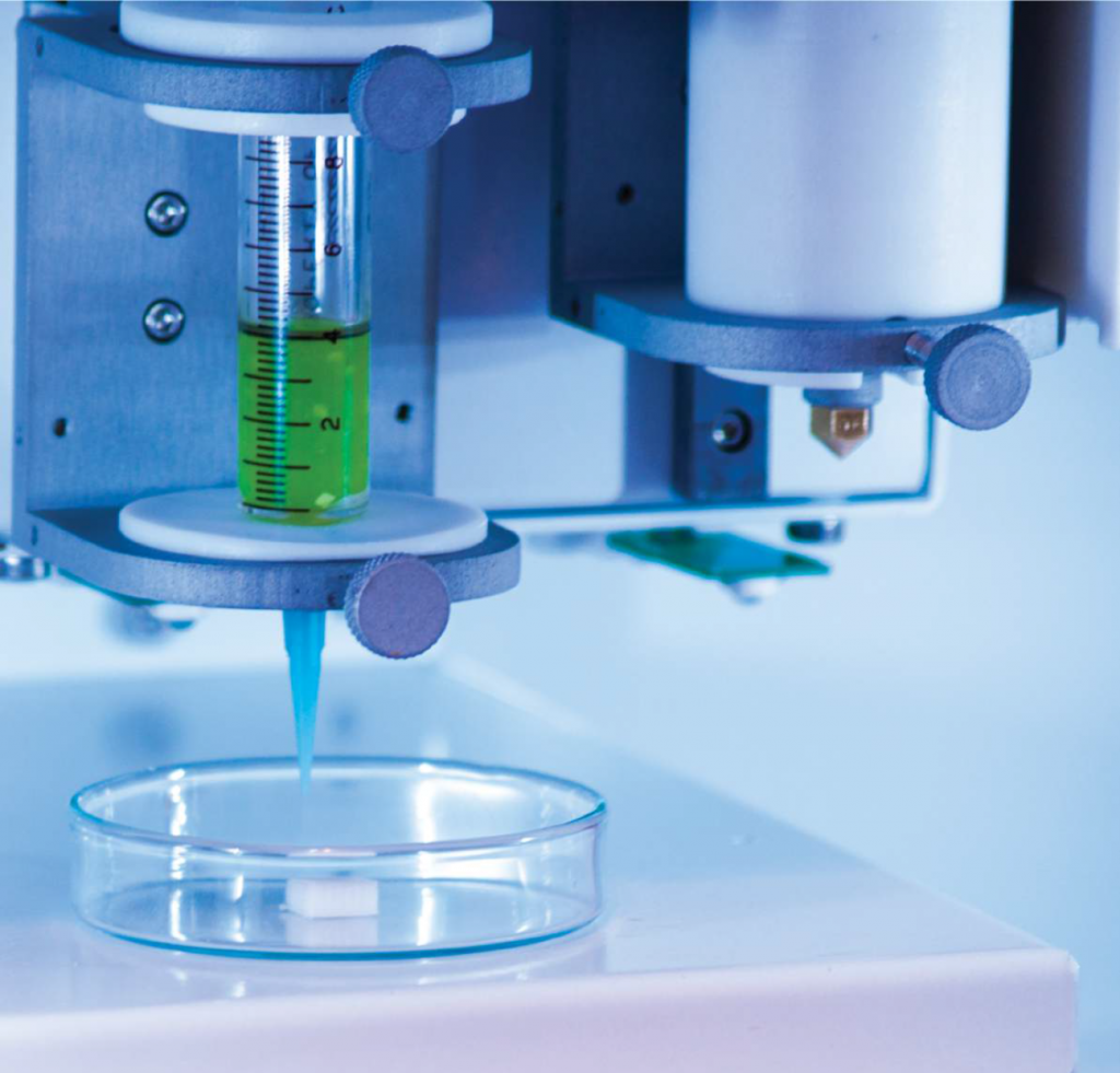
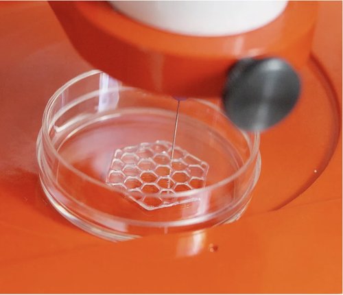
Robust, viable cells are an essential component of any successful cell culture application, and so is 3D bioprinting. A big part of the 3D cell culture workflow involves 3D bioprinting, which is thriving as a powerful technology for engineering complex 3D tissues for in vitro drug discovery research. Cellink is currently providing a wide range of 3D bioprinting systems, like INKREDIBLE, Bio X6 systems, and LumenX, which stems from a collaboration with Volumetric, a startup established by Rice University professor Jordan Miller. Furthermore, since 2016, the company has successfully commercialized the world’s first universal bioink designed to print complex 3D human tissue constructs with any 3D bioprinting system. The biomaterial is useful for scientists as it can be modified with peptides and growth factors to develop a series of customized bioink formulations to meet varying application needs and is used in hundreds of labs around the world.

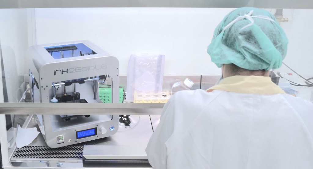
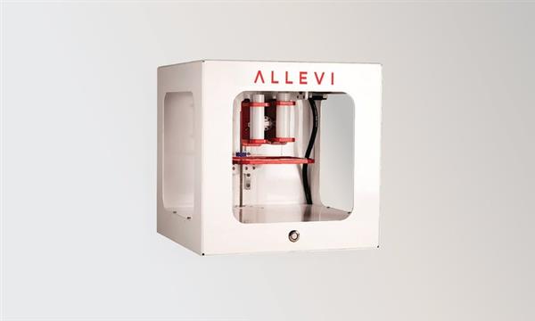
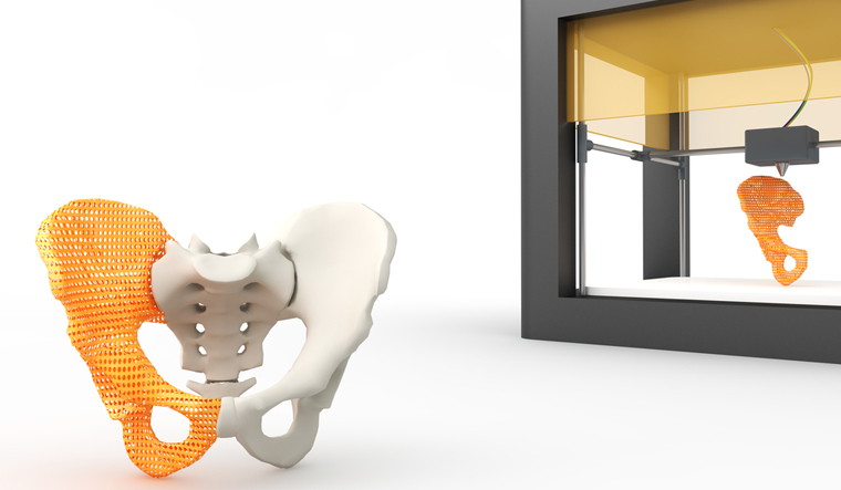
Cellink’s INKREDIBLE system (Credit: Cellink)

“Cell biology laboratories are constantly seeking innovative new technologies to enhance their experimental workflows and help deliver on their promise to drive the next research breakthrough,” explained Katrin Hoeck, head of marketing for Cell Analysis and Testing Solutions at Lonza. “Our broad panel of human-derived primary cells is specifically engineered to enable researchers to develop biological in vitro model systems that more closely reflect disease biology. This new collaboration with Cellink will enable our customers to build physiologically relevant 3D models to accelerate target identification/validation, investigation of mechanisms of action and safety testing in drug discovery.”

Under the agreement, Cellink will provide this complete solution through its global sales channels, supported by Lonza’s well-established logistics processes.

This is not the first partnership with the pharma industry for Cellink. The company also recently announced a collaboration with AstraZeneca, the British-Swedish multinational pharmaceutical and biopharmaceutical company, to utilize Cellink’s 3D bioprinting technology for liver organoid culture for drug discovery purposes in cardiovascular, renal and metabolic diseases.

According to Lonza, “The pharma and biomedical research laboratories are constantly seeking innovative new technologies to enhance their experimental workflows and help deliver on their promise to drive the next research breakthrough.” This collaboration could strengthen the processes and end products of research, offering substantial benefits for more predictive cell models and ultimately increasing the usefulness of 3D culturing cells for many applications in major areas of life sciences.”

The post Cellink Teams with Lonza to Advance Bioprinting Cell Cultures appeared first on 3DPrint.com | The Voice of 3D Printing / Additive Manufacturing.

Aalto University Develops a Novel Bioink for Cardiac Tissue Applications

Finland is one of Europe’s most forested nations. Over 70 percent of the country’s boreal forest is covered with spruce, pine, downy birch, and silver birch. But beyond the splendor of the Finnish woodlands, all these trees have one thing in common, and that is nanocellulose. A light solid substance obtained from plant matter which comprises cellulose nanofibrils (CNF) and is considered a pseudo-plastic that possesses the property of specific kinds of gels that are generally thick in normal conditions. Overall, it is a very environmentally friendly and non-toxic substance that is compatible with the human body and has the potential to be used for a range of medical applications.

In 2018, the Department of Bioproducts and Biosystems at Aalto University, located just outside Helsinki, began searching for new ideas to revitalize one of the country’s traditional economic engines, forests (which are handled sustainably thanks to renewable forest resources). At the time, they noticed that one of the possible applications could be working with nanocellulose. Forward two years and the researchers have come up with a new bioink formulation praising nanocellulose at its basis.

Thanks to the structural similarity to extracellular matrices and excellent biocompatibility of supporting crucial cellular activities, nanocellulose-based bioprinting has clearly emerged for its potential in tissue engineering and regenerative medicine. The qualities of the generally thick and fluid light substance make it an excellent match to develop bioinks that are both suitable and scalable in their production, but also have consistent properties. However, there have been major challenges in processing nanocellulose.

As described by Aalto University researchers in a recently published paper in the science journal ACS Publication, the unresolved challenges of bioink formulations based on nanocelluloses are what stops the substance from becoming one of the preferred components for 3D bioprinting structures. This is why Finnish researchers focused on developing a single-component bioink that could be used to create scaffolds with potential applications in cardiac biomedical devices, while fundamentally dealing with some of the limitations of using nanocellulose-based bioinks.

A co-author of the paper and a doctoral candidate at Aalto’s Department of Bioproducts and Biosystems, Rubina Ajdary, told 3DPrint.com that “other than natural abundance and as a renewable resource, nanocellulose has demonstrated to have an outstanding performance in tissue engineering.” She also suggested that “recent efforts usually consider the use of nanocellulose in combination with other biopolymers, for example, in multicomponent ink formulations or to encapsulate nanoparticles. But we were interested in investigating the potential of monocomponent nanocellulose 3D printed scaffolds that did not require crosslinking to develop the strength or solidity.”

In fact, the Biobased Colloids and Materials (BiCMat) research group at Aalto University, led by Orlando Rojas, proposed heterogeneous acetylation of wood fibers to ease their deconstruction into acetylated nanocellulose (AceCNF). As a unique biomaterial opportunity in 3D scaffold applications, the team considered using nanocelluloses due to the natural, easy to sterilize, and high stability porosity of the substance, and chose to introduce AceCNF for the generation of 3D printed scaffolds for implantation in the human body. The team then went on to evaluate the interactions of the scaffolds with cardiac myoblast cells.
“Most modifications make the hydrogels susceptible to dimensional instability after 3D printing, for instance, upon drying or wetting. This is exacerbated if the inks are highly diluted, which is typical of nanocellulose suspensions, forming gels at low concentrations,” went on Ajdary. “This instability is one of the main reasons why nanocellulose is mainly combined with other compounds. Instead, in this research, we propose heterogeneous acetylation of wood fibers to ease their deconstruction into acetylated nanocellulose for direct ink writing. A higher surface charge of acetylated nanocellulose, compared to native nanocellulose, reduces aggregation and favors the retention of the structure after extrusion even in significantly less concentration.”
This is exactly why it was important for the researches to develop a single component bioink. Nanocellulose has shown promises when combined with other biopolymers and particles. However, Ajdary insists that benefits including similarity to the extracellular matrix, high porosity, high swelling capacity, ease of surface modification, and shear thinning behavior of cellulose, encouraged them to study the potential of monocomponent surface-modified nanocelluloses.

Acetylated nanocellulose (Credit: Aalto University School of Chemical Engineering)

The team at Aalto University used the sustainable and widely available nanocelluloses to make several formulations of bioinks and evaluate them, including unmodified nanocellulose CNF, Acetylated CNF (AceCNF), and TEMPO-oxidized CNF.
To 3D bioprint the hydrogels, researchers used Cellink bioprinters, something Ajdary attributed to the user-friendliness of the device and because it provided a lot of flexibility to test different types of hydrogels and emulsions produced in the research group.
In this new process, the single-component nanocellulose inks were first 3D printed into scaffolds using Cellink’s BIO X bioprinter, which is equipped with a pneumatic print head was used to extrude single filaments and form the 3D structures. Then freeze-dried to avoid extensive shrinkage, and sterilized under UV light. After sterilization the scaffold was ready and cells seeded on the samples.
“3D structures of acetylated nanocellulose are highly stable after extrusion in far less concentrations. The lower concentration in wet condition facilitates the scaffold with higher porosity after dehydration which can improve the cell penetration in the structure and assist in nutrient transport to the cells as well as in the transport of metabolic waste,” specified Ajdary.
The researchers claim that the method was successful as the 3D printed scaffolds were compatible with the cardiomyoblast cells, enabling their proliferation and attachment, and revealing that the constructs are not toxic. Although still in research stages, these bioinks and technique can be used for the inexpensive, consistent fabrication and storage of constructs that can be applied as base materials for cardiac regeneration.
What is novel in this study is the particular focus on single-component nanocellulose-based bioinks that open up a possibility for the reliable and scale-up fabrication of scaffolds appropriate for studies on cellular processes and for tissue engineering. Since this is an ongoing research, we can expect to read more published material from Aalto University researchers as they continue testing their unique technique even further.

Scaffolds corresponding to 3D printed AceCNF (Credit: Aalto University)

The post Aalto University Develops a Novel Bioink for Cardiac Tissue Applications appeared first on 3DPrint.com | The Voice of 3D Printing / Additive Manufacturing.

Texas A&M Researchers Make “NICE” Bioinks to Create Functional Bone Tissues

Managing bone defects and injuries using traditional treatments can be slow and expensive. When bones break, bone cells can usually repair them, unless the break is too large. In that case, clinicians have historically turned to bone grafts using non-essential segments of bone taken from other parts of the same patient; bone from the hips, pelvis, chin, or ribs can do the job. Unfortunately, this requires additional surgeries, which translates into more pain for patients, and basically, there is a limit to how much non-essential bone surgeons can take from a patient.

As in many fields, bioprinting is disrupting the way healthcare specialists think about solving problems. So with an estimated 500,000 annual bone grafting procedures in the United States and more than 2 million around the world, an efficient bone substitute could change millions of lives. In a search to fabricate patient-specific, implantable 3D constructs for regenerative medicine, scientists in the Department of Biomedical Engineering at Texas A&M University have developed a new bioink formulation for 3D bone bioprinting called NICE, which is short for Nanoengineered Ionic–Covalent Entanglement, and have gone on to demonstrate that this bioink can precisely reconstruct large bone structures based upon CT scans that were obtained from actual patients.

Led by Akhilesh K. Gaharwar, an associate professor in the Department of Biomedical Engineering, the research group developed a highly printable bioink as a platform to generate anatomical-scale functional tissues. Their study was recently published in the American Chemical Society’s Applied Materials and Interfaces scientific journal, whereby they state that the NICE bioinks allow precise control over printability, mechanical properties, and degradation characteristics, enabling custom 3D fabrication of mechanically resilient, cellularized structures.

Bioprinting requires cell-laden biomaterials that can flow through a nozzle like a liquid, but solidify almost as soon as they’re deposited. This is why bioinks need to act as both cell carriers and structural components, requiring them to be highly printable while providing a robust and cell‐friendly microenvironment. However, the research group realized that many current bioinks lack sufficient biocompatibility, printability, structural stability, and tissue‐specific functions needed to translate this technology to preclinical and clinal applications.

To address this issue, Gaharwar and his team are leading efforts in developing the more advanced NICE bioinks, essentially a combination of two reinforcement approaches, ionic-covalent entanglement, and nanoreinforcement. In fact, the researchers claim that to design the NICE reinforced bioinks for osteogenic tissue bioprinting, the bioink must be highly printable, mechanically strong, induce osteogenic differentiation, and be biodegradable. However, the difficulty of combining these requirements into a single bioink has been a major obstacle in bioprinting since its inception. So by combining these two distinct reinforcement methods, NICE becomes a robust and superior bioink while providing a highly hydrated and cell-friendly microenvironment for bone bioprinting.

NICE printed structures are highly flexible and resilient, as seen in these 3D printed tube structures that can be completely collapsed and quickly regain their shape (Credit: Texas A&M Engineering)

According to Texas A&M Today, Gaharwar said that developing replacement bone tissues could create exciting new treatments for patients suffering from arthritis, bone fractures, dental infections, and craniofacial defects.

“The next milestone in 3D bioprinting is the maturation of bioprinted constructs toward the generation of functional tissues,” Gaharwar said. “Our study demonstrates that NICE bioink developed in our lab can be used to engineer 3D-functional bone tissues.”

NICE bioinks have three major components: covalently crosslinkable gelatin methacryloyl (GelMA), ionically crosslinkable kappa-carrageenan (kCA), and electrostatically charged nanosilicates (Laponite XLG, obtained from BYK Additives & Instruments).
To bioprint scaffolds that can demonstrate potential clinical uses, the team used their osteoinductive NICE bioinks. Then human mesenchymal stem cells (hMSCs) were encapsulated in the NICE bioink and bioprinted into 3D scaffolds. Once bioprinting was complete, this cell-laden 3D printed NICE structures were crosslinked to form stronger scaffolds. This technique has allowed the lab to produce full-scale, cell-friendly reconstructions of human body parts, including ears, blood vessels, cartilage and even bone segments.

hMSCs are encapsulated in the NICE bioink and cell-laden scaffolds are printed (Credit: Texas A&M Engineering)

Soon after bioprinting, the enclosed cells start depositing new proteins rich in a cartilage-like extracellular matrix that subsequently calcifies to form a mineralized bone over a three-month period. Texas A&M stated that almost five percent of these printed scaffolds consisted of calcium, which is similar to cancellous bone, the network of spongy tissue typically found in vertebral bones.

The scaffolds are initially transparent but turn translucent due to remodeling and deposition of nascent proteins after 60 days (Credit: Texas A&M Engineering)

To understand how these bioprinted structures induce stem cell differentiation, the team worked with Irtisha Singh from the Texas A&M Health Science Center, who served as a co-investigator, to use a next-generation genomics technique called whole transcriptome sequencing (RNA-seq) technology, which takes a snapshot of all genetic communication inside the cell at a given moment.
As for the bioprinting, the researchers modified a commercial ANET A8 3D printer kit to utilize screw extrusion. They replaced the thermoplastic extruder assembly with a 3D printed screw extruder assembly, which holds a stepper motor, guide rail, and a modified clay extruder.

To illustrate the practical utility of NICE bioinks for bone tissue reconstruction, the team demonstrated how to create full-scale bioprinted implants customized for craniofacial defects on real patient CT scans. Relying entirely on open-source software, they used the free 3D modeling software Meshmixer to process the models and create bone defects, and the 3D printing applications PrusaSlicer and Repetier Host to bioprint the scaffolds. After bioprinting, the scaffold was crosslinked and implanted in a thermoplastic model of the lower jaw to demonstrate the closeness of fit. Strength of fit was also demonstrated by injecting and crosslinking NICE bioink between two sections of a full-thickness fracture to prove that NICE is able to quickly adhere surfaces together and resist shearing and delamination forces.

Funded by the National Institutes of Health (NIH)’s Director’s New Innovator Award, a National Science Foundation (NSF)’s Award and an X-Grant from Texas A&M University, the researchers suggest they have discovered a new way to design and produce 3D bioprinted bone tissue to benefit bone regeneration.

Moreover, Gaharwar claims to have demonstrated that the highly printable NICE bioinks can precisely reconstruct large bone structures from CT scans obtained from actual patients. The aim of the research is to enable patient-specific bioprinting of bone scaffolds to precisely match their injuries. The researchers stated their desire to have this technique act as a customizable and easy to work with an alternative to autografts that will provide surgeons with greater options for bone surgery. And with the ultimate goal of getting NICE bioink technology from bench to bedside, Gaharwar’s team plans to establish the in vivo functionality of the 3D bioprinted bone tissue.

3D-bioprinted NICE scaffolds can be used for bone regeneration (Credit: Texas A&M Engineering)

The post Texas A&M Researchers Make “NICE” Bioinks to Create Functional Bone Tissues appeared first on 3DPrint.com | The Voice of 3D Printing / Additive Manufacturing.

CollPlant 3D bioprinted materials business closes $5.5M in funding

3D bioprinter material developer CollPlant (NASDAQ:CLGN) has confirmed an investment of $5.5 million. Provided by a group of investors with special interest in the 3D printing industry and Ami Sagi, the company’s largest shareholder, the deal was confirmed September 9, 2019.  $3.5 million in convertible loans was provided by the anonymous investor group, whereas the remaining $2 […]

Omid Afarinan is Bioprinting in Iran

When we were assembling our bioprinting world map, we omitted some companies. We are adding to that map and will continue to do so until we have everyone. One of the firms that we did not initially find is Omid Afarinan, also known as 3D Bio. This is an Iranian firm that makes bioprinters and bioinks. We have thus far seen comparatively little 3D printing and bioprinting activity in Iran, so we were more than happy to do an interview with the firm to find out more (as well as apologies for forgetting them in the world map!).

What does your company do?

Our company works on the whole bioprinting chain from tissue design to application stage. The focal point of our efforts in this chain is design, fabrication and development of commercial bioprinters. All research and development attempts are defined based on the targeted tissue or in other words the tissue need and market trend. Based on this fact, the target tissue determines the customization of the bioprinters, the choice of living cells and bioinks creating a multi-disciplinary ecosystem of scientific fields.

Where do you hope to be in five years?

Our mid-term goal in the next five years is turning into a leading company in bioprinting research and development with several specialized laboratories and the sole bioprinting service hub. Achieving the first transplantable living tissue would be the ultimate goal in this period.

Why should someone choose to work with you?

Our team has the capability of creating a wide range of customizations in bioprinter design both on the hardware systems and software. This capability leads to customer-made versions and tissue-specific printers. Our bioprinters are of high quality and competitive to its foreign counterparts from the accuracy and cost point of view. Our exemplary teamwork among various experts and the existence of a multi-profession environment led to current achievements and also resulted in training talented trainees. Our team welcomes specialized groups and eager students especially in the field of molecular and cellular biology to strengthen its abilities.

What are the differences between the Biofab and the Pioneer series?

The main difference between these series is their printing mechanism. The Pioneer bioprinter is extrusion-based using screws while Biofab utilizes pneumatic actuators for printing. This, in turn, makes Biofab more capable especially in the case of accuracy. The Pioneer version possesses the ability of unparalleled control over print heads and can print a variety of hydrogels. Moreover, the Biofab version supports a wide range of biomaterials and viscous cell suspensions for printing.

Both series provided in two versions, with 2 or 4 printing heads.

What kind of bioinks have you developed?

Omid Afarinan, as the first national company in Iran, has gathered experts from different fields of science with the aim of producing novel bioinks. Omid Afarinan bioinks are cost-effective, have high printability, mimic extracellular matrix (ECM) and provide a suitable environment for cell proliferation, growth, and differentiation. Some of the new unique bioinks of the 3D-Bio Team are PCL, PCL/Starch, Alginate and Alginate/Gelatin bioinks. Soft-Ink is the newest bioink of the company; a biodegradable bioink based on pure Alginate and Gelatin which can support growth and proliferation of any cell type of soft tissues. One of the main features of this bioink is high printability and uniformity with the ability to adjust the stiffness of its printed matrix.

In addition to a variety of bioinks from thermoplastic materials to hydrogels, the company also produces custom-made bioinks for specific applications.

What are customers doing with your printers?

The customers mainly use the printer for conducting state-of-the-art researches especially on creating regenerative tissues for transplantation and studying the behavior of living tissues. In particular, our current customers work on hard tissue as bone scaffold especially maxillofacial and soft tissue including cartilage, skin, cornea and heart. The study on drug delivery, cosmetic research and cancer treatment are other aspects of what our customers do with bioprinters.

What short term successes do you see occurring in bioprinting?

Successes in bone and cartilage tissues are promising in recent years. This is due to the fact that such tissues have low cell densities. This is more pronounced in the bone tissue where acellular scaffolds can be used. Skin printing comes next in the list on the soft tissue side, for being a flat and multi-layered tissue. Also, Bioinks are being developed in parallel but at a slower pace. These short-term successes would pave the path for tissues with more complex geometries.

Where is bioprinting challenging?

The challenges exist in both pre- and post-printing stages of bioprinting. At pre-printing stage, the challenge is on the printability of biomaterials and Bioinks. In other words, making the materials printable and the suitability of the printing is of prime importance. Overcoming this challenge leads to printed functional tissues that could mimic real ones. On the post-printing stage, the challenges mainly arise from the complexity in living tissues especially vascularization and post-printing processes such as cell culture and migration that makes the printed tissues ready for their applications. These challenges and issues need close collaborations between different experts to resolve in short and long-term periods.


What advice would you have for a researcher wanting to get into bioprinting?

The first advice is that the researcher should wear iron shoes. In other words, facing many difficulties is unavoidable and gaining achievements may take time. So, patience and being hopeful is the key point. Furthermore, bioprinting requires a multi-disciplinary group including engineers, biologist and doctors and no one can individually succeed in this path. Finally, utilizing a standard and reliable bioprinter could be beneficial and time-saving for any researcher. 

The post Omid Afarinan is Bioprinting in Iran appeared first on 3DPrint.com | The Voice of 3D Printing / Additive Manufacturing.

3D Printing Industry Experts Interview With Ricky Solorzano Co-Founder and CEO of Allevi

Ricky Solorzano

Ricky Solorzano is the Co-Founder and CEO of Allevi. Ricky is looking to combining IoT, organ engineering, and 3D Printing to bring bioprinting solutions to the World, Space, and Beyond. Allevi, Inc. believes 3D tissues will have a huge impact on humanity and create an entire new industry. They want to help scientists create more accurate hearts, lungs, and even brains in the lab. Users are automating the creation of tumor models, printing vasculature within 3D gels, and achieving physiological markers unseen before in tissues.

Explain how you got to your current state in life?

Following my passion got me to this point. It was really about pursuing tissue engineering and thinking it had the possibility to change the world. At first I thought about medical school, but then I had a chance to start a company which was really cool.

What is your educational background?

I went to bioengineering school and worked in a tissue lab. I learned about geometry and how tissue engineering is important. I also took an entrepreneurship class in college. It opened my eyes to different ways one can affect the world around me.

Are startups on the cutting edge vs universities?

Our value is giving the scientist the ability to execute the idea. It is important for us to give people value and for them to bring theirs. It’s the combination of this co-creation that really pushes the fields forward.

What is your value proposition?

We are able to have a lot of power go into a bioprinter. We make them very friendly and easy to use still. We are abstracting the complexity of a bioprinter to simplicity.

Allevi

What are future thoughts on this industry?

More and more tissues will be in the drug testing space. There will be more and more adoptions in investigative PIs (private investigators) to have a bioprinter within their labs. More companies will realize that it is important to have one in their labs. It is important to apply geometry to biology.

A lot of people within the industry of bioprinting are focusing on the importance of geometry applied to biology. Could you explain this a bit more?

Hearts need to be aligned. Shapes need to be consistent when created. It causes an organ or body part to be functional or not. Without geometric properties being maintained, parts will not work as efficiently as they could.

What would you do outside of this if you were not running Allevi?

Tissue engineering product development is probably where I would be. Being able to commercialize things is great way to make an impact. I have been able to learn this really well and continue.

Allevi Dual Extruder

What skills should people be looking into to be in this industry?

Be passionate about a specific direction. If you are passionate about using a bioprinter, work as a bioengineer. It is really important to have a skill set within biology and biomaterials. I think every field can contribute, it just matters what makes you personally excited.

What is the toughest obstacle in terms of work?

Discovery and where do we go. It is hard to figure out what the next step is. We do not have a clear guidance on what to do. It is important to understand the industry as a whole and where it is progressing.

Tel Aviv University: Researchers 3D Print Cardiac Patches & Cellularized Hearts

Researchers at Tel Aviv University continue to try to meet the ongoing challenges in cardiac tissue engineering. In ‘3D Printing of Personalized Thick and Perfusable Cardiac Patches and Hearts,’ authors Nadav Noor, Assaf Shapira, Reuven Edri, Idan Gal, Lior Wertheim, and Tal Dvir outline the steps they took to match technology with tissue.

Cardiovascular disease is the leading killer of patients in the US, and organ donor and transplantation processes can still mean a long wait for those suffering from heart failure. Here, the authors demonstrate the need for alternative ways to treat the infarcted (usually referring to clogging of one of more arteries) heart. And while tissue engineering has pointed the way to freeing many patients from terrible physical suffering and organ donor waiting lists, creating the necessary scaffolds with true biocompatibility has presented obstacles.

The authors have created an engineered cardiac patch meant to be transplanted directly onto the patient’s heart, integrating into the ‘host,’ with excess biomaterials degrading over time. This leaves the cardiac patch, full of live, healthy tissue, regenerating a previously defective heart. Because there is always the threat of rejection when implanting anything into the body though, the authors emphasize the need for appropriate materials:

“Most ideally, the biomaterial should possess biochemical, mechanical, and topographical properties similar to those of native tissues,” state the researchers. “Decellularized tissue‐based scaffolds from different sources meet most of these requirements. However, to ensure minimal response of the immune system, completely autologous materials are preferred.”

The researchers were able to create patient-specific cardiac patches in their recent study, extracting fatty tissue from cardiac patients—and then separating cellular and a-cellular materials.

“While the cells were reprogrammed to become pluripotent stem cells, the extra‐cellular matrix (ECM) was processed into a personalized hydrogel,” stated the researchers.  “Following mixture of the cells and the hydrogel, the cells were efficiently differentiated to cardiac cells to create patient‐specific, immunocompatible cardiac patches.”

In using the patient-specific hydrogel as bioink, the researchers were able to create patches, but ultimately, they were also able to 3D print comprehensive tissue structures that include whole hearts.

An omentum tissue is extracted from the patient and while the cells are separated from the matrix, the latter is processed into a personalized thermoresponsive hydrogel. The cells are reprogrammed to become pluripotent and are then differentiated to cardiomyocytes and endothelial cells, followed by encapsulation within the hydrogel to generate the bioinks used for printing. The bioinks are then printed to engineer vascularized patches and complex cellularized structures. The resulting autologous engineered tissue can be transplanted back into the patient, to repair or replace injured/diseased organs with low risk of rejection.

The authors used two different models in their study, with one serving as proof-of-concept, with pluripotent stem cells (iPSCs)‐derived cardiomyocytes (CMs) and endothelial cells (ECs). The other model relied on:

  • Rat neonatal CMs
  • Human umbilical vein endothelial cells (HUVECs)
  • Lumen‐supporting fibroblasts

One bioink, laden with cardiac cells, printed parenchymal tissue, while the other extruded cells for forming blood vessels. The researchers were successful in 3D printing the patient-specific cardiac patches but found when a higher degree of complexity was necessary for fabrication of organs or other tissues, the hydrogels were not strong enough. They created a new process for organs and more complex tissues where they could print in a free-form manner and cure structures at varying temperatures; they were able to overcome previous challenges and 3D print accurate, personalized structures.

Bioinks characterization. A human omentum a) before and b) after decellularization. c) A personalized hydrogel at room temperature (left) and after gelation at 37 °C (right). d) A SEM image of the personalized hydrogel ultrastructural morphology, and e) a histogram of the fibers diameter. f) Rheology measurements of 1% w/v and 2.5% w/v omentum hydrogels, showing the gelation process upon incubation at 37 °C. g) Stromal cells originated from human omental tissues were reprogrammed to become pluripotent stem cells (red: OCT4, green: Ki67 and blue: nuclei). h) Differentiation to ECs as determined by CD31 (green) and vimentin staining (red). Differentiation to cardiac lineage: i) staining for sarcomeric actinin (red), j) staining for NKX2‐5 (red), and TNNT2 (green). Scale bars: (e) = 10 µm, (g,i,j) = 50 µm, (h) = 25 µm.

This study carries substantial weight, considering the researchers were able to create cellularized hearts with ‘natural architectures.’ This furthers the potential for cardiac transplants after heart failure, along with encouraging the process for drug screening. The authors point out that more long-terms studies and research with animal models are necessary.

“Although 3D printing is considered a promising approach for engineering whole organs, several challenges still remain,” conclude the researchers. “These include efficient expansion of iPSCs to obtain the high cell number required for engineering a large, functioning organ. Additionally, new bioengineering approaches are needed to provide long‐term cultivation of the organs and efficient mass transfer, while supplying biochemical and physical cues for maturation.”

“The printed blood vessel network demonstrated in this study is still limited. To address this challenge, strategies to image the entire blood vessels of the heart and to incorporate them in the blueprint of the organ are required. Finally, advanced technologies to precisely print these small‐diameter blood vessels within thick structures should be developed.”

Imaging of the heart and patch modeling. CT image of a) a human heart and b) left ventricle coronary arteries. c) A model of oxygen concentration profile in an engineered patch. d) Replanning of the model showed better oxygen diffusion, sufficient to support cell viability.

Without good heart health, it is very difficult to survive. Responsible for transporting nutrients, oxygen, and more to cells populating the human body, the heart also removes waste like carbon dioxide and more. 3D printing is assisting scientists and doctors in researching and treating a variety of different diseases and conditions, whether they are using 3D printed metamaterials for fabricating heart valves, creating better cardiac catheters, or experimenting with new types of phantoms.

What do you think of this news? Let us know your thoughts! Join the discussion of this and other 3D printing topics at 3DPrintBoard.com.

Printing thick vascularized tissues. a) A top view of a lumen entrance (CD31; green) in a thick cardiac tissue (actinin; pink). b) A model of a tripod blood vessel within a thick engineered cardiac tissue (coordinates in mm), and c) the corresponding lumens in each indicated section of the printed structure. d) Tissue perfusion visualized from dual viewpoints. e–k) A printed small‐scaled, cellularized, human heart. e) The human heart CAD model. f,g) A printed heart within a support bath. h) After extraction, the left and right ventricles were injected with red and blue dyes, respectively, in order to demonstrate hollow chambers and the septum in‐between them. i) 3D confocal image of the printed heart (CMs in pink, ECs in orange). j,k) Cross‐sections of the heart immunostained against sarcomeric actinin (green). Scale bars: (a,c,h, i,j) = 1 mm, (g) = 0.5 cm, (k) = 50 µm.

[Source / Images: 3D Printing of Personalized Thick and Perfusable Cardiac Patches and Hearts]

Biodiscoveries: CELLINK is bioprinting its way into the future

Back in 2015, Erik Gatenholm realized there was no place to purchase bioink for 3D bioprinting. So, blown away by this gap in the market, he quickly worked with co-founder Hector Martinez to create a universal bioink that anyone working with bioprinting could use. It was quite a high stakes bet, and at the beginning they set up a webshop to see if they got any bites. It only took 24 hours for the first sale. With more orders quickly coming in, they realized the enormous potential of the product they developed, and CELLINK was born, becoming the first company to commercialize a universal bioink for bioprinting of human tissues and organs.

CELLINK co-founders Erik Gatenholm and Hector Martinez

In the United States alone, every 10 minutes another person is added to the growing waiting list for organ transplants, most of them (60%) in need of a kidney, and with over 130,000 organs transplanted every year worldwide, is no wonder how demand certainly outweighs supply almost everywhere. In some countries the wait can take years, making 3D printing of organs one of the most sought after technologies out there. Bioprinting in the future could allow patients and doctors to reduce waiting times, increase compatibility and decrease immunological failure. For this to happen medical researchers will need to design organs using modeling software, and then print them with biomaterials such as polymers and hydrogels, in addition to the patient’s own cells. Although currently focused on growing cartilage and skin cells suitable for testing drugs and cosmetics, the Swedish company founded in Gothenburg in 2016, hopes to progress the technology far enough to create replacement organs for transplant in humans in the next 15 years.

“In the coming decade we would like to continue to push the boundaries of 3D bioprinting until it becomes an established technology in the medical field. We have the vision of becoming the first and number one provider of bioprinters, bioinks, software and technical know-how for the next generation of medical device manufacturers,” co-founder and CTO Hector Martinez told 3DPrint.com.

Their unique bioink is a biomaterial innovation that allows human cells to grow and thrive as they would in circumstances close to their natural environment. The startup has already managed to print human skin and is also working on producing liver tissues, as well as the beta cells that produce the insulin we need to survive. In 2018 it began printing tumors to combat cancer as part of a research project that doesn’t endanger human lives, and just a few weeks ago, it teamed up with Volumetric to develop Lumen X, a digital light processing bioprinter, designed to enhance inventions in creating more substantial vascular structures. Skin care products, topological drugs and medical treatments are all in need of enhanced testing procedures that can increase the transability from in vitro testing to in vivo usage of products. With tissue engineering and 3D bioprinting more representative in vitro models can be constructed, limiting the use of testing in animals.

CELLINK’s bioinks

Actually, academic labs and companies worldwide are trying to bioengineer all kinds of sophisticated creations for regenerative medicine, drug testing, screening, and tissue engineering. So it’s no wonder CELLINK has their research team focused on creating the next generation of bioinks. Their top selling product is making bioprinting much easier than it used to be some 10 years ago, with 30 different types of bioink available, with prices that go from 99 to 900 dollars. So, what makes one bioink more expensive than the other? It’s all about the components. Collagen and laminin are more expensive to produce than gelatin, raising the price of the end-product. According to CELLINK, scientists mix their live cells into the company’s bioink, a kind of gel designed to allow cells to survive and multiply. The ink is then loaded into a 3D printer by the customer, which forms the desired shape layer by layer as the gel solidifies. By the time the lights inside CELLINK’s box turn green, researchers have an object that acts like human tissue, and can then apply their drug and see how the living cells inside respond.

CELLINK team printing liver models at the lab

“Today we are taking the necessary steps to build and expand our technology offering and exploring new methods for bioprinting tissues. Such technologies include multiple contact-less dispensing methods and light-based bioprinting techniques that enable the bioprinting of high resolution tissue constructs. Refining such technologies will take a close collaboration with our customers as we define the best practices for bioprinting different tissues and specific functions. We can already anticipate that the integration of different bioprinting technologies with post-bioprinting, real-time monitoring systems will be of utmost importance as the bioprinted tissue matures and attains a specific function through an active and precise manipulation of its environment.” Chief IT Officer Jockum Svanberg explained to 3DPrint.com.

Creating the raw material for bioprinting processes is no easy task. Cellink has been focusing on process-compatible soft biomaterials loaded with living cells to create its bioinks since September 2015. The process of bioprinting requires a delivery medium for cells which can be deposited into designed shapes acquired from computer-aided design (CAD) models, which can be generated using 3D medical images obtained through MRIs or CT scans. Some important features of an ideal bioink material are bioprintability, high mechanical integrity and stability, insolubility in cell culture medium, biodegradability at a rate appropriate to the regenerating tissue, non-toxicity and non-immunogenicity, and the ability to promote cell adhesion. Some bioink types, like hydorgels, are not always suitable as construction materials which is why CELLINK is working on a study to provide an upgraded version of the current CELLINK BONE bioink by incorporating collagen and hydroxyapatite. The bioink currently offered does not get close to the real stiffness of the natural bone tissue, but finely resembles its chemical composition. The advantage of such a soft material is to be able to incorporate cells and, during the bioprinting process, to locate them at a precise position throughout the scaffold. This is still for research use only and might take a few years until it is compatible for human use.

With CELLINK bioinks, 3D bioprinting of tissues will help hasten bone fracture healing

Since its start the technology firm has grown to become one of the big competitors in the industry. CELLINK had only been in existence for ten months before they decided to pursue their IPO in November of 2016, listing on Nasdaq First North after a 1070% oversubscribed IPO, which means that demand for their shares was ten times what they expected. Since then, shares have risen over 400%, giving the company a present-day market cap of around $257 million. CELLINK’s affordable printers have already been bought by customers in 25 countries around the world, mostly universities, like Stanford, Harvard, Yale, Princeton and MIT, and some private customers, including Shiseido, Roche, Merck, Johnson and Johnson, and Toyota

But it’s not just about bioprinting it’s way into the future of medicine, CELLINK is also working with other disruptive technologies, such as machine learning. CELLINK told 3DPrint.com that “they want to empower our users with better tools to simplify the bioprinting learning process and broaden its adoption”. One example of this is by developing algorithms that analyse printed structures and based on the results can recommend printing parameters to the users. Using this tool in the development, has helped them speed up the bioink development process. They have just launched a new product: CELLCYTE X, a live cell imaging microscope with live monitoring and analysis of cells in the cloud. Traditionally cell studies have involved manual labor and relied on analysis of the images from an expert, but using deep learning models they are automating this process to provide better and more reliable analysis to their users. The system relies on the latest in serverless system architecture to provide the most scalable, reliable and most intuitive system on the market.

What do you think, will CELLINK continue its upward trajectory? Will it become superseded by other larger firms or get passed by newer start ups? Find out more through our series of articles exploring bioprinting, Biodiscoveries.

Colombian Researchers Study Potential for SIS-Based Photocrosslinking in Bioinks

(a) Preparation of the 0.5% (w/v) riboflavin (RF) bioink and (b) its successful extrusion through a 21 G needle. (c) Filament formation during extrusion of the bioink through a 21 G needle. (d) Presumed photo-mediated crosslinking reaction thought to be occurring in the proposed bioinks.

Colombian researchers performed a recent study, outlined in ‘Formulation and Characterization of a SIS-Based Photocrosslinkable Bioink,’ explaining the possible value in crosslinking to create better materials for 3D printing cells. Here, they are using small intestinal submucosa (SIS) with photocrosslinked reactions to manipulate the gelation process, despite some expected challenges.

While the use of natural materials is always preferable, the researchers point out that they can also be difficult to work with due to lack of strength and stability. In the end that leads to inferior printability and further challenge.

“An avenue through which to overcome these issues is to mix them with synthetic polymers such as polyethylene glycol (PEG), polylactic acid (PLA), and polycaprolactone (PCL), which have demonstrated their ability to alter the mechanical response upon blending,” state the researchers. “Additionally, they have been proven able to shorten degradation rates, though at the expense of decreasing their biocompatibility.”

The authors also began to study decellularized extracellular matrices (dECMs) further, as they have the potential to copy the natural cellular environment. dECMs include the following proteins:

  • Collagen
  • Elastin
  • Laminin
  • Glycosaminoglycans
  • Proteoglycans
  • Growth factors

dECMs are not always stable though, and that presents challenges in bioprinting:

“Despite these obstacles, several research groups worldwide have attempted the development of bioinks based on dECMs,” state the researchers. “For inducing gelation, these studies have incorporated thermally-induced or photocrosslinking mechanisms, as well as a combination of the two.”

“Despite the crosslinking strategy implemented, the achieved mechanical stability has been observed to still not be sufficient, thereby requiring the use of synthetic materials as structural improvement supports.”

Shear stress profiles of the bioinks at different nozzle diameters and extrusion pressures: (a) 10 kPa, (b) 20 kPa, and (c) 60 kPa.

UV light has been used previously to increase the bioink stiffness in photocrosslinking, and for this study the authors experimented with the SIS dECM-based materials, using riboflavin (RF) as a photoinitiator. Visible light was used for the photocrosslinking. The research team created four different types of bioinks, with successful printability.

“Our experiments suggest that a successful extrusion can be accomplished while the pressure is maintained in the range 25–45 kPa,” stated the researchers.

(a–c) Viscosity and shear rate as a function of time, measured at different points of the nozzle tip geometry (center, middle, and wall). Structural parameter for the three extrusion nozzles studied with diameters of (d) 0.21 mm, (e) 0.25 mm, and (f) 0.41 mm.

They also went on to state that these bioinks demonstrate strong mechanical properties that could ensure success in bioprinting endeavors—following in line with previous research studies where crosslinking resulted in excellent printability parameters, as well as offering better integrity in shape.

“Further in silico experiments allowed us to calculate a stability parameter that provided conceptual evidence for the aggregation of collagen in times as short as 5 s,” concluded the researchers. “Finally, rheology tests allowed us to recover power law parameters for CFD simulations that confirmed shear stress values low enough to maintain high cell viability levels.

“Future work will be focused on reformulating the bioink with the aid of synthetic polymers and/or thermal processing such that collagen fibers remain in an extended state and are readily accessible to the photoinitiated molecules.”

The study of materials in 3D printing has become a vast realm, and a necessary one for those dedicated to such progressive fabrication techniques. It is also a very serious area of study for scientists engaged in seeking out the best ways to grow tissue in the lab, with the potential for making serious impacts in medicine.

Researchers around the world are on an intense journey to perfect bioprinting, and eventually, reach the pinnacle of success in fabricating human organs. The challenge today, as tissue engineering results in a variety of different implants, is to keep cells alive to serve their function in bioprinting. This means seeking out the best bioprinted structures to build, bio-inks, printers, and techniques. Find out more about photocrosslinkable inks here. What do you think of this news? Let us know your thoughts! Join the discussion of this and other 3D printing topics at 3DPrintBoard.com.

[Source / Images: ‘Formulation and Characterization of a SIS-Based Photocrosslinkable Bioink’]

Biogelx Launching First Product Range of Synthetic Bioinks for Variety of 3D Printing Applications

In 2013, a company called Biogelx was spun out from the University of Strathclyde in Glasgow, Scotland for the purposes of developing tunable, synthetic materials for use in 3D cell cultures and 3D bioprinting applications. Early on, the company worked on creating 3D cell culture scaffolds, which were in the form of synthetic peptide hydrogels so as to support cell growth by behaving as an extracellular matrix environment.

Bioinks are loaded up with cells in order to 3D print biological structures. Then, once the structure is printed, secondary crosslinking mechanisms help retain the shape’s structural fidelity. These bioprinting materials can facilitate cell adhesion, differentiation, and proliferation, as well as exhibit all the characteristics of an extracellular matrix environment. This is what makes it possible to create patient-specific human tissues in a laboratory setting, which is why good bioinks are very important.

Biogelx’s hydrogel bioinks have unique physical and chemical tunability, which means they can successfully replicate specific tissue characteristics so cells can engage with, and experience, a 3D environment that’s pretty close to real life. Early on, the industry recognized how beneficial Biogelx’s hydrogel bioinks could be for researchers in the industry, as the materials claim to offer reproducibility, great printability, an easy crosslinking method, and viscosity control in one package. The company’s bioinks can provide a base modular material where the cells’ chemical and mechanical properties are able to be adapted.

That’s why Biogelx is proud to launch its first product range of novel, synthetic bioinks: Biogelx-INKS. As the company says on its website, the future is synthetic.

“We are excited to announce the commercial availability of Biogelx™-INKs,” said Biogelx CEO, Mitch Scanlan. “Providing versatility and improving research outcomes are the key focuses for our product portfolio. We look forward to supporting researchers in their mission to develop realistic 3D disease modules, tissues, and organs for future pharmaceutical and medical applications.”

Developed with 3D printability at the forefront, Biogelx-INKs have been optimized for use in extrusion 3D printers, and they’re also versatile enough to work in 3D printing applications other than just bioprinting. The bioinks maintain the company’s core self-assembling peptide technology, and just like Biogelx’s hydrogel products, these new Biogelx-INKs form a nanofibrous network in order to mimic an extracellular matrix environment. This supports cell growth, proliferation, and signaling, but in addition, Biogelx-INKs were developed in such a way as to, as the company put it, “ensure the rheological properties are suitable for bioprinting applications.”

According to the Biogelx website, its new bioinks can be printed with good 3D fidelity, and don’t require any support, sacrificial, or curing inks. Biogelx-INKs also offer important shear-thinning behavior, which helps with cell viability, and controllable gelation, which is triggered by adding cell culture media and does not require the addition of reactive crosslinking reagents, UV curing, adjustments in pH, or extreme temperature. This feature also makes the material compatible with many different 3D bioprinters.

The company’s technology is based on a system of two peptides – a hydrophobic ‘gelator’ peptide (Fmoc-diphenylalanine) and a hydrophilic ‘surfactant’ (Fmoc-serine) – which self-assemble to form fibers in aqueous environments. These fibers have surface hydrophilic functionality, which is appropriate for cell adhesion.

Additional features of Biogelx-INKs include:

  • Reproducible – these bioinks are totally synthetic and manufactured under strict quality control, which ensures batch-to-batch reproducibility and consistent prints.
  • Tuneable – it’s possible to tailor the biomimetic functionality of the bioinks to specific cell types.
  • Biocompatibility – composed of amino-acids, Biogelx-INKs are >95% water and produce a nanoscale structure which mimics the natural extracellular matrix environment.
  • Material Biomimicry – this product range includes formulations that incorporate several biomimetic peptide sequences, which increases its biocompatibility with various cell types.

According to the Bioglex website, “With our core technology, our bioinks can be tailored to specific applications, meaning they have huge potential in a range of fields including cell research, toxicology, drug screening, and regenerative medicine.”

If you’re interested in the company’s new range of Biogelx-INKs, you can order the product, in one of three sizes, for £280.

Discuss this story and other 3D printing topics at 3DPrintBoard.com or share your thoughts in the Facebook comments below.

[Images: Biogelx]