Robot Skin 3D Printer Close to First-in-Human Clinical Trials

In just two years a robotic device that prints a patient’s own skin cells directly onto a burn or wound could have its first-in-human clinical trials. The 3D bioprinting system for intraoperative skin regeneration developed by Australian biotech start-up Inventia Life Science has gained new momentum thanks to major investments from the Australian government and two powerful new partners, world-renowned burns expert Fiona Wood and leading bioprinting researcher Gordon Wallace.

Codenamed Ligō from the Latin “to bind”, the system is expected to revolutionize wound repairs by delivering multiple cell types and biomaterials rapidly and precisely, creating a new layer of skin where it has been damaged. The novel system is slated to replace current wound healing methods that simply attempt to repair the skin, and is being developed by Inventia Skin, a subsidiary of Inventia Life Science.

“When we started Inventia Life Science, our vision was to create a technology platform with the potential to bring enormous benefit to human health. We are pleased to see how fast that vision is progressing alongside our fantastic collaborators. This Federal Government support will definitely help us accelerate even faster,” said Dr. Julio Ribeiro, CEO, and co-founder of Inventia.

Seeking to support Australia’s biomedical and medical technology sector, the Australian government announced it will invest AU$1 million (US$723,085) to supercharge the Ligõ 3D bioprinting system for regenerating skin. The project is one of 21 initiatives to receive support from the Federal Government’s BioMedTech Horizons (BMTH) program, operated by MTPConnect, a non-profit organization aiming to accelerate the rate of growth of the medical technologies, biotechnologies, and pharmaceuticals sector in Australia.

Late in July 2020, Australia’s Federal Health Minister, Greg Hunt announced that the program’s funding is expected to move the device faster into first-in-human clinical trials. Separately, the team also received funding from the Medical Research Future Fund Stem Cell Therapies Mission to collaborate with stem cell expert Pritinder Kaur from Curtin University, in Perth, to use the Ligō device to deliver stem cell-based products that could improve skin regeneration.

According to Inventia, the skin is the first point of injury in accidents and some diseases and, when significantly damaged, it heals slowly, usually leaving a scar. Moreover, throughout the regeneration process, it is open to infection, a major problem in the body’s first protective barrier, and a good enough reason to find new ways to speed up the healing process.

Focusing energies on creating a robot capable of printing tiny droplets containing the patient’s skin cells and biomaterials directly on the wound gave Inventia the potential to recreate functional and aesthetically normal skin. Moreover, the researchers behind the Ligõ technology suggest this can be achieved in a single procedure in the operating theatre, reducing treatment cost and hospital stays, and minimizing the risk of infection.

The device uses Inventia’s patented technology, which was already successfully featured in its RASTRUM platform for lab-based medical research and drug discovery. By taking this core technology into the clinic through the Ligō robot, the company expects to break new ground with some of Australia’s leaders in skin regeneration.

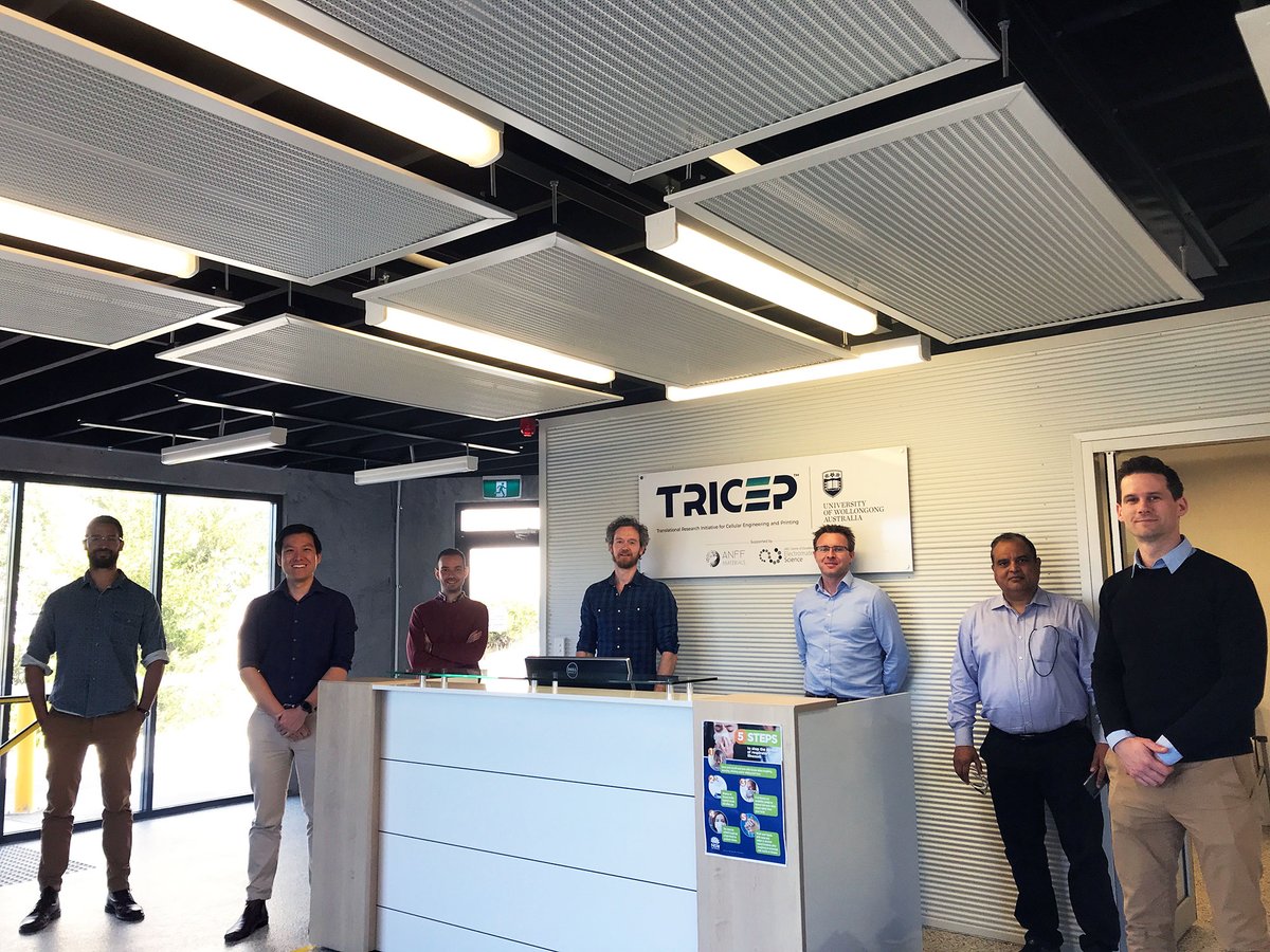
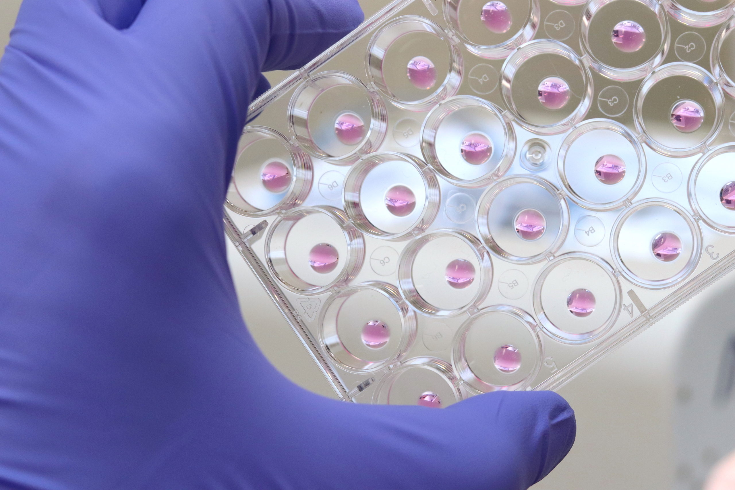
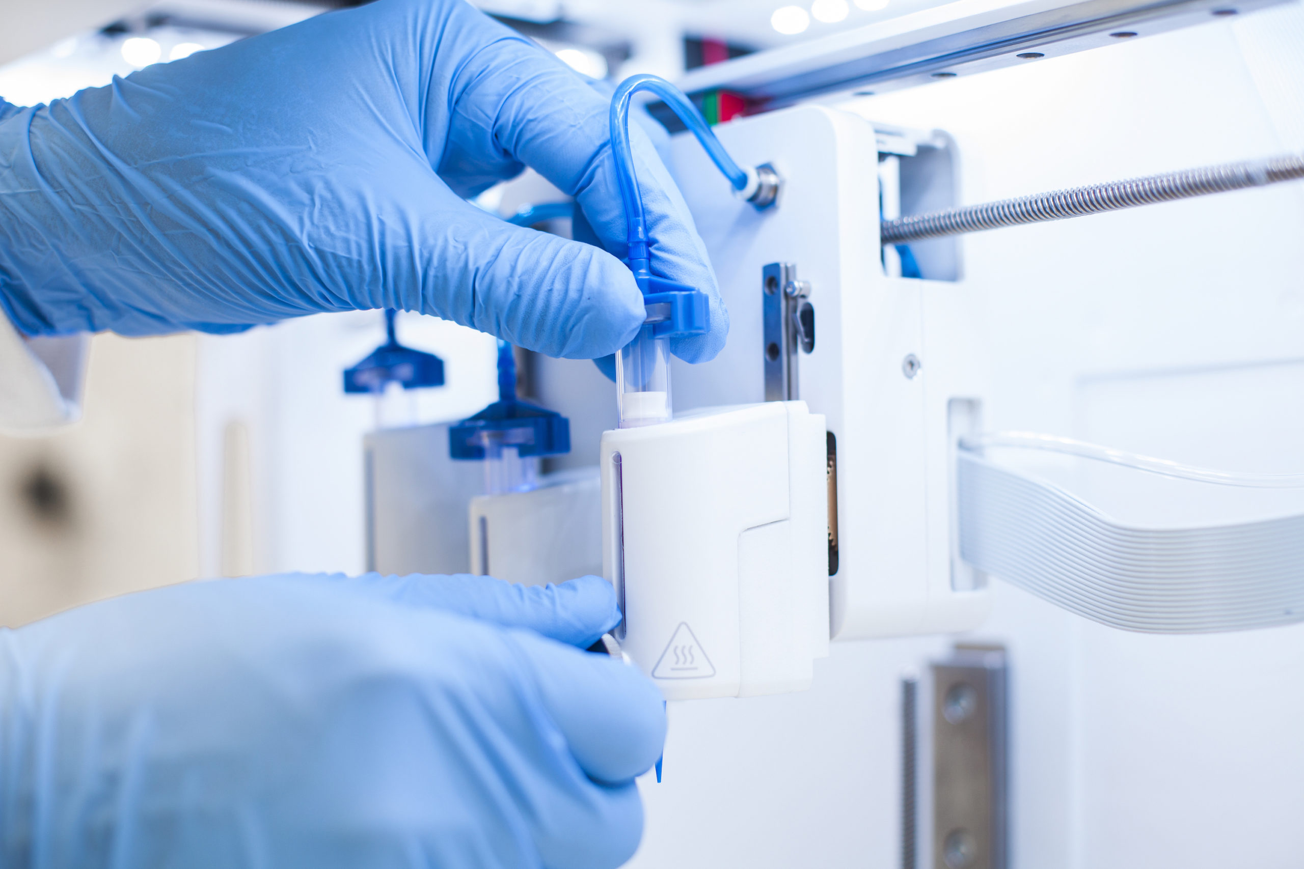
Researchers from Inventia Life Science at the Translational Research Initiative for Cell Engineering and Printing (TRICEP) at Wollongong. (Image courtesy of TRICEP)

Researchers from the ARC Centre of Excellence for Electromaterials Science (ACES) at the University of Wollongong, in Australia, will also lend their internationally renowned expertise in bioinks to develop the new 3D bioprinting system to treat burns during surgery. Led by ACES Director Gordon Wallace, the researchers will provide critical input in the bioprinter and bioink development process. This news comes as no surprise as the ACES team already had a strong working relationship with Inventia.

“ACES is at the forefront of building new approaches to 3D printing, and this project will draw on this significant success we have had in this space in recent years,” Wallace said. “3D printing has emerged as the most exciting advance in fabrication in decades, and I’m excited to continue to build our local capabilities in this area to establish a new, innovative and sustainable industry for the Illawarra [a region in the Australian state of New South Wales]. Being part of this skin regeneration project will help to put Wollongong on the map for the commercial manufacture of bioprinting technologies.”

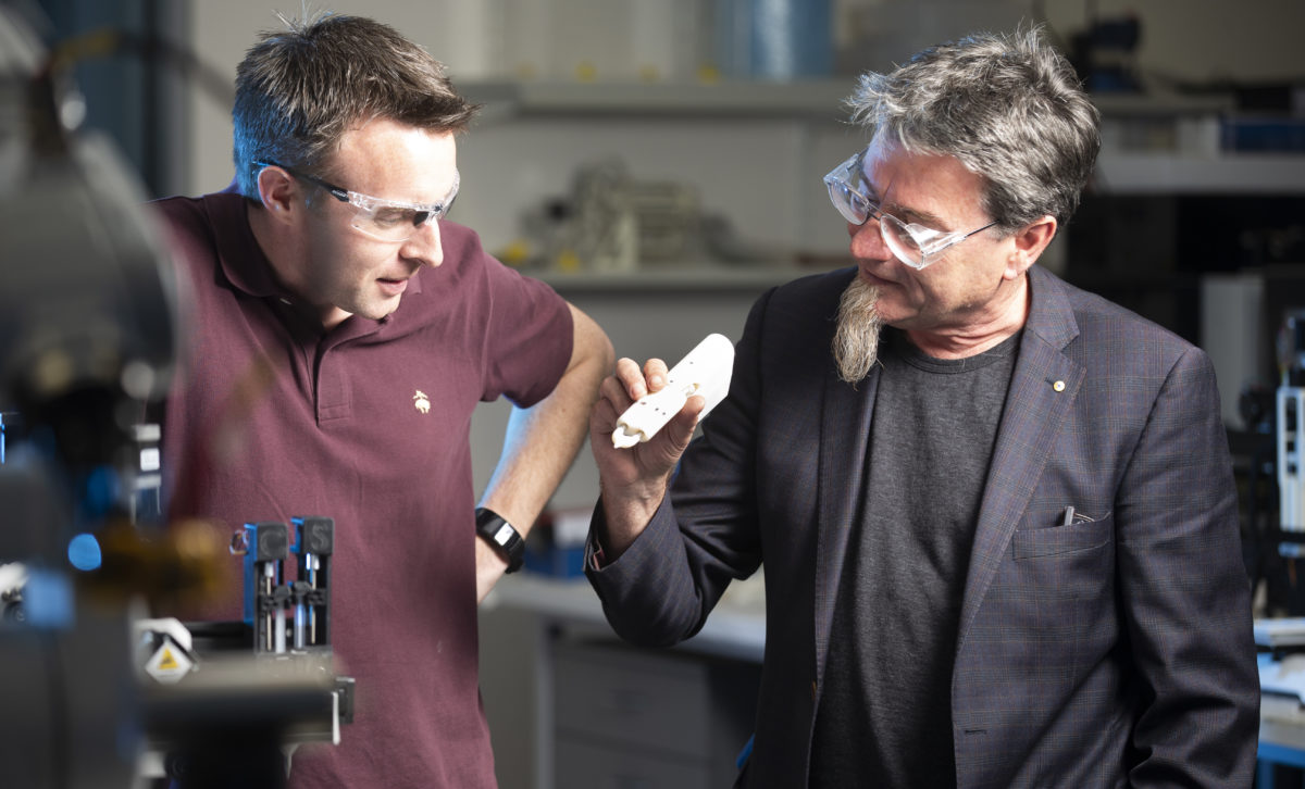
Leading bioprinting researcher Gordon Wallace. (Image courtesy of the ARC Center for Excellence for Electromaterials Science)

For project partner Fiona Wood, a world-leading burns specialist and surgeon, and Director of the Burns Service of Western Australia, this is not the first time that she has looked towards bioengineering to help her patients. In the early 90s, the expert pioneered the innovative “spray-on skin” technique, which greatly reduces permanent scarring in burns victims, and came to notice in 2002, when the largest proportion of survivors from the Bali bombings arrived at Royal Perth Hospital.

“The combination of these grants is an excellent example of the way the Medical Research Future Fund is being applied across the continuum of translational research to commercialization, leading to better patient outcomes,” commented Wood.

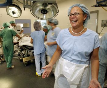
Fiona Wood at the Burns Service of Western Australia. (Image credit Fiona Woods Foundation)

Burns are the fourth most common type of trauma worldwide, with an estimated 11 million burned patients treated every year worldwide, and over 300,000 deaths resulting from serious wounds. In Australia alone Wood’s foundation reported that 200,000 people suffer burns annually, costing the Australian community over AU$150 million per year. Burn injuries are horrific and they present complex problems for both the patient and clinicians to deal with, with a road to recovery beyond easy to tackle. Inventia Skin expects bioprinting technology will be a game-changer in wound medicine. Moreover, the combined expertise of leading specialists in bioprinting and burn wounds, along with funding and support from the local government could lead to one of the most innovative 3D bioprinting systems to treat burns during surgery, and best of all, it could be available in 2022.

The post Robot Skin 3D Printer Close to First-in-Human Clinical Trials appeared first on 3DPrint.com | The Voice of 3D Printing / Additive Manufacturing.

More Efficient Drug Screening with 3D Bioprinting

Taking a drug to market is a competitive, costly and challenging process involving preclinical laboratory and animal testing before the even more time-consuming and expensive four phases of human clinical trials, which can take as many as 7 to 15 years at price tags as high as $5.5 billion. Even if 10 viable drug compounds are identified for human trials, only 1 out of 9 will actually make it to market. Given this high attrition rate, can bioprinting save valuable time and resources by better identifying viable compounds in order to move only the most promising drugs to clinical trials?

The limitations of animal testing

During the initial stages of drug discovery, often referred to as preclinical trials, new chemical entities (NCEs) are monitored to determine the life cycle of the compounds inside and outside of the targeted system (pharmacokinetics) and their chemical reactions (metabolism). Because of the ethical issues surrounding human trials and their high costs, a significant number of these early tests are performed on animals.

While the transition from preclinical animal testing to clinical human trials has improved thanks to better research tools and the rise of artificial intelligence in target identification, there is still a real need for improved preclinical screening because animal testing often fails to recapitulate the complexity of the human metabolism, leading to false positives and negatives that do not accurately reflect the toxicity of drugs to human systems.

3D cell cultures are more relevant

Given the limitations of animal models, it is no wonder that scientists have turned to human organ models. But although human cells have long been cultured in 2D, in recent years, a paradigm shift has led more and more scientists to recognize the importance of working with human cells in the 3D environments afforded by bioprinting in order to produce more physiologically relevant models. Combining the automation of cell culturing in 3D bioprinting with carefully tailored biomaterials, known as bioinks, has made it possible to grow, feed and maintain human organ models in larger quantities and in a lot less time, reducing time and labor spent on these tasks. Laboratory robotics can also now pick and place cell culture reagents or other NCEs and liquid samples in high numbers, enabling higher throughput screening and running a variety of other laboratory tasks more efficiently.

Bioinks better mimic ECM

Bioinks are another powerful tool that help researchers advance their drug discovery research. Tissue-specific bioinks improve cell adhesion and differentiation, helping with the formation of human organoids. Proteins and other biological factors can also be added to more accurately recreate extracellular matrices (ECM), once again better simulating the in vivo microenvironments. Furthermore, with multiple methods of crosslinking (chemical, light, thermal), the stiffness of constructs can be modulated to better serve specific cell types, like cartilage or bone tissue.

Learn more

Bioprinting’s more relevant human organ models can save the drug industry time and money by more efficiently identifying viable compounds in the initial stages of drug development in order to move only the most promising compounds to costly human clinical trials. The technology’s growing influence means that scientists continue to validate more and more applications. Dive deeper into how the bioprinting industry is changing drug screening and development. Watch our webinar on 3D bioprinting for COVID-19 studies or read our application note, which discusses the effectiveness of testing drug efficacy in 2D and 3D.

The post More Efficient Drug Screening with 3D Bioprinting appeared first on 3DPrint.com | The Voice of 3D Printing / Additive Manufacturing.

Bioprinting Biocompatible Hydrogels from Cellulose Inks

Researchers from Italy and Sweden add to the ongoing trend for improving bioprinting techniques and materials. Upon developing bio-based photocurable materials for 3D printing and bioprinting with hydrogels, the authors released the details of their study in ‘DLP 3D Printing Meets Lignocellulosic Biopolymers: Carboxymethyl Cellulose Inks for 3D Biocompatible Hydrogels.’

Modified carboxymethyl cellulose was at the center of this experiment for bioprinting with digital light processing (DLP). While more commonly used as a filler, cellulose has been used in other inks. Beginning the research with the study of lignocellulosic biopolymers, the authors explained that they present a range of options for printing with DLP, while carboxymethyl cellulose (CMC) is often used in food, paint, and detergents. For this reason, it is a sustainable material with particular utility in bioprinting.

Approved by the FDA, and deemed biocompatible, CMC is water-soluble, versatile, and considered “an ideal candidate for the preparation of novel photocurable resins for DLP.” These types of formulations can also imitate cell microenvironments because of their similar makeup to glycosaminoglycan found within the extracellular matrix.

“Due to its versatility, its advantageous properties, water-solubility, and susceptibility to further functionalization, we also expected CMC would be an ideal candidate for the preparation of novel photocurable resins for DLP,” explained the authors. “However, the use of light-assisted printing techniques requires reactive photocrosslinkable functional groups, which means CMC needs functionalization to produce ink formulation for the production of 3D photocured hydrogels.”

“CMC was therefore methacrylated and its photorheology and DLP printability was investigated in two formulations, namely, M-CMC/Dulbecco’s Modified Eagle Medium (DMEM) and M-CMC/water, in presence of a fixed amount of lithium phenyl-2,4,6-trimethylbenzoylphosphinate (LAP) photoinitiator.”

(A) Scheme for the methacrylation of carboxymethyl cellulose (CMC). The presented product only presents one of the possible reaction products. (B) FTIR and (C) 1H NMR spectra for methacrylated CMC (M-CMC, red) and neat CMC (CMC, black).

During evaluation, the authors also investigated compatibility for hydrogels, with M-CMC solubilized in a culture medium (DMEM). Rheological properties (storage modulus, G′, and loss modulus, G″) were evaluated during UV curing for CMC/DMEM/LAP and M-CMC/water/LAP:

“Although the formulation M-CMC/DMEM/LAP showed a slight delay with respect to the onset of the curing process, the DMEM medium still allowed sufficient light penetration for the photocuring process in view of 3D printing,” said the researchers.

Both the CMC/DMEM/LAP and M-CMC/water/LAP formulations proved to be stable after 90 s of UV irradiation. Hydrogels were created from both formulations, and deemed “extremely promising” in comparison with other DLP biocompatible materials.

(A) Photorheology of methacrylated carboxymethyl cellulose (M-CMC) 20 mg/mL (2 wt% lithium phenyl-2,4,6-trimethylbenzoylphosphinate (LAP)) solubilized in water (black) or in culture medium (pink). (B) Gel point. Film thickness 300 m. (C) Frequency sweep. Strain rate 1% and oscillation frequency from 0.01 to 10 Hz.

The researchers created a variety of 3D printed samples, to include cylinders, parallelepipeds, and other complex structures—all stemming from the M-CMC/DMEM/LAP and M-CMC/water/LAP formulations. On further evaluation, the hydrogels were stable, flexible, and the photocrosslink reaction was completed. Although dyes can be helpful in limiting light diffusion, there is also the risk of cytotoxicity, leading the authors to avoid such use in this study.

3D printed M-CMC hydrogels. (A) Simple cylinders and parallelepipeds (solvent: water). (B) The hydrogel exhibited good flexibility and handleability. (C) SEM analysis performed on the freeze-dried hydrogel. (D–F) 3D objects printed from water (D) and from culture medium solution (E,F).

Crosslinking and reactivity were further evaluated, along with compression tests, assessment of swelling ability, and cytotoxicity testing to investigate lack of cell death due to release of LAP photoinitiator or unreacted polymer chains. Ultimately, the team of researchers reported that there were no signs of cytotoxicity, and overall, their work was successful with cells exhibiting viability similar to control samples.

3D-printed M-CMC hydrogels. (A) Simple cylinders and parallelepipeds (solvent: water). (B) The hydrogel exhibited good flexibility and handleability. (C) SEM analysis performed on the freeze-dried hydrogel. (D–F) 3D objects printed from water (D) and from culture medium solution (E,F).

[Source / Images: ‘DLP 3D Printing Meets Lignocellulosic Biopolymers: Carboxymethyl Cellulose Inks for 3D Biocompatible Hydrogels’]

The post Bioprinting Biocompatible Hydrogels from Cellulose Inks appeared first on 3DPrint.com | The Voice of 3D Printing / Additive Manufacturing.

regenHU CEO: Bioprinting Will Strengthen OrganTrans Project to 3D Print Liver Organoid

The European consortium OrganTrans is preparing to develop a tissue engineering platform capable of generating liver tissue. The proposed automated and standardized disruptive alternative solution to organ donation for patients with liver disease will stand on 3D bioprinting know-how from Swiss biomedical firm regenHU. Coordinated by Swiss research and development center CSEM, the eight partners and two transplantation centers engaged in the consortium will be using regenHU’s 3D bioprinters to produce organoid-based liver constructs with organoid laden bioinks.

In April 2020, we reported that OrganTrans would tackle the important healthcare challenge of end-stage liver disease (ESLD) by capitalizing on advancements in the regenerative medicine field, like using biofabricated liver tissue, to develop an entire value chain from the cell source to tissue engineering, biofabrication, post-processing and testing, and liver transplantation under the “compassionate use exemption” regulation (which provides an important pathway for patients with life-threatening conditions to gain access to unproven human cells and tissue products). To understand the key role of biofabrication in this innovative project, 3DPrint.com asked regenHU’s new CEO, Simon MacKenzie, to tell us more about the challenges that lie ahead for the European consortium and his company.

regenHU CEO Simon MacKenzie (Image courtesy of regenHU)

The project officially began in January 2020, what can we expect when it ends in December 2022?

The current goal of this project is to create a functional biofabricated liver construct that can be implanted into a mouse model. I consider that the OrganTrans team will accelerate new solutions for patients with liver failure. It is challenging, but we do envision successful in vivo trials. Of course, this major achievement will not be the end of the story; significant work and research will still be required to transfer these results to human clinical trials. The major remaining challenges will probably be the process scale-up to produce larger tissue and regulatory aspects.

Will this research be groundbreaking to treat liver disease in the future?

Demonstrating the feasibility of the approach in a mouse model will be groundbreaking for the disease because it will demonstrate its potential as an alternative to transplantation. Diseases like NASH [nonalcoholic steatohepatitis, an aggressive form of fatty liver disease] are increasing dramatically, and likely to be a leading cause of death within the next few years. Moreover, the difficulty of detecting the disease until it is potentially too late leads to significant challenges for therapeutic intervention, meaning transplantation will remain the main option for severely affected patients. This well-recognized need, along with the lack of donor organs will ensure bioprinted livers will continue to be well funded. But the value of the project goes beyond liver disease, as the new technologies developed in the frame of OrganTrans will not be limited to liver applications. They relate to the challenges of biofabrication of any organoid-based tissue, which can potentially be beneficial for a large variety of indications.

Can you tell me more about the role of regenHU within the OrganTrans consortium?

Such a complex and ambitious endeavor needs very different and complementary knowledge and competences. Teamwork will be a central element, first to enable, then to accelerate, these new solutions. With this in mind, we have been reorganizing regenHU to bring better project collaborative capabilities to this project, and others like it that we are engaged in. regenHU is a pioneer and global leader in tissue and organ printing technologies converging digital manufacturing, biomaterials, and biotechnology to lead transformational innovations in healthcare. We focus on delivering advancements in the instruments and software required for tissue engineering, and our technology evolving along with the biological research of our partners. We, therefore, consider these partnerships with the scientific community critical for our development.

An outline of the OrganTrans project (Image courtesy of OrganTrans)

regenHU is one of the largest contributors to this project, is this part of the company’s commitment to regenerative medicine?

We can see the need for biotechnology solutions for a wide range of disease states. Our strengths are in engineering the instruments and software necessary to allow the producers of biomaterials and the suppliers of cells to combine their products to achieve functional tissues and organs. Our commitment is to provide disruptive technologies that will enable the community to make regenerative medicine a reality, with precision and reproducibility in mind, for today’s researchers and tomorrow’s industrial biofabrication needs. One of the key challenges is the current limitation in the scale and volume of bioprinting which is linked to the reproducibility of the print. To progress into the manufacture of medical products, bioprinters will need to operate at a scale beyond current capabilities. We design our instruments with these goals in mind and have assembled a team to solve the many challenges to achieve this.

How advanced is the bioprinting community in Europe?

The 3D bioprinting field is several years behind mainstream 3D printing, with the industrialization of the instruments, biomaterials, and cells required before bioprinting can progress to commercial-scale biofabrication. However, as with continued development seen in 3D printing, the technology convergence required for tissue and organ printing that changes medical treatments will become a reality through the efforts of engineering companies like regenHU, biomaterial developers, and human cell expansion technologies, being combined in projects such as OrganTrans.

As the newly appointed CEO of the company, how do you feel taking on this project?

Successfully entering the OrganTrans consortium is just one part of the company. regenHU investors see my arrival as the catalyst to bring regenHU to the next stage in its evolution. Our goal remains the production of industrial biofabrication instruments capable of delivering the medical potential of bioprinting, novel bioinks, and stem cells. To achieve this, we are enhancing the team and structure of the company, bringing forward the development of new technologies and increasing our global footprint to better support our collaborative partners. I have spent many years in regenerative medicine and pharma and can see the potential of bioprinting to revolutionize many areas of medical science, so joining regenHU was an easy choice. As CEO, my main role is to provide the right support structure to enable our entrepreneurial engineering teams to thrive and be brave enough to push boundaries. Additionally, as we cannot achieve our end goal on our own, I am here to nurture the important connections with our user community. Only by listening to their valuable insights and solving problems with them, we will push the technology onward.

The post regenHU CEO: Bioprinting Will Strengthen OrganTrans Project to 3D Print Liver Organoid appeared first on 3DPrint.com | The Voice of 3D Printing / Additive Manufacturing.

KFC Russia Explores 3D Printing Chicken

Considering the amount of time that scientists have spent lately engineering human tissue in the lab, it’s not surprising to hear that restaurant chains want to get in on the action to replace the expense of raising animals for meat. And while you may be trying to imagine how 3D printed meat would be displayed on a KFC menu board, the “powers at be” at KFC are still busy at the drawing board—collaborating with 3D Bioprinting Solutions.

3D printed food is always a shoo-in for garnering excitement, feeding our appetites with potential for the future, whether targeting meat, breakfast, cake, or other items that can be easily extruded.

A variety of different companies have been interested in redefining meat, but often with a plant-based concept. 3D printing enthusiasts, technology buffs, and curious consumers may be interested in finding out more about the techniques and the materials, but still balk at becoming anything close to a vegetarian (or god forbid, a vegan!).

3D printing actual meat has historically been more challenging—along with the overall concept of any type of tissue engineering. The KFC announcement could be more gimmick than true intent, but for now they seem to be exploring the benefits of more environmentally friendly production, better nutrition, and the possibility of eliminating chicken farms (and all the associated flak from activists—as well as consumers who are just generally disgusted).

The process of creating chicken in a lab, dubbed “craft meat,” could mean reducing emissions exponentially as well as freeing up large tracts of land currently required to raise animals.

“The production of cell meat products is the next step in the development of our concept of the restaurant of the future,” explained Raisa Polyakova, KFC CEO in Russia.

Only time will tell whether there is a true market for bioprinted chicken, with success in the lab waiting to be seen, as well as popularity with the Russian fast-food palate. Polyakova is confident that they may be able to pioneer new technology that can be shared with the rest of the world, perhaps transforming menus everywhere one day. Savings on the bottom line could play an enormous role too—not only allowing for chains like KFC to save substantially in production, but also passing that down to consumers who are tired of over-paying for sandwiches, burgers, and most items available in contemporary drive-thrus.

“Technologies based on 3D bioprinting, which were initially widely recognized in medicine, today are gaining more and more popularity in the field of food production, such as, for example, animal meat. The rapid development of such technologies in the future will make meat products printed on a 3D bioprinter more affordable,” said Yousef Hesuani, co-founder and managing partner of 3D Bioprinting Solutions.

[Source / Images: popmech]

The post KFC Russia Explores 3D Printing Chicken appeared first on 3DPrint.com | The Voice of 3D Printing / Additive Manufacturing.

3D Printed Medicine Uses Fish Gelatin to Deliver Cancer Treatment

Japanese researchers Jin Liu, Tatsuaki Tagami, and Tetsuya Ozeki have completed a recent study in nanomedicine, releasing their findings in “Fabrication of 3D Printed Fish-Gelatin-Based Polymer Hydrogel Patches for Local Delivery of PEGylated Liposomal Doxorubicin.” Experimenting with a new drug delivery system, the authors report on new potential for patient-specific cancer treatment.

The study of materials science continues to expand in a wide range of applications; however, bioprinting is one of the most exciting techniques as tissue engineering is expected to lead to the fabrication of human organs in the next decade or so. Such research has also proven that bioprinting may yield much more powerful drug delivery whether in using hybrid systems, multi-drug delivery systems, or improved scaffolds.

Here, the materials chosen for drug delivery are more unique as the researchers combined printer ink with semi-synthesized fish gelatin methacryloyl (F-GelMA)—a cold fish gelatin derivative.

In providing aggressive cancer treatment to patients, the use of doxorubicin (DOX) is common as an anti-carcinogen for the treatment of the following diseases:

  • Breast cancer
  • Bladder cancer
  • Kaposi’s sarcoma
  • Lymphoma
  • Acute lymphocytic leukemia

DOX may also cause serious cardiotoxicity, however, despite its use as a broad-spectrum drug. As a solution, PEGylated liposomal DOX, Doxil has been in use for treatment of cancer with much lower cardiotoxity. The nanomedicine has also been approved by the FDA, and is used for targeting local tumors; for instance, this type of drug delivery system could be suitable for treating a brain tumor.

“PEGylating liposomes can prolong their circulation time in blood, resulting in their passive accumulation in cancer tissue, called the enhanced permeability and retention effect,” state the authors.

Using a 3D bioprinter, the authors developed liposomal patches to be directly implanted into cancerous cells.

(a) Synthesis of fish gelatin methacryloyl (F-GelMA). (b) Hybrid gel of cross-linked F-GelMA and carboxymethyl cellulose sodium (CMC) containing PEGylated liposome. The reaction scheme was prepared in previous studies

“We used a hydrogel containing semi-synthetic fish-gelatin polymer (fish gelatin methacryloyl, F-GelMA) to entrap DOX-loaded PEGylated liposomes. Fish gelatin is inexpensive and faces few personal or religious restrictions,” stated the authors.

Fish gelatin has not been used widely in bioprinting, however, due to low viscosity and rapid polymerization. To solve that problem, the authors created a bioink composite with elevated viscosity.

Viscous properties of drug formulations used as printer inks. (a) The appearance of F-GelMA hydrogels containing different concentrations of CMC. (b) The viscosity profiles of F-GelMA hydrogels containing different concentrations of CMC. The data represent the mean ± SD (n = 3).

And while hydrogels are generally attractive for use due to their ability to swell, for this study, the researchers fabricated a variety of different materials—with the combination of 10% F-GelMA and 7% carboxymethyl cellulose sodium (a thickening agent) showing the highest swelling ratio.

Swelling properties of hydrogels after photopolymerization. (a) Swelling ratio of different concentrations of F-GelMA. (b) Swelling ratio of mixed hydrogel (10% F-GelMA with different concentrations of CMC). The data represent the mean ± SD (n = 3).

Design of the different 3D geometries: (a) cylinder, (b) torus, and (c) gridlines.

Patches were printed in three different sample shapes, using a CELLINK bioprinter syringe as the authors tested drug release potential in vivo. Realizing that surface area, crosslinks density, temperature, and shaker speed would play a role, the team relied on a larger surface volume for more rapid release of drugs.

Printing conditions of patches.

While experimenting with the torus, gridline, and cylindrical sample patches, the researchers observed gridline-style patches as offering the greatest potential for sustained release.

Drug release profiles of liposomal doxorubicin (DOX). (a) Influence of shape on drug release. The UV exposure time was set to 1 min. (b) Influence of UV exposure time on drug release. The gridline object was used for this experiment. The data represent the mean ± SD (n = 3).

“These results indicate that CMC is useful for adjusting the properties of printer ink and is a useful and safe pharmaceutical excipient in drug formulations. We also showed that drug release from 3D-printed patches was dependent on the patch shapes and UV exposure time, and that drug release can be controlled. Taken together, the present results provide useful information for the preparation of 3D printed objects containing liposomes and other nanoparticle-based nanomedicines,” concluded the authors.

[Source / Images: ‘Fabrication of 3D Printed Fish-Gelatin-Based Polymer Hydrogel Patches for Local Delivery of PEGylated Liposomal Doxorubicin’]

The post 3D Printed Medicine Uses Fish Gelatin to Deliver Cancer Treatment appeared first on 3DPrint.com | The Voice of 3D Printing / Additive Manufacturing.

Rice Researchers 3D Print with Lasers and Sugar to Build Complex Vascular Networks

A team of researchers from Rice University has uncovered a promising strategy to generate vascular networks, one of the most daunting structures in the human body. Using powdered sugar and selective laser sintering, the researchers were able to build large structures from complex, branching, and intricate sugar networks that dissolve to create pathways for blood in lab-grown tissue.

This is the team’s latest effort to build complex vascular networks for engineered tissues to show that they could keep densely packed cells alive for two weeks. The findings of their study—published in the Nature Biomedical Engineering journal—prove that developing new technologies and materials to mimic and recapitulate the complex hierarchical networks of vessels gets them closer to providing oxygen and nutrients to a sufficient number of cells to get a meaningful long-term therapeutic function.

“One of the biggest hurdles to engineering clinically relevant tissues is packing a large tissue structure with hundreds of millions of living cells,” said study lead author Ian Kinstlinger, a bioengineering graduate student at Rice’s Brown School of Engineering. “Delivering enough oxygen and nutrients to all the cells across that large volume of tissue becomes a monumental challenge. Nature solved this problem through the evolution of complex vascular networks, which weave through our tissues and organs in patterns reminiscent of tree limbs. The vessels simultaneously become smaller in thickness but greater in number as they branch away from a central trunk, allowing oxygen and nutrients to be efficiently delivered to cells throughout the body.”

Overcoming the complications of 3D printing vascularization has remained a critical challenge in tissue engineering for decades, as only a handful of 3D printing processes have come close to mimic the in vivo conditions needed to generate blood vessels. Without them, the future of bioprinted organs and tissues for transplantation will remain elusive. Many organs have uniquely intricate vessels, like the kidney, which is highly vascularized and normally receives a fifth of the cardiac output, or the liver, in charge of receiving over 30% of the blood flow from the heart. By far, kidney transplantation is the most common type of organ transplantation worldwide, followed by transplants of the liver, making it crucial for regenerative medicine experts to tackle vascularization.

Ian Kinstlinger with a blood vessel template he 3D printed from powdered sugar (Credit: Jeff Fitlow/Rice University)

In the last few years, extrusion-based 3D printing techniques have been developed for vascular tissue engineering, however, the authors of this study considered that the method presented certain challenges, which led them to use a customized open-source, modified laser cutter to 3D print the sugar templates in the lab of study co-author Jordan Miller, an assistant professor of bioengineering at Rice.

Miller began work on the laser-sintering approach shortly after joining Rice in 2013. The 3D printing process fuses minute grains of powder into solid 3D objects, making possible some complex and detailed structures. In contrast to more common extrusion 3D printing, where melted strands of material are deposited through a nozzle, laser sintering works by gently melting and fusing small regions in a packed bed of dry powder. According to Miller, “both extrusion and laser sintering build 3D shapes one 2D layer at a time, but the laser method enables the generation of structures that would otherwise be prone to collapse if extruded.”

“There are certain architectures—such as overhanging structures, branched networks and multivascular networks—which you really can’t do well with extrusion printing,” said Miller, who demonstrated the concept of sugar templating with a 3D extrusion printer during his postdoctoral studies at the University of Pennsylvania. “Selective laser sintering gives us far more control in all three dimensions, allowing us to easily access complex topologies while still preserving the utility of the sugar material.”

Assistant professor of bioengineering at Rice University, Jordan Miller (Credit: Jeff Fitlow/Rice University)

Generating new 3D printing processes and biomaterials for vascularization is among the top priorities for the researchers at Miller’s Bioengineering Lab at Rice. The lab has a rich history of using sugar to construct vascular network templates. Miller has described in the past how sugar is biocompatible with the human body, structurally strong, and overall, a great material that could be 3D printed in the shape of blood vessel networks. His original inspiration for the project was an intricate dessert, even going as far as suggesting that “the 3D printing process we developed here is like making a very precise creme brulee.”

To make tissues, Kinstlinger chose a special blend of sugars to print the templates and then filled the volume around the printed sugar network with a mixture of cells in a liquid gel. Within minutes, the gel became semisolid and the sugar dissolved and flushed away to leave an open passageway for nutrients and oxygen. Clearly, sugar was a great choice for the team, providing an opportunity to create blood vessel templates because it is durable when dry, and it rapidly dissolves in water without damaging nearby cells.

A sample of blood vessel templates that Rice University bioengineers 3D printed using a special blend of powdered sugars. (Credit: B. Martin/Rice University)

In order to create the treelike vascular architectures in the study, the researchers developed a computational algorithm in collaboration with Nervous System, a design studio that uses computer simulation to make unique art, jewelry, and housewares that are inspired by patterns found in nature. After creating tissues patterned with these computationally generated vascular architectures, the team demonstrated the seeding of endothelial cells inside the channels and focused on studying the survival and function of cells grown in the surrounding tissue, which included rodent liver cells called hepatocytes.

The hepatocyte experiments were conducted in collaboration with the University of Washington (UW)’s bioengineer and study co-author Kelly Stevens, whose research group specializes in studying these delicate cells, which are notoriously difficult to maintain outside the body.

“This method could be used with a much wider range of material cocktails than many other bioprinting technologies. This makes it incredibly versatile,” explained Stevens, an assistant professor of bioengineering in the UW College of Engineering, assistant professor of pathology in the UW School of Medicine and an investigator at the UW Medicine Institute for Stem Cell and Regenerative Medicine.

The results from the study allowed the team to continue their work towards creating translationally relevant engineered tissue. Using sugar as a special ingredient and selective laser sintering techniques could help advance the field towards mimicking the function of vascular networks in the body, to finally deliver enough oxygen and nutrients to all the cells across a large volume of tissue.

Miller considered that along with the team they were able to prove that “perfusion through 3D vascular networks allows us to sustain these large liverlike tissues. While there are still long-standing challenges associated with maintaining hepatocyte function, the ability to both generate large volumes of tissue and sustain the cells in those volumes for sufficient time to assess their function is an exciting step forward.”

The post Rice Researchers 3D Print with Lasers and Sugar to Build Complex Vascular Networks appeared first on 3DPrint.com | The Voice of 3D Printing / Additive Manufacturing.

Cellink Teams with Lonza to Advance Bioprinting Cell Cultures

For over four years biotech company Cellink has been helping researchers pursue creative ways to promote organ regeneration, test novel drugs, develop vascularized bioconstructs, and more. Through collaborative partnerships and pioneering research, the firm has quickly become a market leader for cell-based applications. Along that road, they have partnered with several startups and multinationals to open up a broader range of possibilities in the field. Their latest partnership with Lonza—a leading suppliers to the pharmaceutical, biotech, and specialty ingredients markets—will provide researchers and scientists with even more options to enhance bioprinting of complex 3D human tissue constructs.

The companies have joined forces to offer a comprehensive 3D bioprinting solution designed to optimize and increase access to complete 3D cell culture workflows. The solution integrates Cellink’s 3D bioprinting instruments and pioneering commercial bioinks with Lonza’s broad selection of human-derived primary cells and supporting culture media.

Credit: Lonza

“Everything we do at CELLINK, from live cell imaging to our innovative bioprinting systems and bioinks, is meant to support our customers with the products and services needed for them to more effectively and efficiently research solutions to some of the most important challenges of our time. Challenges such as cancer therapeutics, regenerative medicine and the testing and development of drugs, to name a few,” said Ginger Lohman, Biodispensing Product Manager at Cellink’s Gothenburg headquarters in Sweden. “When it came time to expand our portfolio into complete 3D cell culture workflows, we knew it was critical that we brought the right partner onboard. We’re confident that Lonza is that partner.”

According to Cellink, cell biologists will now be able to rely on a high-performing product portfolio to successfully execute some of the most demanding work on 3D bioprinting and boost their scientific research. The proposed solution under the partnership combines Cellink’s 3D bioprinting instruments and bioinks with Lonza’s primary cells and culture media, to meet the needs of cell biologists for enhanced bioprinting of complex 3D human tissue constructs.

Since the origins of cell culture more than a century ago, cells have been cultured in two-dimensions; however, 3D cell culture has proven to be a better model for representing in vivo conditions, offering a more accurate and reliable means of predicting and analyzing cell behavior. In fact, it has proven to be a great enabler for scientists to handle cells in vitro while obtaining results that are closer to the in vivo environment without relying on animals, thereby avoiding many ethical issues.

With so many benefits, 3D cell cultures are being widely adopted in numerous laboratories—even more so as researchers look to create increasingly complex 3D constructs and find solutions to structural and material engineering challenges. For years, Swiss-based Lonza has been developing the building blocks needed to create 3D cell culture models, offering an extensive array of human-derived primary cells and culture media, ethically sourced and authenticated thorough quality control testing.

Robust, viable cells are an essential component of any successful cell culture application, and so is 3D bioprinting. A big part of the 3D cell culture workflow involves 3D bioprinting, which is thriving as a powerful technology for engineering complex 3D tissues for in vitro drug discovery research. Cellink is currently providing a wide range of 3D bioprinting systems, like INKREDIBLE, Bio X6 systems, and LumenX, which stems from a collaboration with Volumetric, a startup established by Rice University professor Jordan Miller. Furthermore, since 2016, the company has successfully commercialized the world’s first universal bioink designed to print complex 3D human tissue constructs with any 3D bioprinting system. The biomaterial is useful for scientists as it can be modified with peptides and growth factors to develop a series of customized bioink formulations to meet varying application needs and is used in hundreds of labs around the world.

Cellink’s INKREDIBLE system (Credit: Cellink)

“Cell biology laboratories are constantly seeking innovative new technologies to enhance their experimental workflows and help deliver on their promise to drive the next research breakthrough,” explained Katrin Hoeck, head of marketing for Cell Analysis and Testing Solutions at Lonza. “Our broad panel of human-derived primary cells is specifically engineered to enable researchers to develop biological in vitro model systems that more closely reflect disease biology. This new collaboration with Cellink will enable our customers to build physiologically relevant 3D models to accelerate target identification/validation, investigation of mechanisms of action and safety testing in drug discovery.”

Under the agreement, Cellink will provide this complete solution through its global sales channels, supported by Lonza’s well-established logistics processes.

This is not the first partnership with the pharma industry for Cellink. The company also recently announced a collaboration with AstraZeneca, the British-Swedish multinational pharmaceutical and biopharmaceutical company, to utilize Cellink’s 3D bioprinting technology for liver organoid culture for drug discovery purposes in cardiovascular, renal and metabolic diseases.

According to Lonza, “The pharma and biomedical research laboratories are constantly seeking innovative new technologies to enhance their experimental workflows and help deliver on their promise to drive the next research breakthrough.” This collaboration could strengthen the processes and end products of research, offering substantial benefits for more predictive cell models and ultimately increasing the usefulness of 3D culturing cells for many applications in major areas of life sciences.”

The post Cellink Teams with Lonza to Advance Bioprinting Cell Cultures appeared first on 3DPrint.com | The Voice of 3D Printing / Additive Manufacturing.

3D Printing Webinar & Virtual Event Roundup, May 31, 2020

With so many events going virtual due to the ongoing COVID-19 pandemic, there’s also been an increase in the number of webinars that companies in the additive manufacturing industry are holding. To make things easier for our readers, since there’s so much online content to choose from these days, 3DPrint.com is compiling all of these available webinars, and the virtual events, into a weekly roundup for you, starting today.

Freeman Technology Webinar

Characterization Tools for Evaluating Polymer Powders for Laser Sintering Webinar

This Tuesday, June 2nd, UK-based Freeman Technology, a Micromeritics company that creates systems for measuring the flow properties of powder materials, will host a webinar at 9 am ET titled “Characterization Tools for Evaluating Polymer Powders for Laser Sintering.” Enrico Gallino, Senior Engineer – Material Specialist at Ricoh UK Products Ltd, will speak about evaluating an AM powder characterization methodology, and will also discuss the results of screening the relevant properties, such as flowability, shape, and thermal properties, of a variety of materials.

“As additive manufacturing (AM) technology transitions from the fabrication of prototypes to serial production of end-use parts, the understanding of the powder properties needed to reliably produce parts of acceptable quality becomes critical,” the webinar site states.

“Achieving the optimal quality for parts does not only depend on setting the right process parameters. Material feedstock also plays an important role when aiming for high performance products. In the case of selective laser sintering, polymer powders are used as a raw material. Therefore, controlling the quality and correctly characterizing the particles used in the process is a key step to successfully apply polymer AM techniques and also to expand the range of material that can be process with this technology.”

Click here to register.

Dassault Systèmes Webinar

Dassault Systèmes be will holding a live webinar on Thursday, June 4th at 10 am ET, titled “Intuitive 3D Designs with CATIA® and SOLIDWORKS® on Mobile Devices.” Participants will have the chance to learn how beneficial flexible design workflows can be when delivering products to market, faster, across many different industries. There will be a live demonstration, using tablets and PCs, on how combining CATIA and SOLIDWORKS on the 3DEXPERIENCE platform will allow your business to add engineering details with simple parametric modeling, create organic surfaces with subdivision (Sub-D) modeling, generate complex patterns and shapes quickly, optimize and evolve designs using an algorithmic approach, and more – all from your own device. The demonstration will be followed by a live Q&A session.

“Discover our portfolio of ready-to-go online Design and Engineering applications in action, which enable you to design from your laptop, your smartphone or tablet! Enjoy increased agility without compromising best-in-class design and engineering capabilities,” the webinar site states.

“With its growing app portfolio and secure cloud technology, the 3DEXPERIENCE platform enables you to manage all facets of your product development process while reducing infrastructure costs, IT overhead, software maintenance and complexity. All 3DEXPERIENCE solutions work together seamlessly making data management, sharing and collaboration easy.”

Click here to register.

3DHEALS 2020 Global Summit

The 3DHEALS conference is going virtual this year, as the 3DHEALS 2020 Global Summit runs from 11 am-9:30 pm ET June 5th and 6th. Offering powerful networking and effective programming on a global stage, this popular bioprinting conference – sponsored by Whova and Zoom – brings together influencers and audiences from over nine countries, offering opportunities and insights that can be beneficial to stakeholders. With over 70 speakers, more than four workshops, startup events, simulated in-conference experience, an interview series hosted by Dr. Jenny Chen, and more, this is one you won’t want to miss.

“3DHEALS2020 is designed to cater to a wide range of professionals, ranging from healthcare early adopter, manufacturers, engineers, legal professionals and policymakers, C-Level executives, entrepreneurs, investors, and more. We aim to create an effective program that maximizes the attendee’s experiences and decreases the barriers in communication among stakeholders,” the event site states.

Click here to register.

Will you attend these events and webinars, or have news to share about future ones? Let us know! Discuss this and other 3D printing topics at 3DPrintBoard.com or share your thoughts in the comments below.

The post 3D Printing Webinar & Virtual Event Roundup, May 31, 2020 appeared first on 3DPrint.com | The Voice of 3D Printing / Additive Manufacturing.

Marine Biologist Modifies Bioprinting for the Creation of Bionic Coral

Corals are dying globally. In the face of climate change and global warming, we can expect some severe consequences, which in turn directly affects marine life. In what is panning out to be a mass extinction event, coral reefs have been dangerously threatened by toxic substances and excess carbon dioxide for years, causing the certain death of may of these diverse marine invertebrates. Once the coral is dead, the reefs will also die and erode, destroying important marine life, that would otherwise feed and spawn on it.

Considering that scientists have predicted that nearly all coral reefs will disappear in 20 years, it is crucial that we protect corals and learn from them. For the expanding field of biotechnology, untapped resources like corals hold great potential, as bioactive compounds for cancer research or simply as an inspiration for the production of bioenergy and bioproducts.

In an interview with 3DPrint.com, interdisciplinary marine biologist Daniel Wangpraseurt, from the University of California San Diego (UCSD)’s Department of NanoEngineering, explained how bioprinting technology was a pivotal point in his work to develop bionic 3D printed corals as a new tool for coral-inspired biomaterials that can be used in algal biotechnology, coral reef conservation and in coral-algal symbiosis research. 

“For many years I have been studying how corals optimize light management and discovered that there are lots of interesting evolutionary tricks, such as different growth forms and material properties, so I became interested in copying these strategies and developing artificial materials that could host living microalgae, just like corals do in nature,” revealed Wangpraseurt.

Daniel Wangpraseurt

As one of the most productive ecosystems globally, coral reefs use photosynthesis to convert carbon dioxide into energy that they in turn use for food. Even though light provides the energy that fuels reef productivity, key nutrients such as nitrogen and phosphorus are also required, but are found in very low quantities in warm tropical oceans where coral reefs are generally found, making scientists wonder how these marine animals have managed to create a competitive habitat with such limited resources.

A laser beam is intensely scattered by elastic coral tissue and aragonite skeleton. (Credit: Daniel Wangpraseurt)

Wangpraseurt described that, while different corals have developed a plethora of geometries to achieve such capabilities, they are all characterized by an animal tissue-hosting microalgae, built upon a calcium carbonate skeleton that serves as mechanical support and as a scattering medium to optimize light delivery toward otherwise shaded algal-containing tissues.

“Taking what we learned about corals and biomaterials, we began working on a project to develop a synthetic, symbiotic system using a 3D bioprinting approach. We know corals have both animal cells and algal cells, and, so far, we have mimicked the animal part of the corals, that is, the physical and chemical microhabitat that partially controls the activity of the algal cells.”

At UCSD, Wangpraseurt expects to continue recreating coral-inspired photosynthetic biomaterial structures using a new bioprinting technique and a customized 3D bioprinter capable of mimicking functional and structural traits of the coral-algal symbiosis. Along with fellow researchers from UCSD, the University of Cambridge, the University of Copenhagen and the University of Technology Sydney, and thanks to a grant from the European Union’s Horizon 2020 research and innovation program, and the National Institutes of Health (NIH), the team reported the results of their work on bioinspired materials that was published in the journal Nature Communications earlier this year.

“We want to go further and not just develop similar physical microhabitat but also modulate cellular interactions, by mimicking biochemical pathways of symbiosis. We hope that this allows us to not only optimize photosynthesis and cell growth, but also to gain a deeper understanding of how the symbiosis works in nature. By doing so, we can improve our understanding of stress phenomena such as coral bleaching, which is largely responsible for global coral death.”

Living colonies of Symbiodinium are visible within the 3D bioprinted tissues (Credit: Daniel Wangpraseurt)

So, how did bioprinters become the go-to technology for this project? Wangpraseurt explains that, while working as a researcher at the University of Cambridge’s Department of Chemistry Bio-Inspired Photonics lab, he noticed that scientists were using cellulose as a biomaterial with interesting optical responses. He was wondering how he could use cellulose to develop a material with very defined architectural complexity. 

“In the beginning, the main aim was to develop a coral-inspired biomaterial, that has a similar optical response as natural coral, and then to grow algae on it or within it. Thereby, we started off with simple techniques, using conventional 3D printers; however, it wasn’t very easy to recreate the spatial resolution we needed for corals.”

Inspired by 3D bioprinting research in the medical sciences, Wangpraseurt reached out to scientists at the UCSD NanoEngineering lab that were developing artificial liver models, and who later became collaborators in the project.

A laser beam is intensely scattered by elastic coral tissue and aragonite skeleton (Credit: Daniel Wangpraseurt)

The team went on to develop a 3D printing platform that mimics morphological features of living coral tissue and the underlying skeleton with micron resolution, including their optical and mechanical properties. It uses a two-step continuous light projection-based approach for multilayer 3D bioprinting and the artificial coral tissue constructs are fabricated with a novel bioink solution, in which the symbiotic microalgae are mixed with a photopolymerizable gelatin-methacrylate (GelMA) hydrogel and cellulose-derived nanocrystals (CNC). Similarly, the artificial skeleton is 3D printed with a polyethylene glycol diacrylate-based polymer (PEGDA).

Close up of coral polyps and living photosynthetic biomaterials. Living colonies of Symbiodinium are visible within the 3D bioprinted tissues (Credit: Daniel Wangpraseurt)

Based in San Diego, Wangpraseurt has spent months trying to recreate the intricate structure of the corals with a distinguished symbiotic system that is known to grow as it creates one of the largest ecosystems on the planet. 

“We used a 3D bioprinter that had been developed for medical purposes, which we modulated and further developed a specific bioink for corals. A lot of the work was related to the optimization of the material properties to ensure cell viability. Having the right bioink for our algal strains was crucial as if we were to use mixtures commonly used for human cell cultures, the cells will not grow very well and can die rapidly.”

The implications of the newly developed 3D printed bionic corals capable of growing microalgae are many. Wangpraseurt said he plans to continue working on bionic corals and potentially scale up the process for his startup, called mantaz, as well as for commercial properties; or to develop coral-inspired materials at a larger scale to have a more immediate impact on efforts related to coral reef restoration, and also for biotechnology.

SEM images of the skeleton structure of the coral Stylophora pistillata and the coral-inspired, 3D-printed material (Credit: Daniel Wangpraseurt)

Wangpraseurt is looking to scale the bioprinting system to have a more immediate impact on algae biotechnology, bioenergy, and bioproducts. He claims that he and his colleagues can “customize the environment of the algae and fine-tune the production of a certain bioproduct to potentially tap into the algae bioproduct market and scale the system for bioenergy production.” 

“Another interest of mine is to further develop a 3D bioprinted synthetic coral-algal symbiosis system, which can provide important insight into the mechanisms that lead to coral death, but can also result in the development of future technology for coral reef restoration.”

The researcher talks about coral reefs with a reverent passion that today goes beyond his lab work. When he is not moving the research along at USCD, Wangpraseurt is working with his social enterprise in Panama, as he and his team try to restore coral reef ecosystems to help coastal communities in the tropics, including local fishermen, by harvesting algae biomass that can be sold for different purposes, such as natural fertilizer, which contributes to an organic and sustainable chain of production. Furthermore, the coral-inspired aspects of Wangpraseurt’s research and startup company are really coalescing to enable him and his team to understand how corals work and, in turn, how we can learn from them for the benefit of our planet.

Nutrient sampling at a polluted reef in Panama (Image: Daniel Wangpraseurt)

The post Marine Biologist Modifies Bioprinting for the Creation of Bionic Coral appeared first on 3DPrint.com | The Voice of 3D Printing / Additive Manufacturing.