
At the European Space Research and Technology Centre (ESTEC) in The Netherlands, a two-day workshop on 3D bioprinting was held. The ESTEC is European Space Agency’s (ESA) primary research and test center for space technology. The event brought together bioprinting experts to discuss the possibility of using 3D bioprinting and regenerative medicine for medical treatment of astronauts […]
BioPrinting with Alzheimer’s Stem Cells May Lead to Improved Drug Testing & Treatment
 Alzheimer’s studies give many of us hope, whether we have a relative or friend suffering from the dreaded disease—or simply because we are hoping that a cure will be there if we begin to experience symptoms of the most common and crushing form of dementia. As bioprinting becomes valuable in so many different areas of medical research, it is no surprise to hear that now scientists are turning their attention toward using it to find better ways for treating Alzheimer’s.
3D printing, and the realm within known as bioprinting, allows for scientists to embrace so many benefits, from affordability and expediency, to the ability to keep trying varying iterations until they complete their mission in research or innovation. One of the greatest challenges in dealing with Alzheimer’s has been finding medication that really works. Not only is there pressure to find it for those who have the existing condition, but also for future generations carrying what is thought to be a genetic marker. In ‘Bioprinting neural tissues using stem cells as a tool for screening drug targets for Alzheimer’s disease,’ Stephanie M. Willerth examines the difficulty in both finding, testing, and approving drugs for this trying disease that tends to affect seniors regarding cognition, memory, and language and reasoning.
“The pathology of Alzheimer’s disease includes the presence of plaques containing aggregates of amyloid beta (αβ) proteins and tangles containing neurofibrillary tangles,” states Willerth in her paper. “Certain genetic mutations increase the possibility of developing Alzheimer’s disease.”
“These mutations consist of an altered APP gene responsible for encoding amyloid precursor protein, along with the PSEN1 and PSEN2 mutations cause decreased activity by the γ-secretase complex. These mutations result in improper processing of the αβ proteins. Currently, approved treatments for Alzheimer’s disease include four different types of cholinesterase inhibitors and the drug memantine – a N-methyl-d-aspartate receptor antagonist that targets glutaminergic neurons to preserve their function.”
 Willerth goes on to detail the facts about Memantine, as the only drug approved for Alzheimer’s patients. It has been in use since 2000, and currently there are over 200 other compounds being tested. Memantine is known to be controversial though and Willerth points out that the current drug targets present issues in terms of both successful use in patients, and toxicity, posing a need for improvement before clinical trials commence.
Better testing before trials would diminish costs in drug development, along with decreasing the amount of time it currently takes to create treatments that are successful.
“Current methods for evaluating target compounds consist of animal models of disease and the use of cadaveric human tissues,” states Willerth. “In addition to their lack of predictive capacity, animal models are costly, and the supplies of human neural tissues remain limited. Developing more effective assays for preclinical identification of drug targets will lower the expense of the drug discovery process and increase the likelihood of successful outcomes at the stage of clinical trials.”
The use of human induced pluripotent stem cells (hiPSCs) is certainly not a novel idea today, having been in use since 2007; however, now, scientists may be able to use such technology regarding Alzheimer’s, reprogramming cells from patients into hiPSCs. While this could offer better tools for studying the disease, and screening drug targets, it could also further studies regarding progression of the disease in patients. Along with that—and this is where bioprinting and 3D printing so often come in—greater patient-specific care should be available too.
“The use of hiPSC lines containing the different genetic mutations associated with Alzheimer’s disease also enables the use of personalized medicine for treating different subsets of the disease. While cells are often cultured on 2D substrates in vitro, these conditions do not accurately mimic the microenvironment present in the CNS,” states Willerth, who goes on to mention research at the University of Wisconsin-Madison where scientists have created stem cells that are able to successfully predict toxicity levels in different compounds.
With alternative methods like 3D bioprinting, tissue engineering is possible but there may still be challenges in controlling viability and ‘behavior’ of cells.
“Recent advances in bioprinting have enabled the printing of hiPSCs using microfluidic extrusion, opening the possibility for applying this technology for high-throughput production of hiPSC-derived neural tissues,” state the researchers. “Recent developments in 3D bioprinting technologies make printing physiologically relevant neural tissues derived from hiPSCs a real possibility.”
Both 3D printing and bioprinting offer untapped potential, along with technology using suspended hydrogels to make complex structures.
“My group has successfully developed microspheres for delivery of two small molecule morphogens – guggulsterone and purmorphamine,” states Willerth. “Our data have shown that such microspheres are powerful tools for promoting the differentiation of hiPSCs into neural tissues and being able to place different combinations and concentrations of microspheres into precise locations using 3D printing would enable production of tissues like that found in the brain and spinal cord. These 3D printed neural tissues could then be used for screening potential drug targets for Alzheimer’s disease as an alternative to expensive preclinical animal testing.”
As work progresses in the future, Willerth’s paper details issues that must be resolved, such as the means for more stable vasculature to bioprint arrays for drug testing.
“Such work could also contribute to development of engineered tissues that could be transplanted for regenerating damaged regions of the central nervous system. Overall, 3D bioprinting hiPSC-derived neural tissues hold significant potential as a novel way to generate new tools for screening drug targets for the treatment of Alzheimer’s disease,” concludes Willerth.
This project is funded by the Canada Research Chairs program, the Natural Science and Engineering Research Council, and the British Columbia Innovation Council’s Ignite Program. SM Willerth also has a commercialization agreement with Aspect Biosystems with regards to 3D printing neural tissues and a provisional patent on a bioink.
What do you think of this news? Let us know your thoughts! Join the discussion of this and other 3D printing topics at 3DPrintBoard.com.
Photo credit: UVic Photo Services
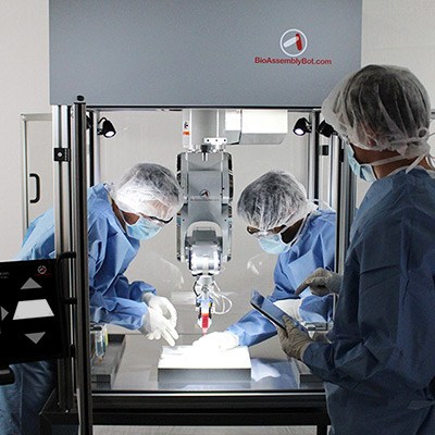
3d.fab’s BioAssemblyBot Wants to 3D Print Skin onto People
3D bioprinting continues to diversify as more and more companies and research organizations join the field, each bringing their own take on the technology to the table. French collaborative platform 3d.fab has an intriguing approach towards bioprinting that involves a freeform robot capable of directly printing on a part of the body. In the video below, the BioAssemblyBot prints what appears to be a bandage directly on an arm:
The “bandage” is actually a bio-ink made from the skin cells of a patient. When applied to the patient’s skin, it forms an autograft that will, within a couple of weeks, create new skin. The BioAssemblyBot is capable of both additive and contour 3D printing, as well as pick and place and assembly thanks to its interchangeable tools. It’s only one of 3d.fab’s bioprinting technologies; the platform has a few other bioprinters in development as well, including another skin printer.
 3d.fab works with other 3D printing technologies as well, including FDM and Polyjet, but everything is geared toward the pursuit of new developments in healthcare. Skin 3D printing is a major priority for the platform, as evidenced by the “Stresskin” project, one of several projects 3d.fab is pursuing. The approach of directly 3D printing on a body part is highly promising; while other organizations have worked on 3D printed skin, the samples generally are too fragile to be sutured, according to 3d.fab. The direct 3D printing concept would eliminate the need for sutures, creating a living bandage that would incorporate itself into the surrounding skin.
This is exciting news for victims of burns, illness or trauma who have lost significant portions of skin. Traditional skin grafts are painful and prone to infection or rejection, and the larger the wound, the more difficult it is to repair with a graft. 3D printing new skin cells directly onto a wound would reduce the risk of rejection, as it uses the patient’s own skin cells to grow new skin, and there would be no limit to the size of the “bandages” that could be applied, thanks to the free-form robot.
3d.fab’s other projects include a 3D printed device for faster and more cost-effective diagnoses of diseases. The goal of the project is to avoid further contributing to antibiotic resistance by thoroughly genetically analyzing pathogens. The platform is also working on improving silicone materials for 3D printed medical models and implants, as well as developing 3D printing technologies that can repair nasal cartilage loss.
Those are just a few of the initiatives 3d.fab is working on to advance 3D printing in the medical field. The platform collaborates with multiple partners and is open to further collaborations; if you are interested in working with 3d.fab, you can contact the organization here.
Discuss this and other 3D printing topics at 3DPrintBoard.com or share your thoughts below.
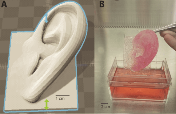
Auricular Reconstruction: Netherlands Researchers 3D Print & Assess New Methods for Making Ears

(A) Schematic of steps in the biofabrication of the implant model. (B) Schematic of the study methods. First, alginate hydrogel beads and 3D‐printed PCL scaffolds were analyzed individually. Next, alginate and PCL were combined in one construct to develop an auricular implant model. PCL: Poly‐ε‐caprolactone.
As users around the world embrace 3D printing, the impacts continue to grow in a wide range of industries—but particularly medicine as researchers make strides in their own labs with bioprinting, as well as creating medical devices, implants, prostheses, and more. And although 3D printing in the realm of hearing devices and bioprinting of ear tissue is not entirely new, researchers in the Netherlands have been working on a novel method for auricular reconstruction.
The Netherlands research team outlines their findings in ‘Design and fabrication of a hybrid alginate hydrogel/poly(ε‐caprolactone) mold for auricular cartilage reconstruction. ‘ With the challenging goal of creating a 3D printed cartilage implant, the researchers assessed whether the bioprinting materials they had in mind were actually viable as they worked to create poly‐ε‐caprolactone (PCL) scaffolds, using alginate as a cell carrier. Success with such technology could mean bypassing more conventional methods that present challenges to include:
- Morbidity at donor site
- Risky exposure of implants
- Difficulty in surgical procedures
“Tissue engineering, in combination with novel biofabrication strategies, is a promising solution to engineer auricular implants with patient‐derived donor cells. These biofabricated auricular constructs could ultimately function as patient‐specific implants for the reconstruction of a deformed auricle,” state the researchers in their paper.
The key for the these scientists was in finding a scaffold strong enough to bear cell growth as well as that of resulting tissue. This type of new scaffold must be durable but also porous and able to break down easily in terms of biodegradability. Bioink composed of synthetic or natural hydrogel could be used for 3D printing cells, or there is also the option of fabricating the support scaffolds and then adding the cell-hydrogel mixture. Poly‐ε‐caprolactone (PCL) is a plastic material used successfully to create scaffolds strong enough for such a purpose.
Scaffolds were created with differences in strands:
- 400 μm
- 600 μm
- 800 μm
- 1000 μm
- 1200 μm
Customized software created G-code, and medical-grade PCL was 3D printed on a 3DDiscovery. Afterward, the molds were cleaned, sterilized, and sealed. The researchers evaluated each sample’s structure using a microscope, digital camera, and fiber optic light. They then assessed cell viability, after which biomechanical analysis examined PCL scaffolds and the alginate hydrogel itself.
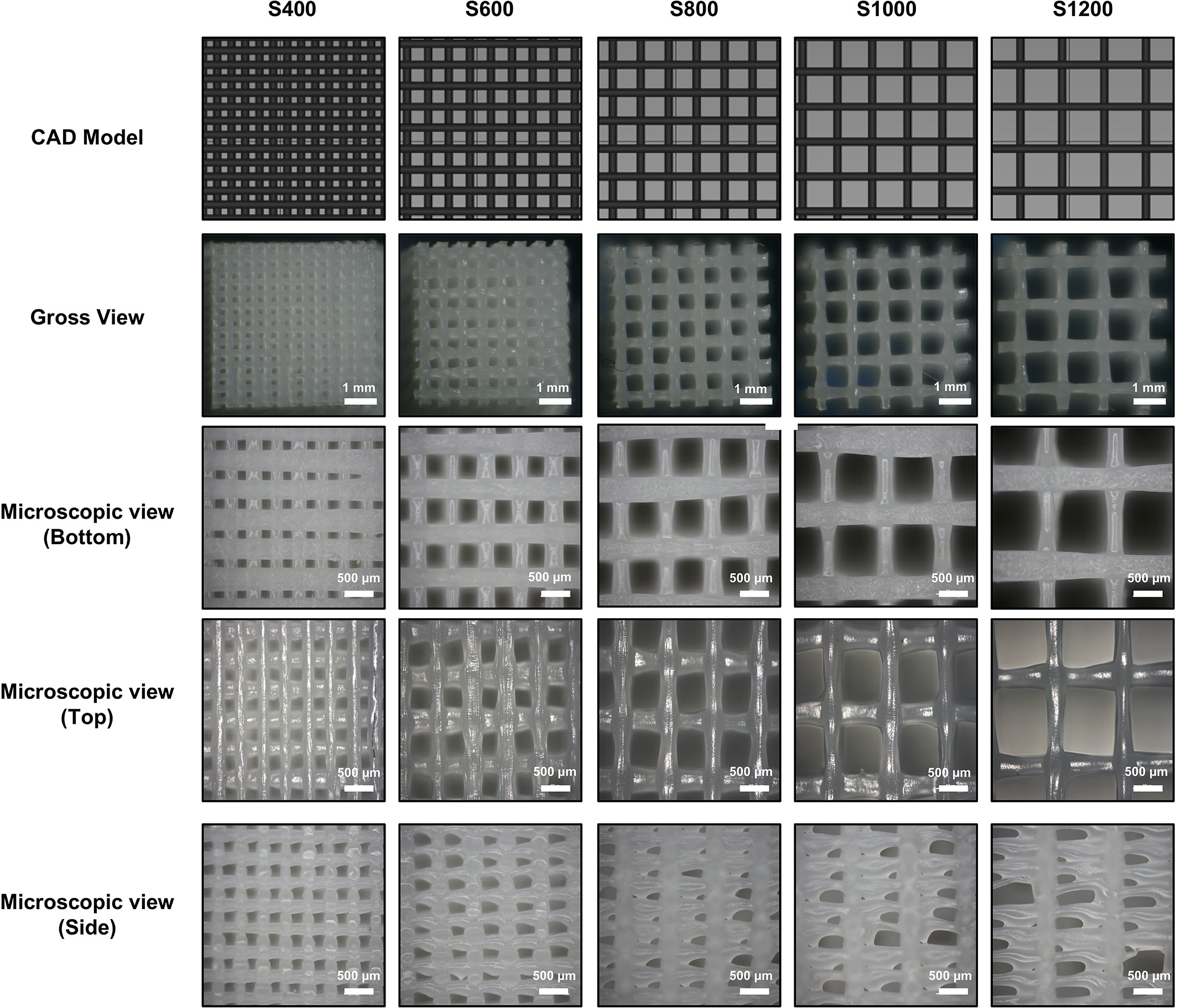
CAD view, gross view, and microscopic views of 3D‐printed PCL scaffolds with varying distances between strands. S represents the sample, with the number representing the distances between strands in micrometers (μm).
They then found that the scaffolds were viable:
“The structural properties of 3D printed PCL scaffolds were determined by examining the surface porosity and mechanical properties. Macroscopic analysis of the PCL scaffolds showed good printing quality,” stated the researchers. “However, microscopic analysis of individual PCL strands showed some variance in strand diameter over a short distance. In addition, the lateral view of the scaffold showed a large variety in pore width. Overall, the smaller the pore width, the more accurate the 3D printed scaffold.”
Overall results were examined further as the researchers considered the following:
- Structural properties of 3D‐printed PCL
- Cell viability and proliferation in alginate hydrogel beads
- Hydrogel biomechanical properties
- Neocartilage formation
- Auricular implant model
The researchers state that the 3D printed cartilage implant does indeed possess the type of mechanical properties required to withstand challenges during in vivo tissue maturation, as well as a natural core that is able to form tissue.
“The mold can be easily printed and assembled, while the design makes it easy to inject any suitable hydrogel for tissue formation,” concluded the researchers. “While long‐term in vivo experiments are required to test its preclinical applicability, the work presented in this study provides a possible strategy for the use of biofabricated tissue constructs in the clinic.”
What do you think of this 3D printing news? Let us know your thoughts! Join the discussion of this and other 3D printing topics at 3DPrintBoard.com.
[Source / Images: ‘Design and fabrication of a hybrid alginate hydrogel/poly(ε‐caprolactone) mold for auricular cartilage reconstruction’]
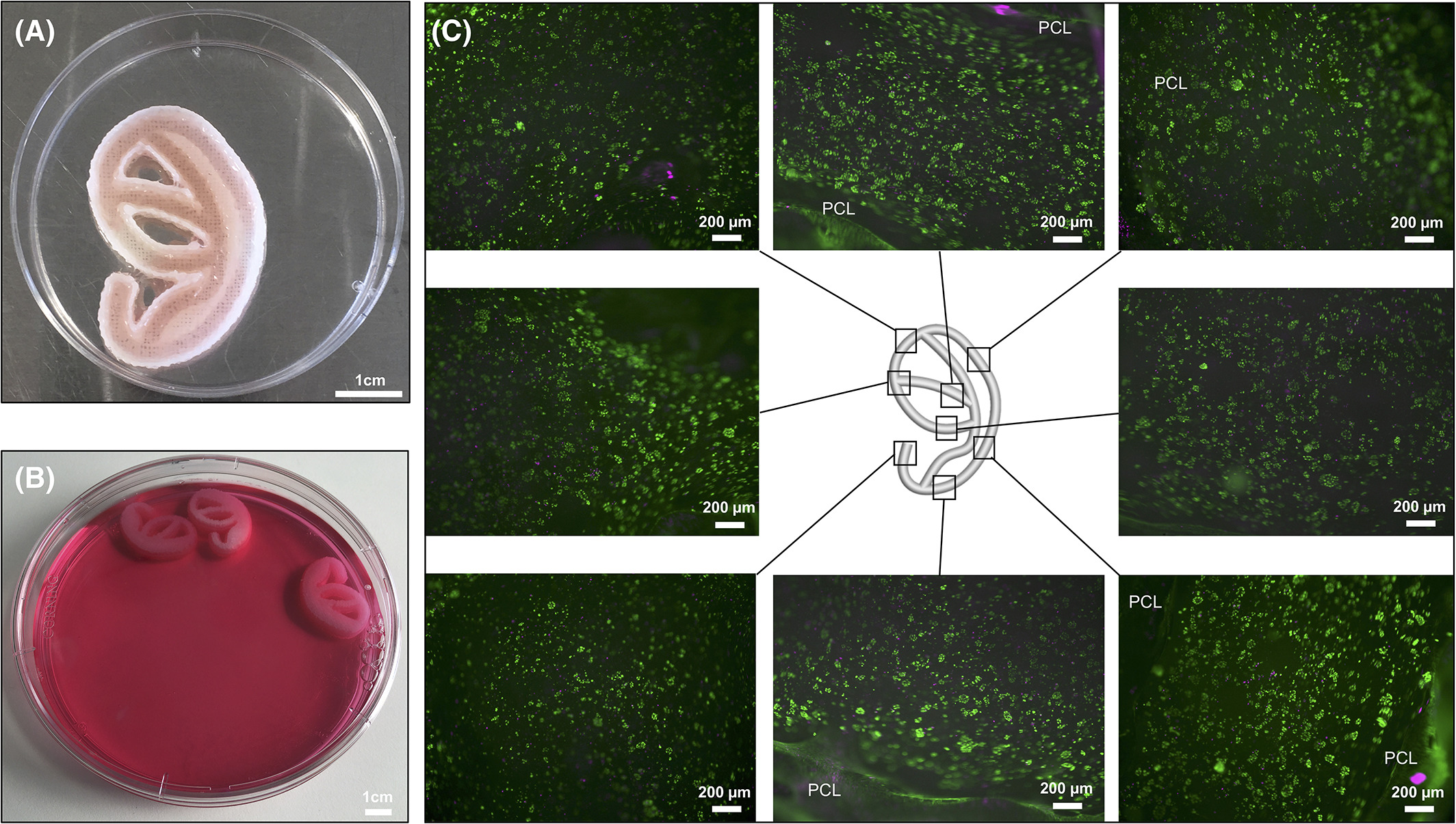
A) Gross view of the PCL‐alginate auricular implant model. Alginate can be found inside the grooves of the PCL mold. Note that one part of the 2‐part mold has been taken off for viewing. (B) in vitro cultured PCL‐alginate auricular implant models. (C) LIVE/DEAD stain of alginate taken out of the PCL mold after 21 days of culture. High‐cell survival was seen throughout the entire implant model.
Interview With Jay Hoying and Michael Golway of Bioprinting Company Advanced Solutions Life Sciences
Some of the biggest impacts 3D printing will have on the world are still quite far away. In labs around the world, people are taking the initial baby steps in bioprinting, tissue printing, using 3D printing in regenerative medicine and making things such as drug loaded implants. We can scarcely conceive of the impacts that bioprinting will have on medicine. We should also especially in this area be careful in distinguishing from the possible to the probable. While researchers see 3D printed organs in a clinical setting to be something like twenty years out most regular consumers see it a something that is bound to happen in a few years. In the middle of this exciting development sits the Advanced Solutions LifeSciences which makes bioprinters, bioprinting software and bioinks and is a part of the larger firm, Kentucky based Advanced Solutions Inc.(ASI). We interviewed Michael Golway the CEO of ASI and the company’s scientific advisor James Hoying to find out what they’re doing in bioprintig.
What is Advanced Solutions LifeSciences?
Michael: “Advanced Solutions Life Sciences (ASLS) exists to democratize and continually improve its BioAssembly 3D Bioprinting Platform, resulting in curative therapies that deliver improved longevity and quality of health while reducing global healthcare costs.”
What does the BioBot Basic do? And how much is it? Who is it intended for?
Michael: “The BioBot Basic is our entry-level bioprinter offering for $4,995. This bundle includes our tissue modeling software (TSIM) and 3D bioprinter which enables research institutions and private companies to rapid prototype with biomaterials.”
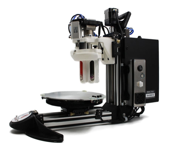
BioBot Basic
Can I adapt the unit for filament, other materials, heating materials etc.?
Michael: “The BioBot Basic is an ambient dispense unit with the ability to 3D print up to 5 different materials in a single print. Our BioAssemblyBot platform enables users the flexibility to adapt temperature control, UV Cure, material movement, etc.”
What is TSIM?
Michael: “TSIM stands for Tissue Structure Information Modeling – it is a 3D tissue modeling software program that enables the user to view both DICOM and 3D solid model constructs within the same workspace to precisely design and prototype simple to highly complex tissue structures.”

TSIM Screenshot
Why do researchers need Tissue Modeling software?
Michael: TSIM integrates many of the software-related tasks needed to generate living 3D tissues into a single workspace. Once a DICOM file is imported, the user can seamlessly navigate and edit the “digital tissue” generated by TSIM from the file to identify regions of interest for design and fabrication. Users can also create tissue models de novo, using whatever 3D design and segmentation tools they prefer. Once created, the digital prototype is sent to our biofabrication platform for production. No other software is required. Upcoming expansions to the TSIM platform will also enable the user to develop automated fabrication and manufacturing processes for tissue production; leveraging the broad manufacturing capabilities of the BioAssemblyBot.
What is the BioAssemblyBot?
Michael: The patented BioAssemblyBot is the world’s first 6-axis robotic arm that 3D prints human tissue structures. The BioAssemblyBot can perform both ‘Additive’ and ‘Contour’ 3D printing. In addition to the 3D printing tasks, the BioAssemblyBot has the flexibility to attach different tools to robotically control the assembly and material movement workflow within the workstation while also interfacing to other agile bioprocessing equipment.
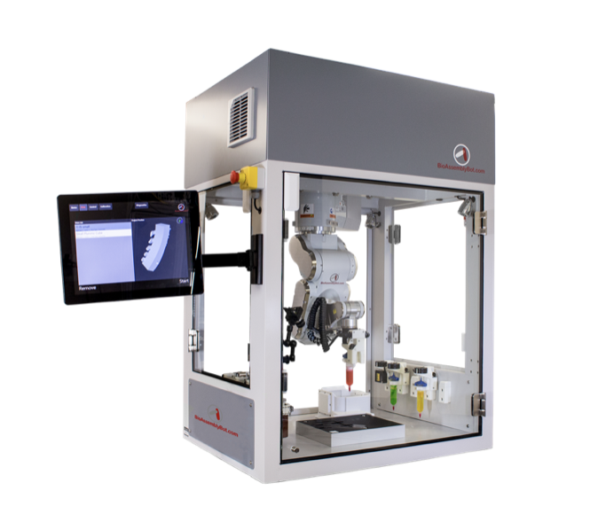
BioAssemblyBot
Why a Six-Axis arm?
Michael: The first industrial robot was invented in 1954. In 60 years, the 6-axis robot has proliferated manufacturing plants across the planet resulting in exponential improvements in assembly and workflow tasks. Today, the technology offers high throughput, exceptional quality, low cost and extreme precision that enables us to now realize manufacturing for patient-matched human tissues. The workflow required to 3D bioprint and assemble complex human tissues are well-suited for a 6-Axis robotic arm and the BioAssemblyBot workstation.
What do you mean with contour 3D printing?
Michael: Contour 3D printing allows a continuous deposition of material along a path in the X, Y and Z plane. Traditional 3D printing is only in the X and Y plane with incremental Z movement. Since the 6-Axis Robot Arm moves with the freedom of a human arm, we are able to 3D print directly onto complex geometric surfaces. These surfaces can pre-exist (e.g. an existing object in the print space) or the result of printing from the 3D Model.

Contour Printing
How does it assemble as well as print?
Jay: While many of these individual subassemblies may be printed, it’s unlikely that an entire organ, with all of its different components will be printed in a single run. Thus, in building complex tissues and organs, we envision the fabrication of sub-assemblies (e.g. valves, vessels, muscle sheets, etc. that make up the heart) which are then assembled into the larger, final product. The robotic arm is designed to perform a variety of manufacturing tasks to enable not just fabrication, but also assembly and bioprocessing. Our BioAssemblyBot does this by automatically switching tool-heads at the end of the arm from fabrication to pick-n-place, to imaging/scanning, and so on. The range of motion of the arm also enables the addition to an existing construct or organ part. In this way, our BioAssembly® Platform enables true tissue and eventually organ manufacturing.
For whom is this intended?
“Michael: Our first suite of products is targeted for research and pharmaceutical applications. We are beginning to release products specifically targeted for clinical applications.”
Why is tool head and motion stage temperature control important? What other tool heads can I add?
Jay: Many of the materials used in regenerative medicine exhibit complex temperature-dependent behaviors that can be leveraged in a tissue fabrication strategy.
- For example, a common preparation of collagen, a native material present in nearly all tissues, requires it to be maintained at cold temperatures. However, at warm temperatures (such as body temperature) the collagen will gel – helping to form the tissue structure. Thanks to our cold bioprinting tool, BioAssemblyBot users can keep the collagen throughout the preparation and entire fabrication process. Our build platform can be heated such that as the cold collagen is printed, it begins to gel immediately as it is added to the structure. The independent temperature control possible with the different aspects of the platform enables flexibility and customization of fabrication protocols.
- The universal adapter at the end of the robotic arm in the BioAssemblyBot® passes power, pneumatics, and data to and from whatever tool head that can be deployed. Thus, the types of tool heads, and therefore manufacturing functionality, is limited by the imagination of our engineering team and users. Everything from 3D scanners to specialized gripper tools to multi-material mixing tool heads are being deployed. We are constantly developing new and custom tool-heads for our customers depending on their applications.
For what kind of an application would I use all eight bioinks on the bot plus pick and place?
Jay: The BAB is capable of working with 8 different tool heads in a single fabrication operation. These could represent 8 different bioinks or a few bioinks plus a pick-n-place, as suggested. Or the operation might include the same bioink in tools fitted with different print nozzle diameters or shapes. One example application involving multi-tool head involves building 3D tissue models in multi-well plates, commonly used in throughput assays and screens. The breadth of tool use depends on how many different cell types, materials, and fabrication approaches are involved. For example, in one application we are developing, the pick-n-place tool is used to move tissue culture plates (before and after fabrication of the tissue) into the work envelope, 3 different tool heads fitted with different caliber nozzles are used to pattern a sacrificial structure, and an additional temperature-control tool is used to dispense cells in matrix. This example highlights the multi-tool use capability and process workflow control of our platform in automating tissue assay production at a high-throughput-like scale.
Can I use other materials than bioinks?
Jay: The BioAssembly Platform can utilize a spectrum of soft materials ranging in operational temperatures from 4oC up to 110oC. As most bioinks fit within this range, the platform is ideal for biomanufacturing. However, nearly any soft material that can be extruded can be employed with the system including silicones, ceramic pastes, glues, paints, biological extracts, food materials, etc. Coupled with our automation controls, the platform is promising great utility in a variety of industries beyond tissue fabrication. Related to this, and reflecting the flexibility of the platform, as our customers identify and develop next generation (bio)materials, we design and create novel printhead technologies for those materials.
Is the pick and place meant for manufacturing or could it be used for mechanized testing?
Michael: Today it is meant to move a tissue through our precision bioprinting workflow. Currently in our development pipeline is a mechanical analytics tool that captures mechanical performance and reliability of a bioprinted tissue or material. Such measurements are critical for assessing the stiffness, elasticity, and viscosity of tissues often employed in load-bearing applications such as menisci, vertebral discs, and bone.
What kind of things have people made with your machines?
Jay: Our Innovations Laboratory, customers, and partners are using our BioAssembly Platform to fabricate a variety of structures, devices, and objects. These include 3D tissue models for research and informative assays, microfluidic platforms for drug discovery and development, tissue patches, small caliber guide tubes, tissue microenvironments for device development, implant systems, organ models, and much more.
Why should I work with you?
Michael: Our organization is founded on the principle of innovating on behalf of the customer to advance the science, and our products are designed so that we can constantly improve them – it likely comes from our roots as a software company. We are obsessed with the promise of regenerative medicine and are working on key partnerships to unlock bioprinting at a therapeutic level, not just research. Not only do our customers get access to the world’s most advanced bioprinter technology platform, but they gain access to a dedicated team of professionals whose sole focus is our customer’s research, commercial and clinical success.
Tunable Bioprinted Tissue Using SLA May Lead to 3D Printed Tissue Engineering Using This Method
Cardiovascular disease such as hypertension and others are frequently caused by hardened blood vessels, and finding a way to replace those vessels has been a challenge in the past. But researchers from the University of Colorado Boulder have developed a 3D printing technique that allows for localized control of an object’s firmness, which could open up new possibilities for the 3D printing tissue. One future possible application of this technology is in the creation of artificial arteries and organ tissue. The research is documented in a paper entitled “Orthoganal programming of heterogeneous micro-mechano-environments and geometries in three-dimensional bio-stereolithography.”
The research features a layer-by-layer printing method with fine-grain, programmable control over rigidity, which allows the researchers to mimic the complex geometry of highly structured yet pliable blood vessels.
“The idea was to add independent mechanical properties to 3D structures that can mimic the body’s natural tissue,” said Xiaobo Yin, an associate professor in CU Boulder’s Department of Mechanical Engineering and the senior author of the study. “This technology allows us to create microstructures that can be customized for disease models.”
To overcome the traditional challenges involved in engineering blood vessels, the researchers found a way to take advantage of oxygen’s role in setting the final form of a 3D printed structure.
“Oxygen is usually a bad thing in that it causes incomplete curing,” said Yonghui Ding, a postdoctoral researcher in Mechanical Engineering and the lead author of the study. “Here, we utilize a layer that allows a fixed rate of oxygen permeation.”
By keeping tight control over oxygen migration and its subsequent light exposure, the researchers can control which areas of an object are solidified to be harder or softer, while keeping the overall geometry the same.
“This is a profound development and an encouraging first step toward our goal of creating structures that function like a healthy cell should function,” Ding said.
The researchers demonstrated their technique by 3D printing three different versions of a simple structure: a top beam supported by two rods. Each structure was identical in shape, size and materials, but varied in rod rigidity: soft/soft, hard/soft and hard/hard. The hard rods supported the top beam while the soft rods allowed it to collapse. The researchers then repeated the exercise with a small Chinese warrior figure, making the outside hard but the inside soft.
The 3D printer used by the researchers is capable of printing biomaterials as small as 10 microns, or one-tenth the width of a human hair. The researchers believe that they can further improve their technique with future work.
“The challenge is to create an even finer scale for the chemical reactions,” said Yin. “But we see tremendous opportunity ahead for this technology and the potential for artificial tissue fabrication.”
One in every four deaths in the United States is caused by heart disease, totaling over 600,000 deaths per year. It’s the leading cause of death for both men and women, but the discovery of a way to 3D print healthy blood vessels could make a tremendous difference. Having said that there are many other tissue printing technologies that are making strides. Tunable geometries and gradient materials are already possible with other technologies and in other materials in bioprinting. We’re saddened that so many media have misreported this story and overstated the claims as expressed by the researchers in their work. This paper gives us a great new path forward to tissue engineering but the media should be careful to read the papers that they write about. For SLA and tissue engineering this is a good step forward but it is wrong to speculate so widely and make unassociated claims especially when the researchers in question express themselves so succinctly and clearly in the paper.
Authors of the study include Hang Yin, Yonghui Ding, Yao Zhai, Wei Tan and Xiaobo Yin.
Discuss this and other 3D printing topics at 3DPrintBoard.com or share your thoughts below.
Real Time Rheology System Used In 3D Printing and Bioprinting
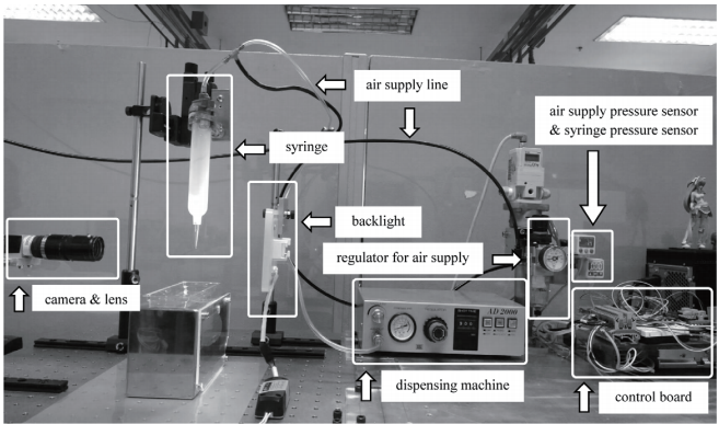
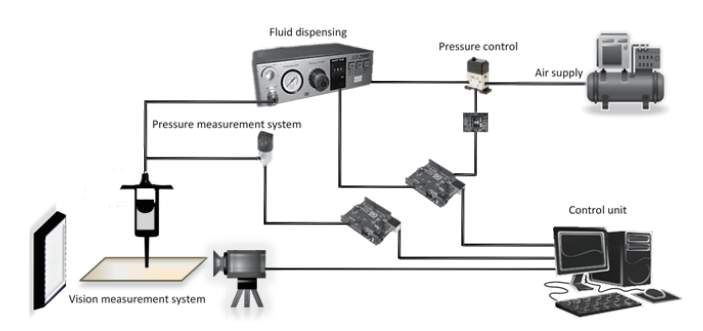
In a paper entitled “A Real-time Rheological Measurement for Biopolymer 3D Printing Process,” a group of researchers develops a method of measuring the rheological properties of solutions for 3D bioprinting. Rheology is the study of the flow of matter, and the flow rate of 3D printing materials is extremely important to the final object, particularly in bioprinting. The typical method for measuring rheological properties is the use of a rheometer before the materials are dispensed.
“However, the rheological properties of biopolymers are time-dependent,” the researchers state. “Therefore, inaccurate parameters may be used under certain processing conditions. It also affects the precise control of flow rate, especially in the case of biopolymers with rapid gelation.”
In the paper, the researchers present a system for the real-time measurement of rheological properties during the dispensing process, rather than before it. The system is a combination of a user interface for setting up measurement and a vision measurement system. An image processing method is applied to measure the volume of dispensed fluid.

“The combination of pressure data in a syringe acquired from a pressure sensor and measured volume flow rates is used to construct a pressure-dependent fluid flow rate curve as a function of time,” the researchers explain. “The rheological properties of fluid materials are then determined by a numerical analysis procedure. The results from the numerical analysis provide a time-dependent power law index (N) of fluids. This index can be used as the flow control parameters of dispensing processes.”
For their experiments, the researchers used both a polymer and a biopolymer: a PVA solution and a PVA solution mixed with chitosan, respectively. The measurement setup included a fluid dispensing system, a system for the measurement of syringe pressure, a compressed air pressure regulator system, and a vision measurement system.
The rheological properties of the solutions gradually evolved after being mixed and put in the syringe during the dispensing process within 50 minutes. The researchers discovered that the increase in syringe pressure increases the fluid droplet volume.
“For different time stamps and recorded applied pressures, the measured flow rates were used to construct pressure-dependent fluid flow rate curves every 10 min for 6 experiments in the case of PVA (within 60 min),” the researchers state. “…According to the pressure-dependent fluid flow rate curve, the fluid flows of both PVA and PVA/CS are significantly sensitive to time changes. With these results, it is clear that the real-time rheological properties measurement system is critically needed to identify fluid flow behavior at a specific time during the fluid dispensing process.”
A time-dependent power law index of the fluids was determined as N, the value of which changed significantly according to the rheological properties of the fluids.
“They are comparable to the actual behavior of dispensed fluids in which they become more viscous (increasing n) after they are mixed and injected from the syringe,” the researchers conclude. “The resulting N can be used to perform automatic fluid flow control in future research.”
This research is very exciting as it could be used to vastly improve the quality of 3D printed parts in the future. This system could let you know more precisely what is being deposited or in real time syringe pressure or other variables could, in the future, be adjusted in order to get more accurate deposition.
Authors of the paper include Anchyza Yokpradit, Teerawat Tongloy, Supranee Kaewpirom and Siridech Boonsang.
Discuss this and other 3D printing topics at 3DPrintBoard.com or share your thoughts below.
3D Printed Ligaments Could Change the Way Common Injuries are Treated
Ligament tears are becoming more common in sports. They’re painful and debilitating, and difficult to treat. The current standard is to replace the torn ligament with tendons, but that can cause additional problems down the line.
“What can happen over time is that the tendon itself begins to kind of stretch and become a little bit relaxed in the joint,” said Christina Salas, PhD, a scientist at the University of New Mexico. “Then (the tendon) becomes deficient again.”
 Dr. Salas is currently working on creating 3D printed ligaments, which she says has never been done before. It’s an area of focus she has been working on for some time, with help from students and professors at the University of New Mexico.
“This is something that hopefully can reduce some of those failures we see,” said Dustin Richter, Assistant Professor of Sports Medicine and Orthopedic Surgery. “And get people back to doing what they enjoy.”
The researchers have developed a special technique involving electrospinning, which uses electric force to create fibers.
“The near-field electrospinning technique that we have added to the bio-printer actually produces really highly aligned fibers that replicate the ligament tissue,” said Dr. Salas.
Doctors could then take a CT or MRI scan of a patient’s damaged joint and create an exact replica using 3D printing. This could allow for less-invasive surgery, and could be a more permanent solution as the synthetic 3D printed ligament would not wear out or weaken.
“We want to make sure that those patients can truly maintain their full function even longer as they grow older in life,” said Dr. Salas.

Dr. Christina Salas
The biggest challenge Dr. Salas and her colleagues are facing is figuring out how to attach the 3D printed ligaments to the bone. Dr. Salas recently received a two-year, $150,000 grant for the research.
3D printing is changing the way doctors and scientists look at the treatment of common injuries, such as torn ligaments. Recently, Queensland researchers revealed a new method of 3D printing joint cartilage, which could greatly shorten recovery time after surgery for arthritis and joint injuries – and that’s only one example of the extensive research that is taking place around the world involving the use of 3D printing for the repair of damaged bones and muscles.
Dr. Salas’ research involves artificial ligaments, but other work is taking place that involves 3D printing new tissue using the patient’s own stem cells. Many people automatically jump directly to talking about 3D printing organs when 3D bioprinting is mentioned, but major progress is being made in other areas, like the treatments of degenerative illnesses and common injuries. With 3D printing, athletes have the potential to stay in the game for much longer than they would have otherwise. There may also be less need for things like walkers and wheelchairs as people age, as diseases like arthritis are healed with new cartilage.
When 3D printed organs eventually become realized, they will potentially enable people to live longer than ever before. Until then, bioprinting has the ability to help people live with better quality of life for their natural lifespans.
Discuss this and other 3D printing topics at 3DPrintBoard.com or share your thoughts below.
[Source: KRQE]
3D Bioprinting: Comparing Lattice Scaffolds with Traditional Rectangular Sheets
Bioprinting is not a simple endeavor – if it were, we would likely be transplanting 3D printed organs by now. It’s a delicate process that requires a number of factors to be in place, including bioinks that are both printable and biocompatible, and proper scaffolds. In a paper entitled “A Comparative Study of a 3D Bioprinted Gelatin-Based Lattice and Rectangular-Sheet Structures,” a group of researchers compares scaffolds with lattice mesh geometries to more traditional flat rectangular sheets.
“We hypothesised that the experiments performed as a part of this study would help us to observe considerable differences between the two structures, i.e., lattice and rectangle, and also open up the possibility of significantly enhancing the design of a 3D bioprinted construct for engineering cardiac tissue-on-a-chip, using bioprinting,” the researchers state.
The researchers used furfuryl gelatin (f-gelatin) as a base for their bioink, which they seeded with mouse mesenchymal stem cells. They used an ALLEVI 2 bioprinter to print the ink into two different structures – a lattice and a rectangular sheet. Rheological characterization of the bioink was conducted, and the bioprinted structures were cultured in an incubator. A live/dead cytotoxicity assay was performed, and the texture of the lattice was analyzed by scanning electron microscopy.
“The SEM cross-sectional image of the gelatin lattice revealed a highly organized, striated, patterned, and networked structure in comparison to the loosely networked and largely porous rectangular-sheet cross-section SEM, as reported in our previous study,” the researchers explain. “Porosity and pore-size are crucial to ensure cell colonization of the scaffold, deposited using bioprinting. Likewise, SEM micrographs showed a homogeneous distribution of equal sized pores within the entire area scanned and imaged….The average apparent porosity of this lattice structure was estimated to be about 50% compared to 21% for the rectangular-sheet. The results led us to conclude that although the mean pore size was significantly reduced by printing in the form of a lattice, the inherent design of the lattice allowed pores to be of a similar size and to be homogenously distributed throughout the entire structure, compared with the rectangular-sheet.”
Swelling behavior of the gels was monitored to study the hydration dynamics of the crosslinked hydrogel structure. Cell proliferation was assessed and flow cytometry was analyzed.
Results of the testing showed that the lattice structure was more porous than the flat rectangular sheet. It also exhibited a lower degradation rate.
“Further, the lattice allowed cells to proliferate to a greater extent compared to the rectangular-sheet, which initially retained a lower number of cells,” the researchers state. “All of these results collectively affirmed that the lattice poses as a superior scaffold design for tissue engineering applications.”
A scaffold is literally a foundation to build upon in bioprinting, and having an effective scaffold is key in any bioprinting application. Cells rely on a strong scaffold in order to survive and proliferate. A printable, biocompatible ink is also crucial for cells to be able to grow into tissue. The researchers find in this study that lattice structures are superior to rectangular sheets, which could mean the difference between success and failure in future applications.
Authors of the paper include Shweta Anil Kumar, Nishat Tasnim, Erick Dominguez, Shane Allen, Laura J. Suggs, Yoshihiro Ito, and Binata Joddar.
Discuss this and other 3D printing topics at 3DPrintBoard.com or share your thoughts below.
3D Bioprinting Makes Progress as Lab-Grown Bladder Helps Young Man to Lead a Normal Life
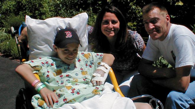
Luke Massella, aged 10 in 2001 with his mother and uncle
Luke Massella was born with spina bifida, a condition that left a gap in his spine. By the age of 10, he had undergone a dozen surgeries and, contrary to doctors’ expectations, was able to walk. But then his bladder malfunctioned, causing his kidneys to fail.
“I was kind of facing the possibility I might have to do dialysis for the rest of my life,” he says. “I wouldn’t be able to play sports, and have the normal kid life with my brother.”
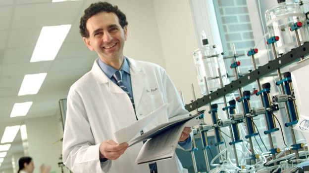
Dr. Anthony Atala [Image: Wake Forest Institute for Regenerative Medicine]
Dr. Anthony Atala of Boston Children’s Hospital had a better idea. He took a small piece of Massella’s bladder and, over the course of two months, grew a new organ in the lab. The new bladder was transplanted into the patient in a 14-hour surgery and, according to Massella, he has been able to live a normal life since then. Now 27, Massella underwent 17 surgeries before he was 13, but has not had to have a single operation since then.
Dr. Atala is an expert in bioprinting, and he and his team have developed eight cell-based tissues that they have transplanted into patients. These include skin, urethras and cartilage grown in the lab. The organs are currently going through clinical trials for approval by the US Food and Drug Administration.
“You need to know how to make these organs by hand, then the bioprinter is really a scale-up tool,” said Dr. Atala, now Director of the Wake Forest Institute for Regenerative Medicine in North Carolina.
According to Dr. Atala, the easiest structures to 3D print are flat structures like skin, followed by tubular structures like blood vessels and urethras. Hollow, non-tubular structures like bladders are more difficult – Massella is one of 10 people who currently has a bladder grown from his own cells. The most difficult of all are solid organs like hearts, lungs and kidneys, which have “so many more cells per centimeter,” Dr. Atala said.
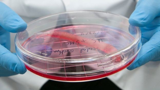
A 3D printed urethra in progress [Image: Wake Forest Institute for Regenerative Medicine]
For a patient like Massella, technology like that Dr. Atala is working with allows a safer transplant, with much less risk of rejection. Massella, who feared he would never be able to have a normal life, went on to become a wrestling coach and now runs events in the jewelry industry.
Massella’s case was an early example of what tissue engineering can accomplish, but advances in bioprinting are making such cases more common.
“A lot has happened in the last couple of years,” said Steven Morris, Chief Executive of bioprinting startup BIOLIFE4D.
BIOLIFE4D has become known as the startup that plans to be the first to 3D print a functioning human heart – and it is getting closer to its goal. Initially, the company will be printing smaller versions of hearts to be used for pharmaceutical testing purposes, but its ultimate goal is to be able to print organs that can be transplanted into patients. That is the dream of most bioprinting companies and research institutions, and while some still doubt that it can be done, the technology is getting closer every day.
Patients like Massella are evidence that functioning organs are possible to create in the lab, and one day – perhaps sooner than anyone expects – cases like his may become much more commonplace. Will we ever be able to eliminate organ donor waiting lists thanks to bioprinting? It’s not nearly as impossible as it once seemed. Generally most practicioners in the field see true 3D printing of organs as something that will happen some 20 to 25 years from now. Approvals, developing all of the tools and assessing long term risk will all take time. We’re still very far from this becoming a day to day procedure for many people. The signs are encouraging however that bioprinting will lead to improvements in patient’s lives.
Discuss this and other 3D printing topics at 3DPrintBoard.com or share your thoughts below.