Indianapolis VA Medical Center and NASA To Explore 3D Bioprinting for Healthcare

Exposure to space radiation, gravity fields, contaminated atmospheres, are all part of the hostile environment astronauts encounter beyond Earth’s orbit. As they transition from one gravity field to another, the fluids in their body can shift upwards, which could lead to pressure on the eyes causing vision problems, among other pretty scary diseases that the National Aeronautics and Space Administration (NASA) has been working to solve for decades as they prepare to send humans on three-year missions to Mars.

NASA wants to make sure that tissue replacement beyond the repairing ability of the organism can be done in space. However, such a scheme would also mean that space agencies will have to plan way ahead of departure on how to leverage innovative technologies, like 3D printing, to aid astronauts on their journey, while directly contributing to their respective development efforts. That is what several United States government agencies are doing, including the Veterans Administration and NASA. More particularly, scientists at the Indianapolis Richard L. Roudebush Veterans Affairs (VA) Medical Center and NASA investigators are now jointly exploring new approaches to 3D bioprinting. The two teams are collaborating in the conversion of human and animal eye fundus images into virtual renderings of the retinal vascular networks for bioprinting. Exploring this field, will not only serve NASA’s interest in space, but also the VA’s concerns here on Earth since tissue replacement is often needed to treat combat, accident or disease-induced damage to veterans.

One of the most serious limitations for the success of tissue engineering, and in particular of the bioprinting approaches to generate artificial tissues, is the difficulty to perfuse the constructs with oxygenated fluids and nutrients. To tackle this issue, Patricia Parsons-Wingerter, senior scientist at NASA’s Space Biosciences Research Branch, and her NASA-wide team joined forces with Nicanor I. Moldovan, founding director of the 3D Bioprinting Core (3DBPC) laboratory at the Richard L. Roudebush VA Medical Center. 

Patricia Parsons-Wingerter (Credit: NASA)

Such combined efforts could bring unprecedented success for the bioprinting of vascular networks. In this instance, the NASA team will use their innovative Vessel Generation Analysis (VESGEN) 2D software application for the design of anatomically realistic vascular patterns for bioprinting, while the Indianapolis-based team will provide their know-how on bioprinting vascular models.

In fact, Parsons-Wingerter and her colleagues developed VESGEN 2D, an image analysis program that performs branching quantification of vascular networks, in order to assist in the early detection of changes in vascular patterns of the eye, which may indicate microgravity-induced retinopathy, also the major blinding disease of working-aged adults. However, vascular-dependent diseases include cancer, diabetes, coronary vessel disease, and major astronaut health challenges in the space microgravity and radiation environments, especially for long-duration missions. In this sense, VESGEN 2D maps and quantifies vascular remodeling for a wide variety of quasi-2D vascularized biomedical tissue applications

Parsons-Wingerter’s concern with eyesight problems related to space travel has been historically well-founded. According to one study sponsored by NASA several years ago, space flights that last six months or longer can cause changes in astronauts’ eyes and vision. At the time, this discovery had a major impact on plans for a manned flight to Mars, making this a top priority for NASA’s Space Medicine research team. During the study, seven astronauts that were examined had eye structure and vision abnormalities, most commonly the flattening of the back of the eyeball, changes in the retina (the light-sensitive area at the back of the eye) as well as the optic nerve. Knowing now that vision can be severely compromised in space now demands a lot of attention from researchers, especially, since astronauts, like airline pilots, must have 20/20 vision to carry out most of their work in orbit.

According to Parsons-Wingerter, VESGEN is in fact, currently being used to help understand and ameliorate vision impairments in astronauts and terrestrial adults diagnosed with diabetic retinopathy. And it’s no surprise that VESGEN 2D will be utilized to aid in the design of anatomically realistic vascular patterns for bioprinting since this image analysis program offers insightful read-outs (or biomarkers) of dominant molecular signaling targeted by drug and therapeutic development. 

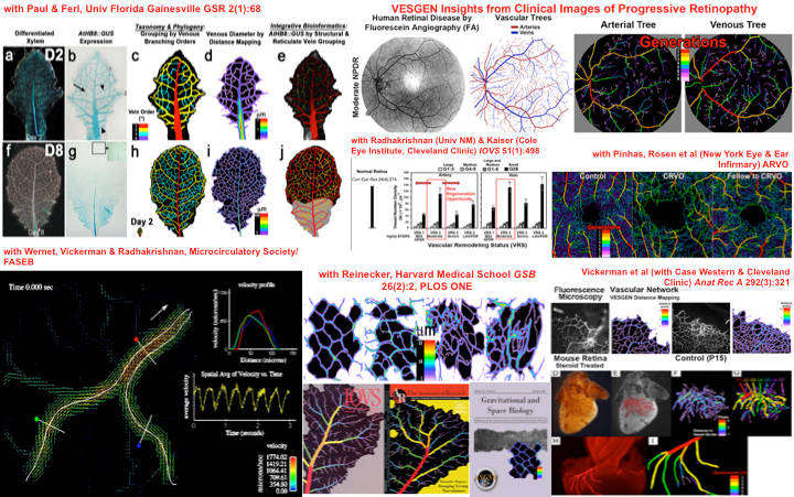
Composite images and journal cover illustrations of vascular patterning collaborations with other scientists for VESGEN research (Credit: NASA)

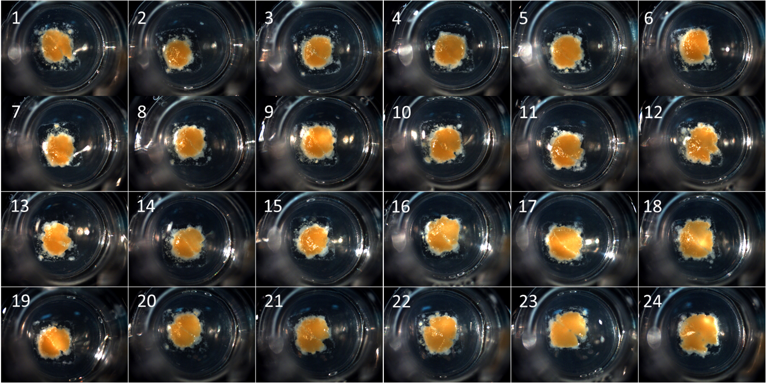
Meanwhile, Moldovan’s 3DBPC laboratory (also known to many as the “Core”) primarily serves the VA investigators’ needs, helping with workflow design and implementation from project conception through funding and execution. More specifically, the Core provides assistance with hydrogels preparation, bioinks characterization, cell cultivation, samples mixing, 3D model printing, cell-containing constructs bioprinting, incubation, perfusion, and characterization (by fluorescence microscopy, micro, and macro photography), up to data analysis and preparation for publication. Current activities in the Core include the generation of bone, cartilage, retinal, neural, and other 3D models.

Nicanor Moldovan (Credit: 3D Tissue Bioprinting Core at VAMC)

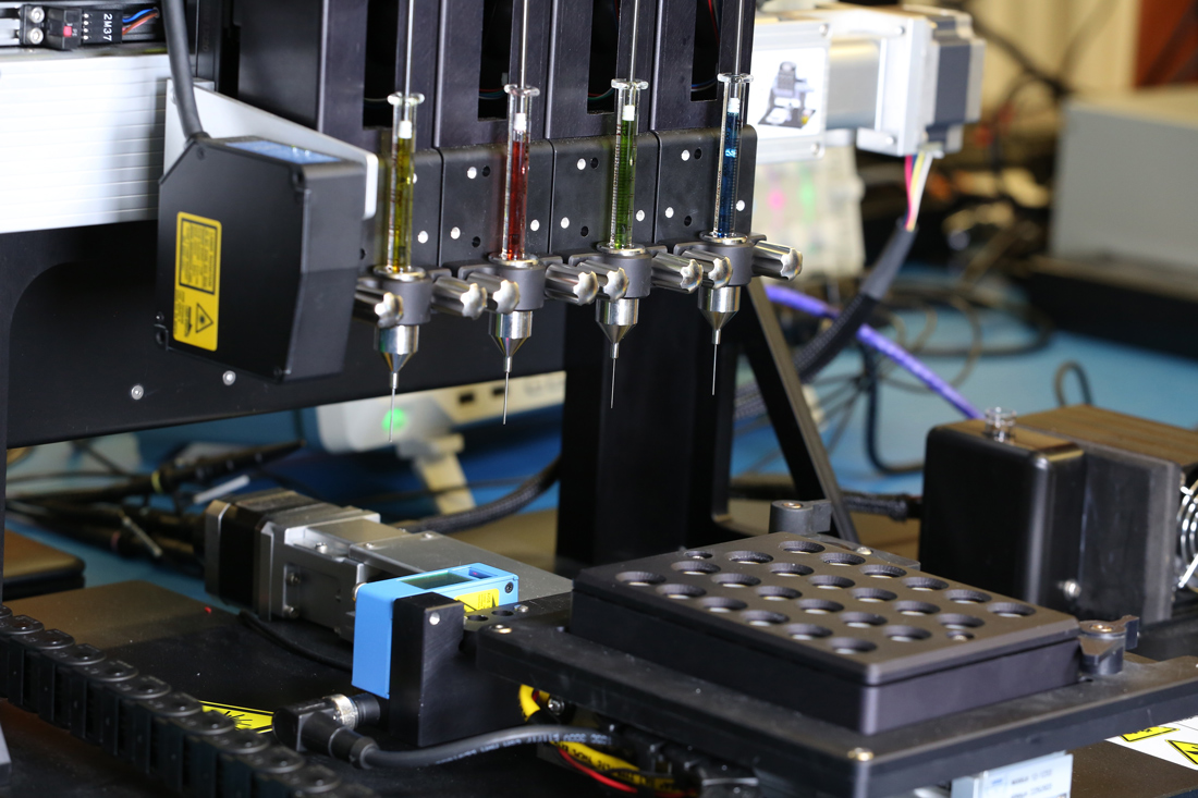
Funded and administered by the Indiana Institute of Medical Research (IIMR) and supporting the overall research mission of the Veterans Affairs Medical Center in Indianapolis, the Core features the bioprinter 3DDiscovery, which was purchased from Swiss company regenHU through a VA Shared Equipment Evaluation Program (ShEEP) grant. 

Another example of the commitment of government agencies to work together to develop healthcare solutions for the future, the Richard L. Roudebush VA Medical Center has also recently joined NASA’s centennial Vascular Tissue Challenge, a project that rallies scientists worldwide to produce viable thick-tissue assays that can be used to advance research on human physiology. NASA began the Vascular Tissue Challenge in May 2019, in collaboration with the Methuselah Foundation and thanks to the support from the New Organ Alliance (NOA), a non-profit organization aiming to catalyze the tissue engineering field. Roudebush VA Medical Center’s Moldovan is also participating in this venture as chair of the NOA’s In Vitro Tissue Models Sub-Committee.

Considering that one of the main projects in the Core is the bioprinting of vascular models, this new joint effort with Parsons-Wingerter is considered by them as a natural step to move forward the research for both teams. In fact, the same approach with VESGEN 2D can be used for the realistic representation, and the adaptation to the printable format of vascular networks of other organs, such as rat mesentery or mouse colon, to be incorporated as region-specific cellular compositions in actual bioprinted tissue constructs. The teams claim that these constructs will be useful both as in vitro models for mechanistic studies and drug discovery and for the eventual replacement of damaged tissues or organs. After all, that is exactly what NASA and the VA are aiming for, both here and in space.

Considering that the US space agency has recently asserted its goal of sending astronauts to Mars once again, researchers, like Parsons-Wingerter and Moldovan, are crucial to the future mission’s, success. This is especially relevant since such Mars missions would last from seven months to two or more years. As they continue to solve many of the health-related issues attached to long-duration space expeditions, we will surely find out more about their work as they combine creative talents to develop more bioprinting innovation. 

The post Indianapolis VA Medical Center and NASA To Explore 3D Bioprinting for Healthcare appeared first on 3DPrint.com | The Voice of 3D Printing / Additive Manufacturing.

Denmark: 3D Printing Conductive Hydrogels for Medical Applications

In the recently published ‘Electrically Conducting Hydrogels for Health care: Concept, Fabrication Methods, and Applications,’ Shweta Agarwala of the Department of Engineering at Aarhus University in Denmark researchers 3D printing techniques in the medical realm, offering a review of conductive hydrogels.

As 3D printing began to infiltrate the mainstream, industries such as automotive, aerospace, and construction have been positively impacted—and now, medical applications have gone beyond 3D models and devices as scientists continue to make huge strides in bioprinting. Scaffolds are a common structure used in tissue engineering featuring a variety of different hydrogels; however, they can also be used for a wide range of applications today, from smart wearables to biosensors, implants, and avenues to wound management.

Hydrogels are attractive for use in research and other applications due to:

  • Ideal extracellular matrix (ECM)
  • Cell support
  • Biocompatibility
  • Natural and synthetic hydrophilic polymer chains offering high absorption of water

“Although hydrogels have found niche application in tissue engineering, they are inherently insulating by nature. Recent research has shown that hydrogels not only possess necessary characteristics to support biological species but can also interface with electrical circuitry if modified,” states Agarwala. “Hence, research on conducting hydrogels have gained widespread interest for applications such as health recording electrodes.”

Schematic illustration of conducting hydrogels, their components and applications.

Agarwala notes that in general, the conductivity associated with hydrogels is ionic conductivity.

Summary of the material composites and their electrical conductivities achieved to make conducting hydrogels.

“The contribution from additives materials to overall conductivity in such cases is small. However, recent research efforts in this direction have shown promise in inducing electrical conductivity from the additive materials,” states Agarwala.

While the method most often used for aqueous compatible conducting materials is to use ultrasonic energy or heating, five other approaches are available:

  1. Hydrogel monomers with cross-linkers and nanoparticles are gelated together.
  2. Nanoparticles are physically embedded into the hydrogel matrix after gelation.
  3. Nanoparticle precursors are loaded into the gel.
  4. Cross-linking using nanoparticles forms hydrogels.
  5. Hydrogels are formed using nanoparticles, polymers, and other molecules.

Schematic diagram depicting various approaches to synthesize conducting hydrogel: (A) hydrogel monomers with cross-linkers and nanoparticles gelated together; (B) physically embedding nanoparticles into hydrogel matrix after gelation; (C) reactive nanoparticle formation aided by the hydrogel network where nanoparticle precursors are loaded in the gel; (D) cross-linking using nanoparticles to form hydrogel; and (E) hydrogel formation using nanoparticles, polymers, and other molecules.

One of the greatest benefits in 3D printing is that users are able to create much more complex geometries, along with enjoying enormous latitude in both design and customization, as well as being able to make projects faster, and make changes to them on demand. All of these benefits apply to why there has been such an increase in 3D printing conducting hydrogels.

Techniques usually rely on shear thinning, causing them to flow as pressure is applied, using a piezoelectric head.

“A piezoelectric material deforms on applying voltage or current. Thus, the orifice opening can be controlled by varying the voltage applied to the printer head. Inkjet printing creates small droplets (sub-micron volume), which are deposited on the surface,” states Agarwala. “Small volume of material deposition, as against large material ejection through extrusion, helps to print high-resolution constructs and scaffolds.”

“Ink development is considered one of the most important aspects of 3D printing. Hydrogel inks need to have the right rheological properties to fulfill the physical and mechanical needs of the orienting process.”

Sketch of (A) 3D bioplotting system (Reproduced with permission [65]) (B) digital light projector (DLP) 3D printing system to 3D print conducting hydrogel scaffolds (Reproduced with permission [74]), and (C) stereolithography process (Reproduced with permission [75]).

Such hydrogels have the potential to be used in sensor technology, drug delivery systems, and tissue engineering. A variety of composites have been used too, from graphene-chitosan to silica nanoparticles, silica alumina, but the author points out that commercialization of such manufacturing is ‘still far away,’ due to the many challenges involved.

“These materials are unable to follow the original design models, as the printed construct does not retain the original shape. Achieving functional gradients and hierarchical properties have also been challenging and new design approaches are being developed to tackle them,” concludes Agarwala. “

“The area of conducting hydrogels is still full of unresolved technological challenges, and thus provides researchers with opportunity for development, as this field is growing fast beyond its early stage. Improvement in the conductivity of the hydrogels may be one research direction, while incorporating new functionalities such as biodegradability and mechanical strength can open new avenues for applications. Innovation is also required in fabrication methods to allow varied composition of hydrogels to be laid down in desired fashion.”

What do you think of this news? Let us know your thoughts! Join the discussion of this and other 3D printing topics at 3DPrintBoard.com.

[Source / Images: ‘Electrically Conducting Hydrogels for Health care: Concept, Fabrication Methods, and Applications’]

The post Denmark: 3D Printing Conductive Hydrogels for Medical Applications appeared first on 3DPrint.com | The Voice of 3D Printing / Additive Manufacturing.

Inventia Life Science Empowers Researchers to Rapidly Scale 3D Cell Culture

No disease has ever been as overwhelming as cancer, not only does it kill close to 10 million people every year, but even though we still fail to understand how to avoid it, one thing is for sure, researchers are beginning to look at this disease from a different perspective. We have seen lots of research on bioprinting human cells to mimic tumors for testing cancer drugs and now more than ever, new companies are surfacing to create bioprinters for a demand that will surely grow in the future. That is the case with Sydney based start-up Inventia Life Science. Built around digital bioprinting technology for fast, scalable, and reproducible printing of 3D cell constructs, company founders Julio Ribeiro, Aidan O’Mahoney, Cameron Ferris, and Philippe Perzi, expected their creation to remove the need for time-consuming manual labor of medical lab workers.

In 2013, the company’s research led to the development of a proprietary platform encompassing bioprinting technology, an expanding library of printable bioink materials, and custom protocols for specific applications. Their 3D bioprinting platform called Rastrum has been used to rapidly print human cells to help with cancer drug testing and recently, the Coronavirus pandemic motivated the company to produce 3D lung microtissues for researching therapies.

Highly recognizable and easily distinguished from its competitors due to its stand-out pink color, the device was designed with cell biologists in mind, instead of tissue engineers. In the last years, the developers emphasized how this as a great advantage of the printer for researchers who seek better 3D cell models. In that sense, Rastrum creators claim that the machine delivers a platform where hydrogels, printable structures, and printing parameters are pre-validated, enabling a simple and efficient workflow for the creation of 3D cell models. And best of all, no prior bioprinting knowledge is required.

Last year, the hot pink machine won one of Australia’s major design awards, the prestigious Good Design Award of the Year. Designed by two leading medical and science academics from Sydney’s Australian Centre for Nanomedicine (ACN), at the University of New South Wales (UNSW), Justin Gooding and Maria Kavallaris, as the result of a strong collaboration between the university, the Children’s Cancer Institute, and Inventia Life Science, Rastrum is being used extensively throughout Australia as well as other countries.

Rastrum bioprinting platform (Credit: Inventia Life Science)

The printer uses ink-jet technology to print human cells at a rapid rate, quickly cultivating realistic tumors for testing cancer drugs. The technology focuses on printing high volumes of human cancer cell spheroids so that cancer researchers can try to find better ways to eradicate the disease. At the University of Technology Sydney, researchers are printing ovarian cancer cells, while the Victorian Centre for Functional Genomics (VCFG) at the Peter MacCallum Cancer Center in the Australian city of Victoria, was the first lab to install the Rastrum system and apply the technology to their ongoing cancer research.

The innovative technology allows scientists to print 3D cell models at unprecedented speed, replacing a time-consuming and manual process, expanding the capacity for research and drug development in cell models. According to scientists at VCFG, the machine is able to produce 1,000 3D cell models in less than six hours, a task that would regularly take more than 50 hours using current manual techniques.

One of the first users of the device, Kaylene Simpson, associate professor and head of the VCFG at Peter Mac said that “this is a novel and exciting platform for cancer research,” with “the ability to create realistic three-dimensional cell models through an automated and scalable process [that] will vastly accelerate our research progress and advance therapeutic target discovery.” She also revealed that “we have a very clear vision of the clinical applications of the technology.”

However, Inventia is also moving beyond its focus on cancer cells and is now claiming that the versatility of the Rastrum platform can also rapidly print 3D lung microtissue for COVID-19 therapy development. This is not the first biotechnology company that has chosen to focus its efforts to aid researchers in accelerating procedures and seeking cures for the newly discovered infectious disease caused by severe acute respiratory syndrome coronavirus 2 (SARS-CoV-2).

In fact, Inventia revealed in April that their Rastrum 3D bioprinting platform could become a powerful tool for the
production of 3D lung microtissues and that the team of company researchers could move their expertise and capacity to develop these cell models, which can be tailored for therapy development. Furthermore, the Australian-based team proposed to even adjust the 3D cell models for the specific requirements of individual research laboratories and are already working with sites in Australia to explore the potential to accelerate in vitro research into COVID-19 therapies using advanced 3D lung microtissues.

Considering that to date there are currently no effective vaccines, antiviral treatments, or therapeutic agents against COVID-19, Inventia claimed that there is a very high need for multi-cellular in vitro microtissues to understand and assess treatments against this new virus to fend off the global pandemic. Previous lung alveolar research, they say, has shown that in vitro models that recapitulate the original tissue arrangement can be valuable tools both for lung toxicity studies and important therapeutic drug development.

Just like with scalable cancer models, Inventia claims that their cell model platform is capable of reliably producing several hundred 3D alveolar cell models per day, composed of the essential cell types and their native extracellular environment, to enable and accelerate the discovery and validation of novel treatments.

Rastrum regents ((Credit: Inventia Life Science)

Based on proprietary digital bioprinting technology, Rastrum includes hardware, software, and printable biomaterials that together enable a robust drop-on-demand bioprinting approach, as opposed to the common extrusion-based bioprinter.

The Rastrum platform is basically being used by biomedical researchers to print advanced 3D cell culture models. However, the company has indicated that it is also working with world-leading scientists and clinicians on longer-term regenerative medicine programs.

The fast-growing, venture capital-backed startup also sought to transform the medical research sector by providing Rastrum hydrogels, which are the only validated hydrogels that the machine will work with. Inventia chose to provide their own library of natural and synthetic printable hydrogel bioinks, as well as their own custom software-embedded printing protocols, to help users focus on the biology.

A prominent feature of the Australian bioprinting community is how fast it’s growing. From research institutions to universities, companies, and government-funded projects, the field is amassing a lot of followers, mainly students determined to find the next boom in life science occupations. The field is opening up opportunities for young innovators to create new machines and push the boundaries of the technology. And startups, like Inventia Life Science, are doing just that, upgrading their machines to create versatile and robust instruments that are easy to handle and cost-effective for researchers and labs. As one of the leading firms to supply Australian cancer research labs, we certainly expect to hear more about them in the future.

Rastrum inside view (Credit: Inventia Life Science)

The post Inventia Life Science Empowers Researchers to Rapidly Scale 3D Cell Culture appeared first on 3DPrint.com | The Voice of 3D Printing / Additive Manufacturing.

Viscient Biosciences is Leveraging 3D Bioprinting for Drug Discovery

Biotechnology companies are uncovering new methods to develop drugs across a range of therapeutic areas. Looking to demonstrate a commitment to drug discovery and moving away from traditional laboratory animal models, many companies are taking on new technologies that could prove more relevant to human physiology. In recent years, numerous reports have emerged spurring a change within the industry, like last year’s published article by Gail Van Norman, a professor at the Department of Anesthesiology and Pain Medicine, at the University of Washington, suggesting that the dramatically rising costs and extremely high failure rates in drug development have led many to re-evaluate the value of animal studies.

There is an extremely high failure of drugs that enter clinical trials, with some studies acknowledging a 90% failure rate, while others establishing it closer to 86%, mainly due to efficacy and safety issues  – the worst-case scenario being adverse effects in humans or toxicity. In 2017, two successful CEOs had a common vision, to create a new company that would focus on using 3D tissue technology and multi-omics to discover and develop drugs across a range of therapeutic areas with significant unmet medical needs. And so, Keith Murphy, former CEO and founder of Organovo, along with Jeffrey Miner, previously a scientist and senior director of Ardea, founded Viscient Biosciences.

The versatile biotechnology company, created a more faithful picture of specific human diseases by relying on human cells and a 3D reconstruction of the tissue to show the truest possible biology in vitro. Then, their expertise in transcriptomics and other novel methods allowed them to detect genes and pathways that drive disease progression. These driver genes became critical targets for the company’s drug discovery programs.

For the last two years, the duo, along with a team of researchers and scientists, focused on conducting discovery and development work in serious forms of fatty liver disease called non-alcoholic fatty liver disease (NAFLD) and non-alcoholic steatohepatitis (NASH).

Deteriorating liver function is a growing and serious public health concern. Across the United States, millions of people of all ages suffer from this silent killer that slowly morphs from nonalcoholic fatty liver disease, a condition that according to the Center for Disease Analysis affects 89 million individuals. Even more troubling is the National Institutes of Health estimate that 12 percent of U.S. adults now have NASH, that’s as many as 30 million people. With a total economic burden that could grow to $40 billion, if left unchecked, the disease can progress to cirrhosis and cancer, and can further progress to the need for a liver transplant.

According to Viscient, this is considered the second leading cause of liver transplants in the U.S. and the co-founders of the company have indicated that despite decades of intense research worldwide, the understanding of NASH progression and the development of novel therapeutic approaches have been limited by the lack of advanced systems that mimic human liver biology over an extended period of time.

Viscient has made strides forward using 3D liver tissue to discover drug opportunities for NASH. Since 2017, Viscient has built a platform that gives powerful insight into the biology of NASH and is currently at the lead target stage, while entertaining business development discussions with pharma around both platform deals and individual target opportunities.

Last year, Murphy described how “Viscient has built a powerful NASH drug discovery platform to a value of tens of millions of dollars in a very short period of time, with strong internal efforts coupled with contract research performed by Organovo.”

Researchers at the lab (Credit: Viscient Biosciences)

The company’s bioprinted disease models are turning out to be more accurate than animal models at detecting drug targets. Also, because researchers at Viscient are leveraging cutting edge technologies, like multi-omics, a new approach where the data sets of different omic groups are combined during analysis, such as genomics or proteome (the entire set of proteins as expressed in the genome). So that in addition to doing bioprinted disease models, they have pioneering ways to analyze and really see through the disease.

Murphy described to 3DPrint.com how using multi-omics approaches, such as single-cell genomics for their work “leads to a better understanding of the disease at the individual cell level so that it’s not just a one-size-fits-all model, but instead researchers comprehend at the individual cell level what genes are turned on and off, as well as learning how each cell type is acting. Using newly available 3D biotech, we explore biology in a previously unavailable context, leading to a better understanding of the disease and an improved opportunity to impact patients.”

24 Well Tissues (Credit: Viscient Biosciences)

“Our researchers have a special way to look at disease, through accurately bioprinted tissues. At Viscient, we are taking the disease out of the patient’s body and proving that we can reproduce it,” described Murphy. “There are four different cell types in our liver model and it might be the case that if there is a disease signal in one of those four, it could be drowned out inside the big picture as well as the single answer of what genes are turned on; instead, when we look at one cell type and the one cell population that is acting one way, we can more easily detect many differences in cell behaviour that are driving disease.”

Drug discovery at Viscient has the potential to move away from animal models thanks to their accurate bioprinted disease models that help them identify drug targets. Perhaps it is too early to say that we are nearing the end of animal testing in pre-clinical pharma trials, but from Viscient’s experience, we can already observe how non-animal approaches and more biotechnology can aid researchers to create successful substitutes.

The post Viscient Biosciences is Leveraging 3D Bioprinting for Drug Discovery appeared first on 3DPrint.com | The Voice of 3D Printing / Additive Manufacturing.

Keith Murphy: “We Believe We are Discovering the Next Step in Drug Development”

It’s been three years since Keith Murphy and Jeffrey Miner joined forces to create the biotechnology company Viscient Biosciences. In 2017, the co-founders had recently left their previous successful enterprises and were looking to continue working in the field of biotech and pharma, seeking to contribute their know-how to drug discovery. Thanks to their expertise, innovative strategy, and vision, they began tackling a big industry by tentatively trying something different: using bioprinted tissue instead of animals to model disease for drug research and discovery.

During an interview with 3DPrint.com Murphy discussed the potential success of the company’s developments as well as everything that he hopes to accomplish for the future. At Viscient, founding partners Miner and Murphy became interested in driving drug discovery in a previously unavailable context. Viscient’s CEO, Murphy was also co-founder of pioneer bioprinting firm Organovo and was its CEO and Chairman from 2007 until 2017.

After Murphy left Organovo, he began working with Miner, who had just left Ardea Biosciences after a successful buyout by Astra Zeneca for 1.2 billion dollars. During that time, the co-founders were interested in the potential capabilities of using bioprinted models instead of animal ones to predict disease. The core idea for Viscient was to develop tissue that would better represent the native environment for human biology in a way that researchers could use for drug discovery. Murphy considered that the traditional reliance on animal models of disease today often leads to clinical trial failures due to species differences that prevent accurately reproducing human disease. Further inquiry into the specifics of why clinical trials fail where animal models didn’t lead him to understand that they needed to focus on a human-friendly approach.

“For a long time, animal models have been very productive because there are a lot of things that are similar between animals and humans and researchers did a great job of finding those areas that could help us learn,” said Murphy. “Yet, what drives the failures in clinical trial settings is often the difference between animals and humans. Not everything that works in animals works on humans.”

That is why San Diego-based Viscient is working at the intersection of human 3D tissue technology and multi-omics (that is genomics, transcriptomics, metabolomics) analysis to discover and develop drugs across a range of therapeutic areas with a significant unmet medical need, leaving behind animal models.

Miner and Murphy put together a team of scientists that had worked for Ardea during the discovery of the company’s main drug candidate, lesinurad, for the treatment of gout and hyperuricemia, as well as a research contract with Organovo to use its bioprinting tech, they moved forward with their plan. Since then, Viscient has built up the capacity to carry out the research themselves, as well as developing their own internal biprinting and other 3D biology capabilities to the point where they have a fully capable platform for discovery and development of drugs.

Viscient has progressively moved forward using 3D liver tissue to discover drug opportunities for very persistent and expanding fatty liver diseases called non-alcoholic fatty liver disease (NAFLD) and non-alcoholic steatohepatitis (NASH). 

“During the last two years, we have been working with NASH, taking the disease out of a patient’s body, and showing that we can accurately reproduce it. As the next step, we used tools to look at what genes are turned on and off in a bioprinted healthy tissue versus a diseased one. So that at this stage we know which genes are turned on, those are all potential targets that could help us eventually develop a drug.

“The next step after that was to make tissues and attempt to knock those target genes down, and by doing that we have identified a set of validated novel drug targets for fatty liver disease. When we block the gene that is causing the disease, we see the fibrosis go down in the 3D model, which means we have found a way to potentially treat that disease, and the next step is to find a drug that can block the effects of that gene in a patient.”

Bioprinter (Credit: Viscient Biosciences)

Indeed, today Viscient is looking at things such as fatty liver disease. Murphy considered that the results have been consistent with the expectation since their findings using bioprinted tissue are naturally different from the animal models, which is why nobody else is finding them. He claims that this particular disease “is a classic case where animal models are not working, and there have been probably close to 20 drugs go into human trials that have not worked but had previously been successful in animals.”

Viscient’s bioprinted NASH model imitates the disease in humans very accurately. In fact, Viscient’s model matches the liver biopsy from a patient with fatty liver disease NASH. In NASH, the fat that builds up in the liver and collagen is a marker of fibrosis, so the bioprinted tissue actually reflects the fatty droplets and collagen fibers seen in the patient tissue biopsy. Under the microscope, the two tissues look very similar, and hard to distinguish them for the untrained eye.

Biopsy from a patient with NASH (Credit: Viscient Biosciences)

We think we are discovering the next step in drug development. These new 3D cell cultures can be more predictive than animal models. It’s still going to take some time to develop our own drug, but we can speed things up by partnering with established pharma companies. And considering that we have multiple targets, we can run multiple programs. At this stage, there is a big funnel with a broad range of targets that could work or not,” revealed Murphy. “However, I think that our success rates are going to be higher than traditional methods once we get into the clinical phase, but that doesn’t mean that there might be a few surprises on the way that could delay our plans.”

Murphy recently revealed that Viscient will turn its attention to 3D bioprinting lung tissue for infectivity research to assist global efforts to combat SARS-CoV-2, the novel coronavirus that causes COVID-19.

“What we know broadly speaking, is that infectivity – that is, the ability for a virus to get inside a tissue – is higher using 3D models. We haven’t worked extensively with lung cells yet, but we believe that we can make a really compelling model very quickly,” Murphy asserted. “We feel that we have this ability to model bioprinted tissue and that is what the Coronavirus response calls for. Furthermore, we wish to contribute to the global efforts with a model that we believe will help with the selection of the best therapeutics.” 

Thanks to their bioprinted tissue models, Viscient expects to identify which patients benefit from each kind of drug being tested against COVID-19. Since drugs like chloroquine, hydroxychloroquine, the antiviral medication remdesivir, or the antiviral combination of lopinavir and ritonavir are all generic, Viscient can start working with academic and non-profit partners.

“There are researchers worldwide working on vaccines, and we can potentially help make sure that they are correctly getting inside the tissue, and training the immune system the right way,” he went on. “We are working fast, but it takes some time to build these models, somewhere between four and six months. For now, our biggest challenge is the testing turn-around time.” 

Researchers working at the lab (Credit: Viscient Biosciences)

In spite of the current situation that has hundreds of countries and companies under lockdown, Viscient researchers are continuing work under safe operating conditions. For now, Murphy and Miner are restarting lab operations to work on their lung tissue program and will soon continue focusing their efforts on NASH, but they have a lot more ideas for the future and one thing is for sure, they expect to continue working with bioprinted tissue. 

The post Keith Murphy: “We Believe We are Discovering the Next Step in Drug Development” appeared first on 3DPrint.com | The Voice of 3D Printing / Additive Manufacturing.

CLECELL: A New Bioprinted Model Could Aid COVID-19 Vaccine Testing

Global crises can accelerate technology. Disrupting traditional responses with innovation can result in untapped opportunities that become crucial tools for humanity. This year, the Coronavirus pandemic could accelerate the evolution of drug and vaccine testing, as researchers are harnessing new technology to facilitate safety testing in people soon after preclinical work is completed on more than 60 emerging vaccine candidates. In the meantime, a bioengineering startup from South Korea could change the framework behind vaccine testing as we know it.

Founded in 2017, CLECELL has focused on the research and development of artificial tissue and has gone on to create a respiratory epithelium model earlier this year using its proprietary 3D bioprinter, the U-FAB, as well as other bioprinting technology. What is so interesting about this model is that it is expected to become a testbed for the severe acute respiratory syndrome coronavirus (SARS-Cov-2), as well as for research on the mechanisms of various other viruses.

According to a company statement, this is especially important since SARS-Cov-2 is markedly less infectious towards animals, so this new method is being considered as a potential alternative to more traditional ones that require the use of fertilized eggs to create vaccines. For more than 70 years, egg-based vaccine manufacturing has been used to make both inactivated (killed) vaccine (usually called the flu shot) and live attenuated (weakened) vaccine. Scientists would inject a fertilized egg with the virus, incubating it, and then extracting, diluting, and refining it into an antigen. Instead, CLECELL’s innovative bioprinting technique has the potential to become a testbed for various viruses.

Respiratory epithelium is a type of tissue found lining most of the respiratory tract, the role of this unique type of epithelium is to function as a barrier to pathogens and foreign particles; however, it also operates by preventing infection and tissue injury via the use of the mucociliary elevator.

The respiratory epithelium (Credit: Blausen.com staff, Medical gallery of Blausen Medical 2014, WikiJournal of Medicine)

In fact, the company’s respiratory epithelium model for in vitro testing was saught out by a team of researchers at Harvard University’s Medical School. CLECELL claims that on April 10, they received a formal letter of correspondence from Choi-Fong Cho, an assistant professor of neurosurgery at Harvard Medical School and instructor at Brigham and Women’s Hospital. The message centered around an urgent request for information on the respiratory epithelium model created with bioprinting technology.

According to the company, Cho, who is an expert in neurovascular research, the development of new drugs and neurovascular organoids, along with her research team are interested in methods to create a respiratory epithelium model through 3D bioprinting and has supposedly expressed that CLECELL’s solution will be of great assistance in conquering SARS-Cov-2.

CLECELL also suggests that Cho has professed a desire to research SARS-Cov-2’s effect on vascular structure, the virus’s infection routes, and creating an in vitro testing platform that mimics human lung tissue via CLECELL’s bioprinting solution.

Up until now, the high number of casualties from the COVID-19 pandemic has spiraled a frenetic interest in a cure, skyrocketing a profound interest from governments, research institutes, companies, and societies in drug and vaccine development, disease control, and all branches of healthcare. As a result, experts are seeking alternate methods of research that could bypass the limitations of contemporary and traditional methods for the creation of vaccines, that could take months.

CLECELL has plans to collaborate with researchers around the world to offer a testbed for the research of viruses and the development of cures, with plans to carry out research not only on virus infection, but also drug delivery, toxicity, and inflammation.

Furthermore, CLECELL claims that its proprietary 3D bioprinter, the U-FAB, is slated for tissue engineering research at the Boston Bioprinting Consortium, which is comprised of world-class scholars from Boston’s universities and hospitals and consists of eight joint-research teams.

“Despite the various existing methods of testing respiratory viruses in vitro, we require a more effective platform for testing,” indicated Young-Jae Cho, a professor at the Department of Pulmonology at Seoul National University Bundang Hospital. “The creation of precise artificial respiratory models through 3D bioprinting technology offers a potential alternative.”

The startup’s bioprinting technology could provide solutions for tissue engineers and life scientists to research and develop biomimetic human tissues and organs. Since its origin, the company has been researching and developing various reconstructed human skin models using scalable 3D bioprinting technology, focusing on building and implementing transplantable biomimetic human skin in the future.

Along with the U-FAB, the company has two other printing platforms: U-Printer for the development of artificial tissues and organs and U-Skin for reconstructing artificial human skin models. Through their U-Printer prototype, they have created artificial skin models, which they consider superior to the existing commercial ones as they are able to retain the shape and dimension without shrinking throughout the culture period and pigmentation, and this was realized by 3D bioprinting melanocytes without UV or chemical stimulation.

A need for hastening change has been at the center of many technological revolutions, and in these uncertain times, it seems imperative to rely on bioprinting technology that can accelerate results. CLECELL’s revolutionizing respiratory epithelium model could become a fundamental resource for vaccine testing. Never before have so many of the world’s researchers focused so urgently on a single topic. With so many minds mobilizing to understand the disease, this emerging powerful technology developed by the South Korean startup could reduce the time towards finding a cure for COVID-19.

The post CLECELL: A New Bioprinted Model Could Aid COVID-19 Vaccine Testing appeared first on 3DPrint.com | The Voice of 3D Printing / Additive Manufacturing.

Fabricating Bionic Corals Could Improve Bioenergy and Coral Reefs

Replicating structures with live cells have the potential to create environments to study processes and cell development that could become very beneficial to scientists and one of Earth’s largest ecosystems, coral reefs. These structures are complex and interesting for researchers, most reef-building corals have a mutualistic relationship with the algae that live in their tissues. The coral provides the algae with a protected environment and compounds they need for photosynthesis and in return, the algae produce oxygen and supply the coral with glucose, glycerol, and amino acids, which are the products of photosynthesis. The coral uses these products to make proteins, fats, and carbohydrates, and produce calcium carbonate.

This intricate relationship between coral and algae, that began 160 million years ago, can enlighten researchers that seek to provide a source of bioenergy and bioproduct generation. Earlier this month, a group of researchers from the University of Cambridge and the University of California San Diego has developed bionic 3D printed corals as a new tool for coral inspired biomaterials that can find use in algal biotechnology, coral reef conservation and in coral-algal symbiosis research.

The coral-inspired photosynthetic biomaterial structures were fabricated using a rapid 3D bioprinting technique capable of mimicking functional and structural traits of the coral-algal symbiosis and their results were reported in the journal Nature Communications, opening a new door to bioinspired materials and their applications for coral conservation.

The first author of the paper and a Marie Curie Fellow from Cambridge’s Department of Chemistry, Daniel Wangpraseurt, indicated that “corals are highly efficient at collecting and using light, in our lab, we’re looking for methods to copy and mimic these strategies from nature for commercial applications.”

Wangpraseurt along with his colleagues have found a way to 3D print coral structures and use them as incubators for algae growth. They claim to have tested various types of microalgae and found that growth rates were one hundred times higher than in standard liquid growth mediums.

To create the intricate structures of natural corals, the researchers have developed a bioprinting platform capable of reproducing detailed structures that mimic the complex designs and functions of living tissues, like the photosynthetic matter mimicking coral tissue and skeleton source geometries. This method can print structures with micrometer-scale resolution in just minutes.

To scan living corals and use the models for their 3D printed designs, the team used an imaging technique that uses low-coherence light to capture micrometer-resolution called an optical coherence tomography (OCT), that is usually used for medical imaging and industrial nondestructive testing. The OCT data were imported into MATLAB and converted so that the images could be sliced into 2D image sequences for bioprinting.

The bioink for the hybrid living bionic coral constructs capable of cultivating high algal cell densities was made up of final concentrations of a green microalga; a combination of polyethylene glycol diacrylate (PEGDA) with gelatin methacrylate (GelMA) to make a mechanically robust and tunable hydrogel; the photoinitiator lithium phenyl-trimethyl-benzoyl phosphinate (LAP); a food dye; cellulose-derived nanocrystals (CNC), and artificial seawater.

“We developed an artificial coral tissue and skeleton with a combination of polymer gels and hydrogels doped with cellulose nanomaterials to mimic the optical properties of living corals,” stated co-senior author Silvia Vignolini, also from Cambridge’s Department of Chemistry. “Cellulose is an abundant biopolymer; it is excellent at scattering light and we used it to optimise delivery of light into photosynthetic algae.”

The final bionic coral was designed in CAD software and was then sliced into hundreds of digital patterns with a custom-written MATLAB program. The digital patterns were uploaded to a digital micromirror device (DMD) in sequential order and used to selectively expose the prepolymer solution for continuous printing.

The custom-made 3D bioprinter uses light to print coral microscale structures in seconds. So that the printed coral copies natural coral structures and light-harvesting properties, create an artificial host-microenvironment for the living microalgae.

Chlorophyll fluorescence of bionic tentacles (Credit: Daniel Wangpraseurt)

Cambridge University suggested that the coral-inspired structures were highly efficient at redistributing light, just like natural corals, since only biocompatible materials were used to fabricate the 3D printed bionic corals.

This is critical for replicating structures with live cells, said co-senior author Shaochen Chen, also a professor from UC San Diego. “Most of these cells will die if we were to use traditional extrusion-based or inkjet processes because these methods take hours. It would be like keeping a fish out of the water; the cells that we work with won’t survive if kept too long out of their culture media. Our process is high throughput and offers really fast printing speeds, so it’s compatible with human cells, animal cells, and even algae cells in this case,” he went on.

The technique allows replication of any coral architecture, providing a variety of design solutions for augmenting light propagation. The team of researchers claims that their work “defines a class of bionic materials that is capable of interacting with living organisms and can be exploited for applied coral reef research and photobioreactor design.”

Wangpraseurt explained that “by copying the host microhabitat, we can also use our 3D bioprinted corals as a model system for the coral-algal symbiosis, which is urgently needed to understand the breakdown of the symbiosis during coral reef decline.”

Undoubtedly, the decline of coral reefs has been well documented and is a great concern to conservationists and should be a pressing matter for society at large. A 2017 study by UNESCO claims that the world’s coral reefs, from the Great Barrier Reef off Australia to Seychelles off East Africa, are in grave danger of dying out completely by mid-century unless carbon emissions are reduced enough to slow ocean warming.

As part of a growing concern to aid this fragile underwater ecosystem, Wangpraseurt, along with other colleagues has created a company, called mantaz, that uses coral-inspired light-harvesting approaches to cultivate algae for bioproducts in developing countries, as well as restore coral reefs with the help of local fishermen.

A colony of the coral Stylophora pistilla growing on Watakobi Reef, East Sulawesi, Indonesia (Credit: University of Cambridge)

“We hope that our technique will be scalable so it can have a real impact on the algal biosector and ultimately reduce greenhouse gas emissions that are responsible for coral reef death,”

The study, funded by the European Union’s Horizon 2020 research and innovation program, as well as the European Research Council, the David Phillips Fellowship, the National Institutes of Health, the National Science Foundation, the Carlsberg Foundation, and the Villum Foundation, appears promising. By mimicking the coral’s light management strategies and designing a bionic coral made out of sustainable polymers for enhanced microalgal light absorption and growth, they were able to define a class of bionic materials that is capable of interacting with living organisms and can be exploited for coral reef research and energy production.

The post Fabricating Bionic Corals Could Improve Bioenergy and Coral Reefs appeared first on 3DPrint.com | The Voice of 3D Printing / Additive Manufacturing.

New Imaging Technique Helps to Understand Bioprinted Glioblastoma Tumors

With cancer as the second leading cause of death globally, hundreds of researchers continue their efforts to fight the disease. Years of oncology investigations have suggested that each person’s cancer has a unique combination of genetic changes – such as mutations in DNA – yet some types are considered to be more combative, uncontrollable, and fatal than others. In the case of glioblastomas, a very aggressive brain tumor, the median survival time is between 15 to 16 months in people who get surgery, chemotherapy, and radiation treatment. However, a median means that only around half of all patients with this tumor survive to this length of time. Experts suggest that since glioblastoma is the deadliest form of brain tumor, less than 10 percent of people who are diagnosed with it will survive more than five years.

Glioblastomas grow very fast inside the brain. The National Cancer Institute indicates that its cells copy themselves quickly, and a lot of blood vessels feed these tumors. We have reported quite a few researchers in the last months that are developing bioprinting techniques to work on different ways to tackle the disease. Now a team of scientists has created a new imaging technique that enables the study of 3D printed brain tumors.

In a recently published paper in Science Advances, Xavier Intes, a professor of biomedical engineering at Rensselaer Polytechnic Institute, New York, joined a multidisciplinary team from Northeastern University, in Boston, and the Icahn School of Medicine at Mount Sinai, New York, to demonstrate a methodology that combines the bioprinting and imaging of glioblastoma cells cost-effectively that more closely models what happens inside the human body.

“There is a need to understand the biology and the complexity of the glioblastoma,” said Intes, who is also the co-director of the Center for Modeling, Simulation and Imaging for Medicine (CeMSIM) at Rensselaer. “What’s known is that glioblastomas are very complex in terms of their makeup, and this can differ from patient to patient.”

To create their 3D tumor cell model, a team, led by Guohao Dai, an associate professor of bioengineering at Northeastern University and corresponding author on the study, made bioinks out of patient-derived tumor cells and printed them along with blood vessels. That vasculature allowed the printed tissue to live and mature, enabling researchers to study it over a matter of months.

As detailed in the paper, an integrated platform enabled generating an in vitro 3D bioprinted glioblastoma multiforme (GBM) tumor model with perfused vascular channels that allow long-term culture and drug delivery, as well as a 3D imaging modality – a second-generation mesoscopic fluorescence molecular tomography (2GMFMT) imaging system – that enables researchers to noninvasively assess longitudinal fluorescent signals over the whole in vitro model. And according to Northeastern University, this work could help medical professionals better understand how the tumor grows and to speed up the potential discovery of new drugs to fight it.

The study indicated that each imaging session exposes laser light on samples, and cells undergo stressful conditions during these long imaging processes, which reduces the cell viability. Thereby they selected an imaging modality not only for the shortest possible image acquisition time but also without potential photodamage. The 2GMFMT offers the least stress on cell culture allowing frequent imaging sessions without compromising tissue integrity.

“This is a very difficult brain tumor to treat,” said Dai. “And it’s also difficult to do research on the brain tumor, because you cannot really see what’s happening.”

Associate professor Guohao Dai (Image: Matthew Modoono/Northeastern University)

Dai also described that animal studies (typically done in mice or rats) to understand a tumor’s development, are expensive, time-consuming, and don’t allow for day-to-day observations of the same tumor in living tissue. Dai’s lab, specializing in 3D printing live tissue, grew a three-dimensional model to act as brain tissue for tumor cells to infiltrate so that they would be able to study glioblastomas more directly.

“We use human brain blood vessel cells, and connect them with all the neurons, pericytes, astrocytes, the major cell types in the human brain,” Dai said. “A water-based substance known as a hydrogel serves as a matrix to hold these cells in place. Then we use 3D printing to stack them in three-dimensional fashion.”

In the middle of the structure, which is only a few millimeters thick, the researchers place glioblastoma tumor stem cells. The cells are derived from brain tumor patients thanks to Hongyan Zou, a neurosurgeon and professor of neuroscience at Mount Sinai’s medical school and head of the Zou Lab at the Icahn School of Medicine.

“We can observe how the brain tumor cells aggressively invade, just like what we see in patients,” Dai went on. “They invade everywhere. We treated the tumor with the same kind of drug you give to a patient when they undergo chemotherapy. We monitored this chemotherapy over two months, and what we found was that the chemotherapy was not able to kill the tumor.”

To get an accurate picture of what’s happening inside the 3D model without disrupting it, Intes used a laser to scan the sample and quickly create a 3D snapshot of the cellular structure, an imaging technique developed in his lab. This combination of techniques allowed them to evaluate the effectiveness of a commonly used chemotherapy drug, temozolomide (TMZ). Initially, the tumor shrank in response to the drugs, but then it grew back swiftly and aggressively. This indicates that the drug did not work in the long term, which seems to line up with the experience of patients with glioblastoma.

The TMZ chemotherapy treatment traveled through the channels provided by the bioprinted blood vessels. The team claims that in the body, drug delivery to glioblastoma cells is especially complicated because of the blood-brain barrier, a wall of cells that blocks most substances from reaching the brain. It appears that the team’s method provides a more accurate evaluation of a drug’s effectiveness than directly injecting the therapy into the cells.

Moreover, Dai suggested that they need to develop and screen other chemotherapy drugs, and this model may be able to speed up that process, since this method could be used to weed out unsuccessful drugs early, ensuring that only the most promising ones move to animal, and eventually human, trials.

Dai considered that “you have a tremendous amount of time and cost associated with animal research,” but “with our 3D glioblastoma model and imaging platform, you can see how the cells respond to radiation or chemotherapy very quickly.”

They conclude that beyond the necessity to guide the development of new drugs, efficient model systems that enable fast and predictive evaluations of candidate drugs are a critical need. To provide biological relevant experimental settings in which drug efficacy can be assessed, a suitable tumor growth environment and long-term culture capabilities are required.

The publication offers a detailed recount of the new technique that according to Intes, could allow researchers to evaluate the effectiveness of multiple drugs at the same time. According to Rensselaer’s School of Engineering, Intes pointed out that it is not yet realistic though for studying the effectiveness of certain therapeutics on a person’s tumor because of the short time that clinicians often have to provide treatment.

“We developed a new technology that allows us to see, first, if the cells are growing, and then, if they respond to the drug,” conveyed Intes.

If glioblastomas are the most common malignant brain tumor and one of the hardest to treat, then this team is certainly moving in the right direction. Glioblastoma tumor growth is considered to almost always outpace chemotherapy and radiation treatments, so the remaining available treatments are primarily experimental. With so much uncertainty with regards to a successful treatment, new techniques, like this one, offer an encouraging message. Researchers are hurrying to mimic the conditions of tumors thanks to 3D bioprinting and the ability to generate bioinks out of patient-derived tumor cells. Moving from lab research to actual clinical trials might take a long time, but at least the technology is providing the strong foundations needed to understand the true nature of these malignant tumors.

The post New Imaging Technique Helps to Understand Bioprinted Glioblastoma Tumors appeared first on 3DPrint.com | The Voice of 3D Printing / Additive Manufacturing.

Techshot’s Bioprinter Successfully Fabricates Human Menisci in Space

Current Bioprinting in space could become a pathway that guides future decisions for biofabrication on Earth as well as in orbit. Astronauts have already used two bioprinters on the International Space Station (ISS), experimenting with human bone tissue and even heart tissue. Interest in creating these machines arose as Earth’s gravity was making printing functional organ-like structures quite challenging, making the space environment a feasible alternative. Techshot, a commercial space company, chose to develop a BioFabrication Facility (BFF) that has been mounted inside the ISS U.S. National Laboratory and is being used by astronauts on board since last summer. This week the company announced that the space-based 3D bioprinter was used to successfully manufacture human knee cartilage test prints in space.

Techshot’s BFF, which aims to print organ-like tissues that could one day lead to 3D printing human organs in space for transplants, was used to successfully manufacture test prints of a partial human meniscus aboard the ISS last month. The meniscus pattern was manufactured for the company’s customer: the 4D Bioprinting, Biofabrication, and Biomanufacturing (4D Bio3) program, which is based at the Uniformed Services University of the Health Sciences (USU). The program is a collaboration between the university and The Geneva Foundation, a non-profit organization that advances military medical research.

BioFabrication Facility Patch (Image: Techshot)

Manufacturing human tissue in the microgravity conditions of space could ultimately aid in the race to manufacture hearts and other organs using a 3D bioprinter. Although the actual fabrication of functional organs that could finally replace the shortage of donor organs to help patients in need of a transplant could be a decade away – if not more – the team at Techshot was optimistic around this project since research in space might illuminate a lot of the work done on Earth.

In the last six months, astronauts, like NASA’s flight engineer Christina Koch, have tested the ability of the BFF to print cells. Using adult human cells (such as stem or pluripotent cells) and adult tissue-derived proteins as its bioink, the BFF is able to create viable tissue.

Astronaut Jessica Meir is using the BFF at the International Space Station (Image: Techshot)

According to the ISS U.S. National Lab, although researchers have had some success with 3D printing of bones and cartilage on Earth, the manufacturing of soft human tissue (such as blood vessels and muscle) has been difficult. What they claim occurs is that, on Earth, when attempting to print with soft, easily flowing biomaterials, tissues collapse under their own weight, resulting in little more than a puddle; but if these same materials are produced in the microgravity environment of space, the 3D printed structures will keep their shapes.

A meniscus, which is a crescent-shaped disc of soft cartilage that sits between the femur and the tibia, acts as a significant cushion or shock absorber, yet when the meniscus tears, the cushioning effect functions poorly, leading to arthritis and knee pain. Meniscal injuries are one of the most commonly treated orthopedic injuries and have a much higher incidence in military service members and sports players.

Early in March, Techshot sent equipment and samples supporting plant, heart and cartilage research for three of its customers to the ISS on SpaceX mission CRS-20. Astronauts on-board the station used the BFF to manufacture human knee menisci as a test of the materials and the processes required to print a meniscus in space. According to Techshot, the first experiment for 4D Bio3 aboard the ISS U.S. National Laboratory served as a test of the materials and the processes required to print a meniscus in space. Astronaut Andrew Morgan, a medical doctor and graduate of USU loaded biomaterials into BFF, while Techshot engineers uploaded a customer-provided design file to the printer from the company’s Payload Operations Control Center (POCC) located in Greenville, Indiana, from which the devices in space are controlled. The success of the print was evaluated via real-time video from inside the unit.

“Some of our criteria for mission success, such as the ability to work with customer-specified print materials and customer-supplied design files, were met before we even launched back on March 6,” said Techshot Senior Scientist Carlos Chang. “But commanding BFF to print from here at Techshot, and watching it all literally come together in real-time, provided the confirmation we needed that we’re on the right track.”

Founded more than 30 years ago, Techshot operates its own commercial research equipment in space and serves as the manager of NASA-owned ISS payloads – such as the Advanced Plant Habitat and two materials-science research furnaces. The company provides its catalog of equipment and services for a fee to those with their own independent research programs – serving as a one-stop resource for organizations seeking access to space. And launched to the station in July 2019 aboard SpaceX CRS-18, the BFF has been tested since. Techshot has even suggested that biomaterials for a second meniscus print, which will be returned to Earth for more extensive testing, will launch on a later SpaceX mission.

As astronauts stationed at the ISS U.S. National Lab continue to advance work with Techshot’s 3D bioprinter and microgravity research, we can expect to hear more about the cutting edge science that is being done that aims to improve patient care. The technology offers a unique opportunity to support bioprinting structures and construct tissues, providing an ideal scenario that will enable remarkable changes to move forth the medicine of the future.

The post Techshot’s Bioprinter Successfully Fabricates Human Menisci in Space appeared first on 3DPrint.com | The Voice of 3D Printing / Additive Manufacturing.

3D Printed Haversian Bone: Biomimicking for Cell Regeneration

Chinese researchers continue to take on the challenge of bone regeneration in tissue engineering, sharing their findings in the recently released ‘3D printing of Haversian bone—mimicking scaffolds for multicellular delivery in bone regeneration.’

The fabrication of scaffolds is at the center of successful bone regeneration, but this is an area of medicine that is still known to be difficult—despite a wide range of research projects performed already, from using variations in nanotubes to structures with controlled antibiotic release, alternative materials and coatings.

As the authors point out, while bioprinting is an area of medical research where scientists have made huge strides, there are several reasons that obstacles remain:

  • Complexity of hierarchical structures
  • Mechanical property requirements
  • Diversity of bone resident cells

Most regeneration occurs in cancellous bone, however, so this is what researchers must focus on for success in treating patients:

“Cancellous bone is a meshwork consisting of plate-like or rod-like structures at about 200-mm thickness,” explained the authors. “By one estimate, 80 percent of bone remodeling processes occur in cancellous bone. However, bone regeneration not only needs to reconstruct bone structure but also involves repairing other tissues like blood vessels or nerves.”

Biomimetic structures are showing great potential as ‘high-performance bone tissue engineering biomaterials,’ however, leading the research team to focus on the use of 3D printed Haversian bioceramic structures which can imitate bone in a ‘simple but versatile design.’

3D printing Haversian bone–mimicking scaffolds integrated with Haversian canals, Volkmann canals, and cancellous bone structure for delivery of osteogenic and angiogenic cells. Osteogenic cells were seeded in cancellous bone structure of scaffolds, and angiogenic cells were seeded on Haversian canals. The Haversian bone–mimicking structure–based multicellular delivery system contributed to the formation of new bone and new blood vessels.

Due to superior osteoconductivity and osteoinductivity, materials composed of akermanite were used in fabrication of five samples with varying measurements in canals and cancellous bone serving as meshwork.

3D printing of Haversian bone–mimicking bioceramic scaffolds with cortical bone and cancellous bone structure. Cortical bone structure contained Haversian canals and Volkmann canals. (A to E) Optical microscope images exhibited different diameters (D) and numbers (N) of Haversian canal indicated by magenta arrows (A) N = 8, D = 0.8 mm; (B) N = 8, D = 1.2 mm; (C) N = 8, D = 1.6 mm; (D) N = 4, D = 1.6 mm; and (E) N = 2, D = 1.6 mm. Scale bars, 1 mm. (a to e) Micro–computed tomography (CT) images show Volkmann canals (blue arrows) connecting Haversian canals in the interior of scaffolds. Scale bars, 1 mm. (F to J) SEM images presented the microstructure of the scaffolds.

“Interconnected Haversian canals were isolated from the cancellous bone structure for noncontact cell coculture,” stated the researchers. “Furthermore, the periphery and bottom of the scaffolds were sealed so that the scaffolds could be used for holding the seeded cells.”

Several different techniques were necessary in creating the scaffolds, to include using electrospinning and twin-screw extrusion, modular tissue engineering, and 3D printing. The team had the most success with DLP 3D printing, designing a series of structures with Haversian canals, Volkmann canals, and cancellous bone.

Haversian bone–mimicking bioceramic scaffolds for the HBMSC-HUVEC coculture system performed better in cell proliferation and angiogenic differentiation than monoculture. (A to D) CLSM images of HBMSCs seeded on the cancellous bone structure (A) and HUVECs seeded on the Haversian canal with different diameters, (B) D = 1.6 mm, (C) D = 1.2 mm, and (D) D= 0.8 mm. Scale bars, 100 m. (E to H) SEM images of (E) HBMSCs seeded on the cancellous bone structure and HUVECs seeded on the Haversian canal with different diameters of (F) 1.6 mm, (G) 1.2 mm, and (H) 0.8 mm. (I and J) The proliferation activity of HBMSC, HUVEC, and cocultured HBMSC-HUVEC seeded on scaffolds with different (I) diameters and (J) numbers of Haversian canals after culturing for 1, 3, 7, and 14 days. n = 6 replicates. (K and L) The osteogenic (K) and angiogenic (L) gene expression of HBMSC, HUVEC, Co-HBMSC (HBMSCs in HBMSC-HUVEC coculture), and Co-HUVEC (HUVECs in HBMSC-HUVEC coculture) for 3 days. n = 3 replicates. *P < 0.05, **P < 0.01, ***P < 0.001, $ P < 0.05, $$P < 0.01.

Haversian bone–mimicking bioceramic scaffold–based rBMSC-rSC coculture system performed better in cell proliferation and neurogenic differentiation
than monoculture. (A to D) The CLSM images of rBMSC in the (A) rBMSC monoculture group and rBMSC-rSC coculture group with the ratio of rBMSC to rSC being (B) 3:7, (C) 5:5, and (D) 7:3 seeded on the cancellous bone of scaffolds. Scale bars, 50 m. (E to H) The CLSM images of rSCs in (E) the rSC monoculture group and rBMSC-rSC coculture group with the ratio of rBMSCs to rSCs being (F) 3:7, (G) 5:5, and (H) 7:3 seeded on the Haversian canal of scaffolds.

“Here, we further established the Haversian bone–mimicking scaffold–based rBMSC-rSC coculture system with rBMSCs grown in cancellous bone structure and rSCs grown on Haversian canals. Our results indicated that the rBMSC-rSC coculture system exhibited a better proliferation and a higher expression of NGF, BDNF, TrkA, and S100 as compared to rSC monoculture,” concluded the researchers. “It was found that BMSCs could promote SC proliferation and were traditionally used to facilitate the recovery of sensory system.”

“Considering the clinical applications of Haversian bone–mimicking scaffolds, there are still some issues to be studied. First, more bone-resident cells such as osteoblasts, osteoclasts, and macrophages should be further considered in the coculture system. The mechanism of multicellular synergistic effects is not fully understood. Further studies are needed to identify the individual effects of coculture cells on the formation of new bone, blood vessels, and nerves in the Haversian bone–mimicking scaffold–based coculture system.”

What do you think of this news? Let us know your thoughts! Join the discussion of this and other 3D printing topics at 3DPrintBoard.com.

[Source / Images: ‘3D printing of Haversian bone—mimicking scaffolds for multicellular delivery in bone regeneration’]

The post 3D Printed Haversian Bone: Biomimicking for Cell Regeneration appeared first on 3DPrint.com | The Voice of 3D Printing / Additive Manufacturing.