Marvel Medtech has developed a revolutionary way to defeat early-stage breast cancer by combining three unlikely counterparts: MRIs, cryotherapy, and the XJet Carmel 1400 Additive Manufacturing system.
Marvel Medtech is a US-based startup that is in business to battle breast cancer. Breast cancer kills more than 500,000 women worldwide each year. In the US alone, one in eight women will be diagnosed with breast cancer in their lifetime. Marvel Medtech’s innovation is a robotic guidance system that will destroy breast cancer cells at the time they are discovered – during breast magnetic resonance imaging (MRI) scans.
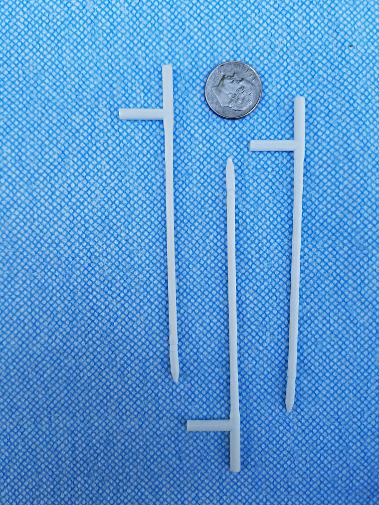
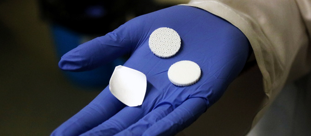
Marvel Medtech’s cryotherapy probe, developed using XJet NanoParticle Jetting technology.
Ray Harter, President of Marvel Medtech, said, “Our new approach preempts the need for many biopsies, surgeries, radiation and chemotherapy treatments. Obviously, the expectation is that it’s likely to save many lives, but it will also dramatically improve the quality of life for patients. In addition, we also know that by eradicating those procedures, it will also reduce overall healthcare costs. And these are not insignificant savings – annually, these could be in the many billions of dollars.”

Ray Harter, Founder and President of Marvel Medtech LLC
After identifying early-stage tumors during breast MRI scans, Marvel Medtech’s technology carefully targets the most dangerous cancer cells and applies cryoablation to freeze and destroy the cells before they could grow.
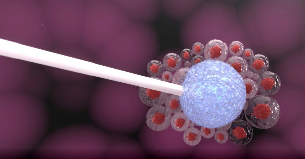
Marvel Medtech’s cryotherapy probe targetting cancer cells.
The technology transforms MRIs from a diagnostic-only tool into an actual treatment device.
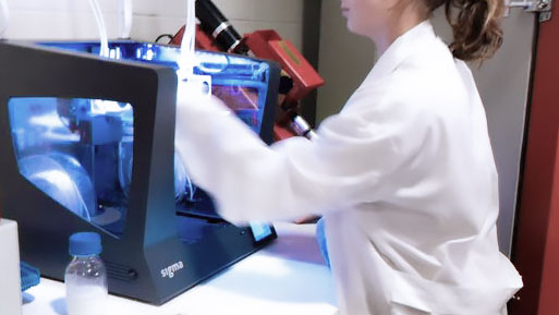
The final challenge for Marvel Medtech was to develop the intricate probe that would work in conjunction with the MRI but not interfere with the machine’s magnetic field. The probe also needed to have very small features and possess complex geometry. 3D printing was the answer, but which printer could manufacture the appropriate material?
According to Harter:
“The tools used inside an MRI scanner must be compatible with strict safety guidelines, and crucially, not disrupt image quality. Because they are one of the most electrically insulating materials, ceramics are an ideal material to achieve this. However, we were unable to find a ceramic-based 3D printer able to accurately and cost effectively produce our ceramic probe. This is why we are adopting XJet’s Carmel 1400 solution.”
With XJet’s NanoParticle Jetting (NPJ) technology and the ability to 3D print zirconia (ceramic), Marvel Medtech was finally able to complete the last piece of their life-saving puzzle. They 3D printed the highly complex, ceramic cryotherapy probe. Now the company and its invention are poised to save thousands of lives, dramatically improve patient care, and save potentially billions of dollars in healthcare spending.
There are untold applications for 3D printing ceramic. Register for AMS 2020 and hear XJet’s Chief Business Officer, Dror Danai, talk about Marvel Medtech’s lifesaving probe at AMS 2020 in Boston, February 12 at 3:20. You will also hear him talk about other potential solutions NPJ technology can provide to industries around the world.
The post Marvel Medtech Uses Additive Manufacturing by XJet To Prevent Breast Cancer appeared first on 3DPrint.com | The Voice of 3D Printing / Additive Manufacturing.