In the last few years, there has been a continuous growth of bioprinting companies around the world, probably because the medical field is one of the most exciting industries taking advantage of 3D printing. After all, health is one of the top priorities for most people and when some of the top biotech startups create soft human tissue-like structures, vascular networks and three-dimensional liver tissue constructs for experimentation, curiosity strikes. Along with some of these technical milestones in 3D printing is Bengaluru-based firm Pandorum Technologies’ successfully engineered cornea tissue. Thanks to their cell-laden hydrogel, they can promote scarless healing of corneal wounds by regeneration. Like most firms in the industry, Pandorum is pushing the technology hoping to eventually become part of the no donor required utopia. We must caution our readers however that from a thing becoming possible in bioprinting and 3D printing for medicine it may take several decades before it is available to you and me.
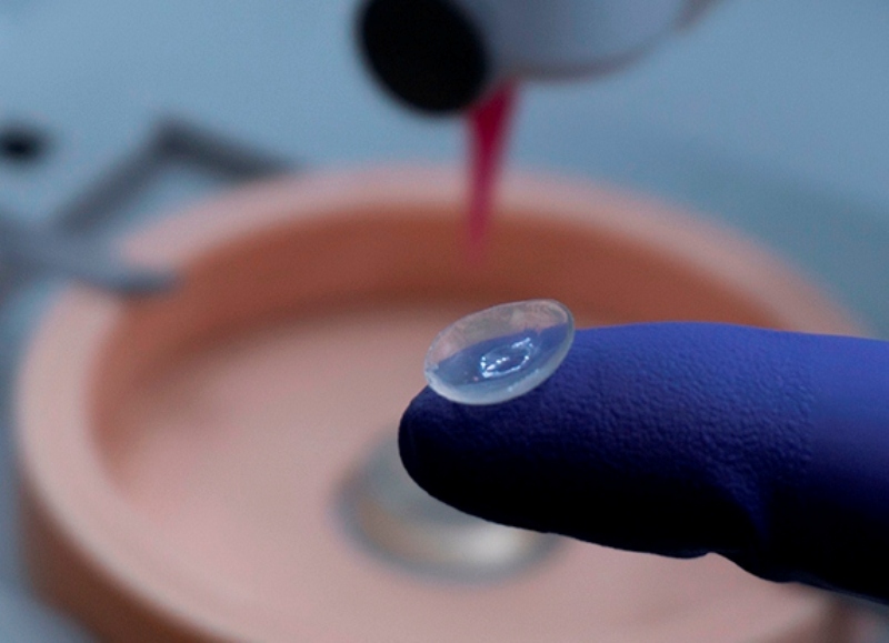
Pandorum’s bio-engineered cornea
The cornea is a thin piece of transparent tissue on the front of the eye that resembles a soft contact lens and is vulnerable to disease and injury. The World Health Organization (WHO) estimates that corneal opacities accounted for 7% of the world’s blind population in 2010, making it the third most common cause of blindness. And over 12 million people are waiting for a corneal transplant globally while 20% of childhood blindness is estimated to be caused by the cornea. In India alone, bilateral corneal blindness affects 1.2 million people and there are not enough donated corneas for transplants. A few years back a survey globally quantified the considerable shortage of corneal graft tissue, with only one cornea available for 70 needed, and although efforts are still strong for cornea donation everywhere, it is also crucial for bioengineering to look for alternative solutions.
 Pandorum’s novel hydrogel uses a combination of specialized stem cells and bio-mimetic that can be directly applied in a minimally invasive manner as ‘Liquid Cornea’ to corneal wounds and perforations and used to print transparent and suturable corneal lenticules embedded with live corneal cells for therapeutic applications and future human implantation to treat visual impairment. The bioengineered extracellular matrix with corneal cells developed as ‘Liquid Cornea’ and Corneal Graft for Human implantation is a regenerative approach towards vision restoration through tissue engineering. It is designed for a simple, minimally-invasive application procedure, to reduce the need for post-operative medication and care.
The tissue engineering and regenerative medicine startup company announced the development of their bioengineered cornea tissue study at the annual meeting of the Association for Research in Vision and Ophthalmology (ARVO) held in Vancouver, Canada, last April. Where Tuhin Bhowmick, co-founder of Pandorum said that “being able to bioengineer critical tissues such as the human cornea is a significant milestone.” And while the work is currently in the animal studies stage, the research team is getting ready for their first pilot human trials in 2020. Founded in 1928, ARVO is the largest eye and vision research organization in the world that includes nearly 12,000 researchers and clinicians from over 75 countries.
“Though surgically replacing the opaque tissue with a clear corneal allograft is usually effective in improving vision, there is an acute shortage of cadaveric human corneas available for transplantation. In India alone, there are over a million people suffering from a bilateral loss of vision due to corneal disorders, and at least a few folds more from unilateral corneal blindness. At Pandorum, we are working to close this gap using a bioengineering approach through stage-wise development of a platform, which is ultimately aimed to liberate us from the dependencies on human donor cornea,” suggested Bhowmick.
It’s quite a big deal for an Indian company to do such groundbreaking work since the Asia Pacific is the fastest growing 3D bioprinting region in the global market due to the presence of a huge patient population and continuously developing economies, including India and China, with growing healthcare industries. According to the Indian Brand Equity Foundation, in 2017 the Indian healthcare sector was one of the fastest growing industries, advancing at an annual growth rate of over 22% since 2015 and is expected to reach 130 billion dollars in 2022.
But Pandorum is not entirely new to bioprinting, it was the first Indian company to 3D print a liver tissue and their work is focused on tissue engineering and regenerative medicine, a much broader field, with the final goal to develop both liver and cornea as lab-grown transplantable tissues. The company can generate tissues that comprise 10,000 to 1 million cells, still it could take up to five years to build the working prototype of a tissue that has billions of cells and can be transplanted into a patient -which will need to have its own blood vessel structure and be able to integrate within the receiver’s body. “Right now, our mini-liver tissue reflects the characteristics of those grown within the human body. So pharma companies can test the hepatotoxicity of drugs, or FMCG companies their products, nixing the need for animal trials,” explained Pandorum co-founder Arun Chandru last year in an interview with Forbes India.

Pandorum scientists working at the lab on tissue engineering
The startup incubated at the Bangalore Bioinnovation Centre (BBC), located at the National Centre for Biological Sciences campus is making tremendous progress in tissue engineering. Since it’s foundation in 2011 by academic entrepreneurs from the Indian Institute of Science, they have focused on research and development of artificial human organs, and have become quite a household name in India’s new bioprinting industry. Thanks to their bio-engineered liver and cornea tissue they are getting ahead of the game fast.
Pandorum is not the first tissue engineering startup that attempts to create a cornea, last October, North Carolina-based bioprinting startup Precise Bio announced plans to advance its research into bioprinted corneas. The company was the first to transplant a 3D printed cornea graft into an animal and plans to start with a human cornea suitable for transplantation soon. While earlier this year, Korean researchers successfully bioprinted tissue for cornea with transparent bioink. On the academic front, Newcastle University researchers successfully 3D printed human corneas from stem cell bioink. With all the startups and university researchers racing to create the most compatible and functional organs that could one day replace organ transplantation, the stakes are high with pressure from the public opinion, anxiously awaiting a change in the way medicine will solve some of the most complex illnesses. The 3D bioprinting field is entering a very exciting time, but some big challenges in bringing these therapies to reality are still out there.