LulzBot Releases It’s First Bioprinter

Bioprinting is revolutionizing the way 3D printed tissues can be used to mimic in vivo conditions. The fields of regenerative medicine, pharmaceutical development, and cosmetic testing are benefiting from this technological disruption, enabling researchers and companies to better predict efficacy and toxicology of potential drugs early on in the drug discovery process. But it’s no wonder this technology is so enticing, since bringing a new drug to market, with current methods, could cost $350 million dollars and can take more than a decade from start to finish. On the North American front, Colorado-based manufacturer Aleph Objects, the developer behind the LulzBot 3D Printers, announced today a new open-source bioprinter: the LulzBot Bio.

After almost ten years of manufacturing 3D printers, LulzBot finally decided to move into the bioprinting market. The new machine, which is now available for pre-order on the site and will begin shipping in November, enables 3D printing with materials such as unmodified collagen, bioinks, and other soft materials, and is the company’s first-ever Fluid Deposition Fabrication (FDF) 3D printer. FDF is a newfangled name for the FRESH process which we wrote about here and here.  According to LulzBot, unlike its pneumatic counterparts, the Bio’s syringe pump system allows for precise stopping and retraction, preventing unintentional extrusion and stringing while printing intricate models, like vasculature.
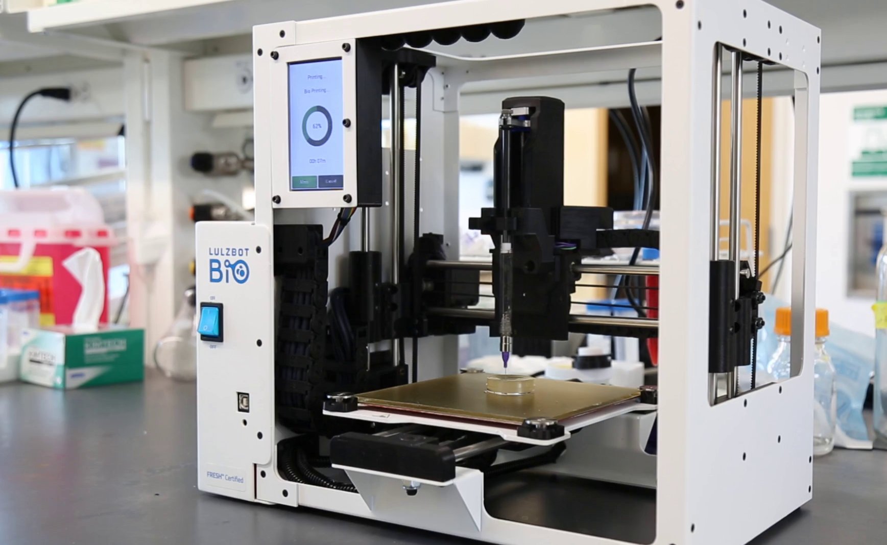
The new LulzBot Bio

The printer has a Free Software design that removes proprietary restrictions, providing, what the company considers, a versatile platform for innovation that grows with everchanging discoveries and advancements. LulzBot reports a commitment to freedom of design in general, developing machines that come with freely licensed designs, and specifications, allowing for modifications and improvements to both software and hardware. In this respect, they have partnered with organizations, such as the Open Source Hardware Association, Free Software, and Libre Innovation. The Bio’s free software and open hardware design give researchers the ability to innovate together, letting the machine be easily adjusted for new materials and processes.

“For researchers, you don’t know what materials or processes you’ll be using in six months, let alone one year from now, so you need hardware that can be adjusted quickly and easily, without proprietary restrictions,” said Grant Flaharty, CEO and President of Aleph Objects.

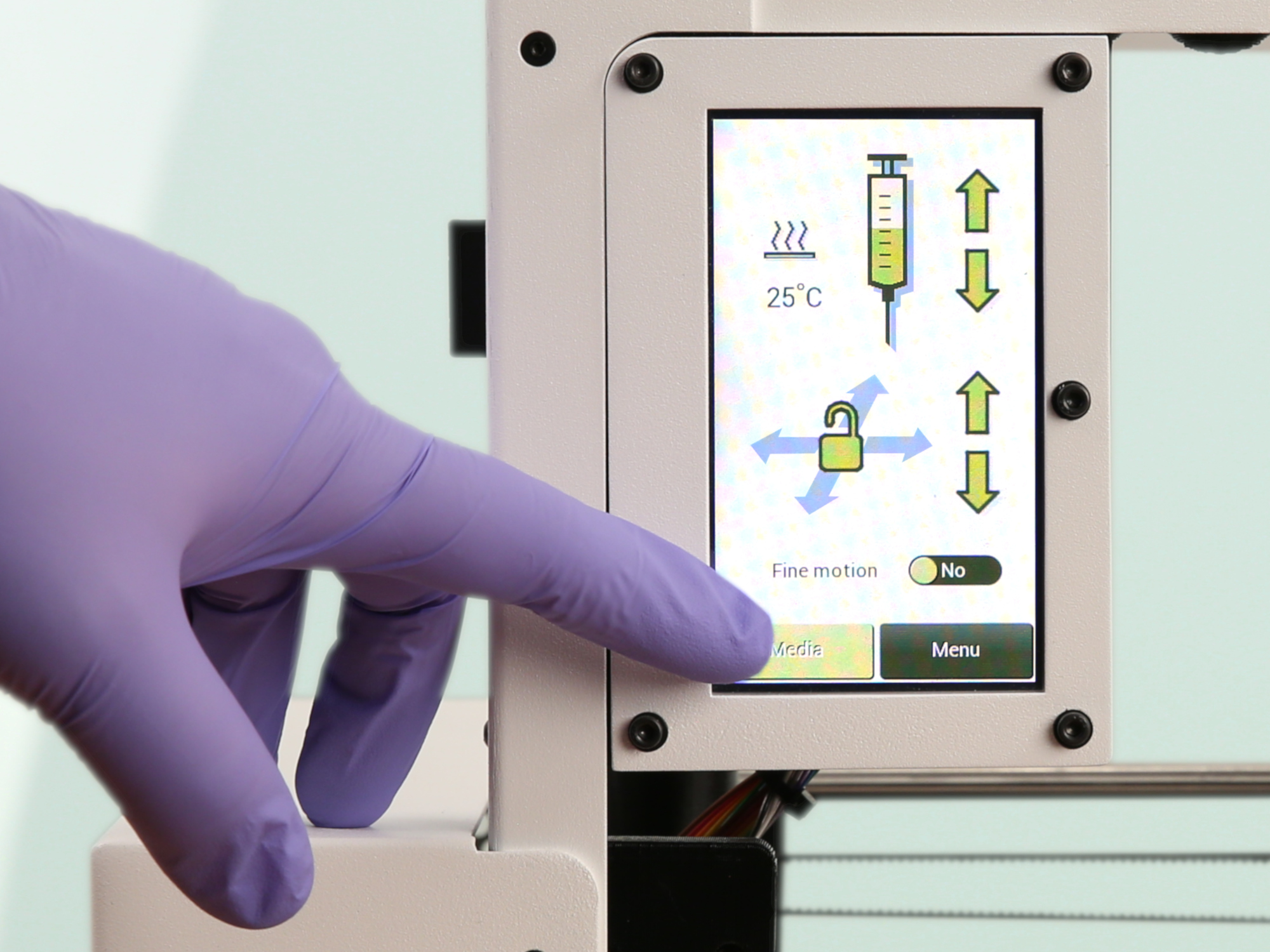
The LulzBot Bio touchscreen for easy control

The LulzBot Bio comes with nearly everything needed to start bioprinting right away, including extensively tested, preconfigured material profiles in Cura LulzBot Edition, the recommended software for the LulzBot printers; Petri dishes; Life Support gel (by FluidForm); alginate, and tools. It also enables printing with unmodified collagen, something that has proven extremely difficult and is considered one of the most promising materials for bioprinting applications, since it is the human body’s major structural protein and is prominent in biological structures.

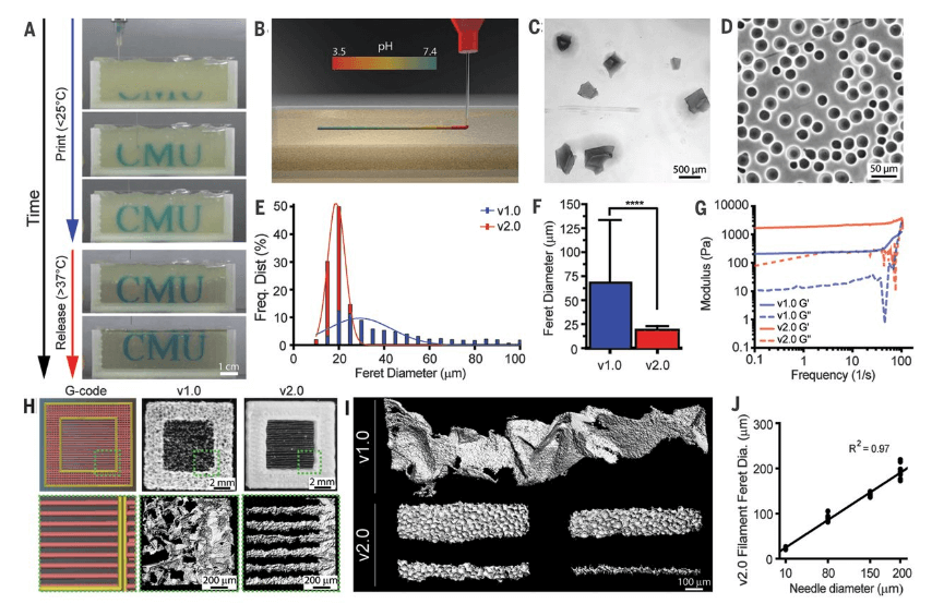
Actually, printing with unmodified collagen is currently done using the FRESH method, short for Freeform Reversible Embedding of Suspended Hydrogels, which was developed and refined by the Regenerative Biomaterials and Therapeutics Group at Carnegie Mellon University, in Pittsburgh. The LulzBot Bio is actually FRESH-certified, which means it uses thermoreversible support gels to hold soft materials during printing. Then, the temporary support gel is then dissolved, leaving the print intact.

“Other bioprinting techniques often require materials to be chemically altered or mixed with other materials to make them 3D printable,” explained Steven Abadie, CTO of Aleph Objects. “Because of the excellent biocompatibility of collagen, being able to 3D print with it in its original form brings us that much closer to recreating models that mimic human physiology.”

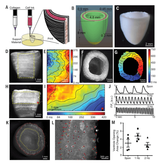
As stated by the company, the LulzBot Bio has already been instrumental in 3D printing some of the first-ever fully functional human heart tissue. This was achieved by a team of researchers at Carnegie Mellon, led by Adam Feinberg, that used the new device to 3D print heart tissue containing collagen and producing parts of the heart at various scales, from capillaries to the full organ.

“What we’ve shown is that we can print pieces of the heart out of cells and collagen into parts that truly function, like a heart valve or a small beating ventricle. By using MRI data of a human heart, we were able to accurately reproduce patient-specific anatomical structure and 3D bioprint collagen and human heart cells,” inidcated Adam Feinberg, principal investigator of the Regenerative Biomaterials and Therapeutics Group at Carnegie Mellon and co-founder of FluidForm.

FluidForm, powered by Carnegie’s research, has been working on the science behind the FRESH technology for quite some time. Now, Aleph Objects has taken the concept straight to the hardware, manufacturing this new machine, which they expect will be the first step to open up bioprinting to the broader market for exponential innovation.

Last June, LulzBot had already announced its collaboration with FluidForm, to combine their expertise and offer new bioprinting solutions. The LulzBot Bio has also been used by Newell Washburn, professor of biomedical engineering and chemistry at Carnegie, and a team of his colleagues to demonstrate how a new machine-learning algorithm could optimize high quality, soft material 3D prints.

According to company execs, the LulzBot Bio will satisfy the needs of many industries, for example, biotechnology, pharmaceuticals, cosmetics, medical devices, and life sciences. It could be ideal for producing bioprinted tissue for pre-clinical testing or used to recreate physiology to study diseases. It certainly seems like a great start to a new printer and perhaps the beginning of the company’s immersion in the bioprinting world.

[Images: LulzBot]

The post LulzBot Releases It’s First Bioprinter appeared first on 3DPrint.com | The Voice of 3D Printing / Additive Manufacturing.

FluidForm & Carnegie Mellon: Closer Than Ever to Bioprinting a Human Heart

While the realm of bioprinting has continued to expand significantly in the past few years, allowing for better sustainability of cells in the lab—and the bioink used to print them—the end goal is almost always to get just that much closer to 3D printing organs, meaning the potential for enormous change in the current medical culture of patients suffering and often dying while waiting for transplants. Now, with a collaboration between startup FluidForm and Carnegie Mellon University, scientists may actually be much closer than ever to bioprinting a human heart.

Findings on the subject were published recently in the August 2nd edition of Science regarding work by the Carnegie Mellon University research team, made up of co-first authors and FluidForm co-founders Andrew Lee and Andrew Hudson, and the nine members of the Carnegie Mellon team. They have just developed an advanced version of Freeform Reversible Embedding of Suspended Hydrogels (FRESH) technology which allows them to 3D printed collagen. With these new advancements, the scientists can fabricate cardiac components, including tiny blood vessels, valves, and beating ventricles.

(A) Time-lapse sequence of 3D bioprinting of the letters “CMU” using FRESH v2.0. (B) Schematic of acidified collagen solution extruded into the FRESH support bath buffered to pH 7.4, where rapid neutralization causes gelation and formation of a collagen filament. (C and D) Representative images of the gelatin microparticles in the support bath for (C) FRESH v1.0 and (D) v2.0, showing the decrease in size and polydispersity. (E) Histogram of Feret diameter distribution for gelatin microparticles in FRESH v1.0 (blue) and v2.0 (red). (F) Mean Feret diameter of gelatin microparticles for FRESH v1.0 and v2.0 [N > 1200, data are means ± SD, ****P < 0.0001 (Student t test)]. (G) Storage (Gʹ) and loss (Gʺ) moduli for FRESH v1.0 and v2.0 support baths showing yield stress fluid behavior. (H) A “window-frame” print construct with single filaments across the middle, comparing G-code (left), FRESH v1.0 (center), and FRESH v2.0 (right). (I) Single filaments of collagen showing the variability of the smallest diameter (~250 μm) that can be printed using FRESH v1.0 (top) compared to relatively smooth filaments 20 to 200 μm in diameter using FRESH v2.0 (bottom). (J) Collagen filament Feret diameter as a function of extrusion needle internal diameter for FRESH v2.0, showing a linear relationship.

The patent for this important work belongs to FluidForm, headquartered in both Pittsburgh and Boston.

 “We now have the ability to build constructs that recapitulate key structural, mechanical, and biological properties of native tissues,” said Prof. Adam Feinberg, CTO and co-founder, FluidForm, and Principal Investigator, Regenerative Biomaterials and Therapeutics Group, Carnegie Mellon, where the research was done. “There are still many challenges to overcome to get us to bioengineered 3D organs, but this research represents a major step forward.”

Created from MRI data, the FRESH bioprinted hearts were comprised of small cardiac ventricles printed with human cardiomyocytes demonstrated the following:

  • Synchronized contractions
  • Directional action potential propagation
  • Wall-thickening up to 14 percent during peak systole

“We found that FRESH 3D-bioprinted hearts accurately reproduce patient-specific anatomical structure as determined by micro–computed tomography. Cardiac ventricles printed with human cardiomyocytes showed synchronized contractions, directional action potential propagation, and wall thickening up to 14% during peak systole, state the researchers in their paper, 3D bioprinting of collagen to rebuild components of the human heart.”

Other obstacles are still present too though as scientists wrestle with generating the billions of cells needed for 3D printing larger tissues.

“Although the 3D bioprinting of a fully functional organ is yet to be achieved, we now have the ability to build constructs that start to recapitulate the structural, mechanical, and biological properties of native tissues,” stated the researchers in their recently published paper.

While previously scientists have projected bioprinting of organs in the very near future, it has proven to be much more difficult than anticipated, obviously. Milestones continue to be surpassed, but challenges continue to erupt—mainly in keeping cells alive in coordination with the proper technology—meaning materials, software, and hardware continue to be a significant focus. With FRESH, a temporary support gel is used to 3D print collagen scaffolds, driven by a rapid pH change that drives collagen self-assembly. There is promise not only for 3D printing hearts, however, but a range of different tissues and organs in the future.

“FluidForm is extraordinarily proud of the research done in the Feinberg lab” said Mike Graffeo, CEO, FluidForm. “The FRESH technique developed at Carnegie Mellon University enables bioprinting researchers to achieve unprecedented structure, resolution, and fidelity, which will enable a quantum leap forward in the field. We are very excited to be making this technology available to researchers everywhere.”

(A) FRESH-printed collagen tube construct. (B) C2C12 cell and collagen gel mixture cast around the collagen tube and static-cultured for 5 days. (C) Cross section of the tissue from (B) stained for live (green) and dead (red) cells. (D) C2C12 cell and collagen gel mixture cast around the collagen tube and perfused for 5 days. (E) Cross section of the tissue from (D) stained for live and dead cells. (F) Percent cell viability as a function of depth from the top surface of the tissues from the static and perfused collagen tube constructs [N = 3, data are means ± SD, *P < 0.05 (two-way ANOVA followed by Bonferroni multiple-comparisons posttest)]. (G) Multiphoton imaging showing microscale porosity in FRESH-printed collagen constructs after removal of the gelatin microparticle support bath. (H and I) Collagen constructs cast (H) and FRESH-printed (I) without VEGF 7 days after subcutaneous implantation. (J and K) Masson’s trichrome staining to visualize cells (red) and collagen (blue) in cast (J) and FRESH-printed (K) collagen disks after 7 days of subcutaneous implantation. (L) Cell density after implantation as a function of depth for the cast and FRESH-printed collagen disks. (M and N) Collagen constructs cast (M) and FRESH-printed (N) with VEGF (100 ng/ml) 10 days after subcutaneous implantation. (O and P) CD31 staining (brown) and cells (blue) of cast (O) and FRESH-printed (P) collagen disks doped with VEGF (100 ng/ml) after 10 days of subcutaneous implantation. (Q) Host vascular infiltration of vessels (diameter 8 to 50 μm) in the FRESH-printed collagen disk labeled by lectin tail vein injection (red). (R) Multiphoton image 70 μm into the FRESH-printed construct, showing red blood cells within the lumen of the microvasculature.

FluidForm is unveiling its first product, LifeSupport™ bioprinting support gel, commercializing FRESH technology and giving scientists and researchers everywhere the opportunity to 3D print collagen, cells, and biomaterials.

(A) FRESH-printed collagen tube construct. (B) C2C12 cell and collagen gel mixture cast around the collagen tube and static-cultured for 5 days. (C) Cross section of the tissue from (B) stained for live (green) and dead (red) cells. (D) C2C12 cell and collagen gel mixture cast around the collagen tube and perfused for 5 days. (E) Cross section of the tissue from (D) stained for live and dead cells. (F) Percent cell viability as a function of depth from the top surface of the tissues from the static and perfused collagen tube constructs [N = 3, data are means ± SD, *P < 0.05 (two-way ANOVA followed by Bonferroni multiple-comparisons posttest)]. (G) Multiphoton imaging showing microscale porosity in FRESH-printed collagen constructs after removal of the gelatin microparticle support bath. (H and I) Collagen constructs cast (H) and FRESH-printed (I) without VEGF 7 days after subcutaneous implantation. (J and K) Masson’s trichrome staining to visualize cells (red) and collagen (blue) in cast (J) and FRESH-printed (K) collagen disks after 7 days of subcutaneous implantation. (L) Cell density after implantation as a function of depth for the cast and FRESH-printed collagen disks. (M and N) Collagen constructs cast (M) and FRESH-printed (N) with VEGF (100 ng/ml) 10 days after subcutaneous implantation. (O and P) CD31 staining (brown) and cells (blue) of cast (O) and FRESH-printed (P) collagen disks doped with VEGF (100 ng/ml) after 10 days of subcutaneous implantation. (Q) Host vascular infiltration of vessels (diameter 8 to 50 μm) in the FRESH-printed collagen disk labeled by lectin tail vein injection (red). (R) Multiphoton image 70 μm into the FRESH-printed construct, showing red blood cells within the lumen of the microvasculature.

3D printing has been used to help a wide range of patients with heart issues, from cardiac patches to cardiac phantoms, catheter devices, and more. What do you think of this news? Let us know your thoughts! Join the discussion of this and other 3D printing topics at 3DPrintBoard.com.

[Source / Images: FluidForm press release]


The post FluidForm & Carnegie Mellon: Closer Than Ever to Bioprinting a Human Heart appeared first on 3DPrint.com | The Voice of 3D Printing / Additive Manufacturing.

Aleph Objects enters 3D bioprinting with Fluidform, hardware coming summer 2019

Aleph Objects, the manufacturer of LulzBot open source 3D printers, has confirmed its entry into the 3D bioprinting market. In partnership with Massachusetts based 3D bioprinting technology developer FluidForm, Aleph plans to launch LulzBot Bio hardware later this year. For Grant Flaharty, Aleph Objects CEO and President, this undertaking presents a chance for LulzBot to […]