
Researchers from the University of Minnesota (UMN) have worked with medical technology firm Medtronic to develop realistic customizable 3D printed aortic models. Using tailor-made silicone-based polymeric inks, the UMN team fabricated lifelike duplicates of a human aortic root, the vascular section which is attached closest to the heart. The patient-specific replicas could enable surgeons to […]
CLECELL: A New Bioprinted Model Could Aid COVID-19 Vaccine Testing
Global crises can accelerate technology. Disrupting traditional responses with innovation can result in untapped opportunities that become crucial tools for humanity. This year, the Coronavirus pandemic could accelerate the evolution of drug and vaccine testing, as researchers are harnessing new technology to facilitate safety testing in people soon after preclinical work is completed on more than 60 emerging vaccine candidates. In the meantime, a bioengineering startup from South Korea could change the framework behind vaccine testing as we know it.
Founded in 2017, CLECELL has focused on the research and development of artificial tissue and has gone on to create a respiratory epithelium model earlier this year using its proprietary 3D bioprinter, the U-FAB, as well as other bioprinting technology. What is so interesting about this model is that it is expected to become a testbed for the severe acute respiratory syndrome coronavirus (SARS-Cov-2), as well as for research on the mechanisms of various other viruses.

According to a company statement, this is especially important since SARS-Cov-2 is markedly less infectious towards animals, so this new method is being considered as a potential alternative to more traditional ones that require the use of fertilized eggs to create vaccines. For more than 70 years, egg-based vaccine manufacturing has been used to make both inactivated (killed) vaccine (usually called the flu shot) and live attenuated (weakened) vaccine. Scientists would inject a fertilized egg with the virus, incubating it, and then extracting, diluting, and refining it into an antigen. Instead, CLECELL’s innovative bioprinting technique has the potential to become a testbed for various viruses.
Respiratory epithelium is a type of tissue found lining most of the respiratory tract, the role of this unique type of epithelium is to function as a barrier to pathogens and foreign particles; however, it also operates by preventing infection and tissue injury via the use of the mucociliary elevator.

The respiratory epithelium (Credit: Blausen.com staff, Medical gallery of Blausen Medical 2014, WikiJournal of Medicine)
In fact, the company’s respiratory epithelium model for in vitro testing was saught out by a team of researchers at Harvard University’s Medical School. CLECELL claims that on April 10, they received a formal letter of correspondence from Choi-Fong Cho, an assistant professor of neurosurgery at Harvard Medical School and instructor at Brigham and Women’s Hospital. The message centered around an urgent request for information on the respiratory epithelium model created with bioprinting technology.
According to the company, Cho, who is an expert in neurovascular research, the development of new drugs and neurovascular organoids, along with her research team are interested in methods to create a respiratory epithelium model through 3D bioprinting and has supposedly expressed that CLECELL’s solution will be of great assistance in conquering SARS-Cov-2.
CLECELL also suggests that Cho has professed a desire to research SARS-Cov-2’s effect on vascular structure, the virus’s infection routes, and creating an in vitro testing platform that mimics human lung tissue via CLECELL’s bioprinting solution.
Up until now, the high number of casualties from the COVID-19 pandemic has spiraled a frenetic interest in a cure, skyrocketing a profound interest from governments, research institutes, companies, and societies in drug and vaccine development, disease control, and all branches of healthcare. As a result, experts are seeking alternate methods of research that could bypass the limitations of contemporary and traditional methods for the creation of vaccines, that could take months.
CLECELL has plans to collaborate with researchers around the world to offer a testbed for the research of viruses and the development of cures, with plans to carry out research not only on virus infection, but also drug delivery, toxicity, and inflammation.
Furthermore, CLECELL claims that its proprietary 3D bioprinter, the U-FAB, is slated for tissue engineering research at the Boston Bioprinting Consortium, which is comprised of world-class scholars from Boston’s universities and hospitals and consists of eight joint-research teams.
“Despite the various existing methods of testing respiratory viruses in vitro, we require a more effective platform for testing,” indicated Young-Jae Cho, a professor at the Department of Pulmonology at Seoul National University Bundang Hospital. “The creation of precise artificial respiratory models through 3D bioprinting technology offers a potential alternative.”
The startup’s bioprinting technology could provide solutions for tissue engineers and life scientists to research and develop biomimetic human tissues and organs. Since its origin, the company has been researching and developing various reconstructed human skin models using scalable 3D bioprinting technology, focusing on building and implementing transplantable biomimetic human skin in the future.
Along with the U-FAB, the company has two other printing platforms: U-Printer for the development of artificial tissues and organs and U-Skin for reconstructing artificial human skin models. Through their U-Printer prototype, they have created artificial skin models, which they consider superior to the existing commercial ones as they are able to retain the shape and dimension without shrinking throughout the culture period and pigmentation, and this was realized by 3D bioprinting melanocytes without UV or chemical stimulation.
A need for hastening change has been at the center of many technological revolutions, and in these uncertain times, it seems imperative to rely on bioprinting technology that can accelerate results. CLECELL’s revolutionizing respiratory epithelium model could become a fundamental resource for vaccine testing. Never before have so many of the world’s researchers focused so urgently on a single topic. With so many minds mobilizing to understand the disease, this emerging powerful technology developed by the South Korean startup could reduce the time towards finding a cure for COVID-19.
The post CLECELL: A New Bioprinted Model Could Aid COVID-19 Vaccine Testing appeared first on 3DPrint.com | The Voice of 3D Printing / Additive Manufacturing.
Aerosint and Aconity3D develop multi-material metal 3D printer
The specialist recoater developed by Belgium-based Aerosint will be deployed in a new 3D printer made by Aconity3D, capable of laying dual-powder layers for laser powder bed fusion systems. The Formnext Startup challenge winner previously announced its partnership with German metal processing specialist, Aconity3D. A new joint sales agreement between the two companies has now been initiated whereby […]
Researchers develop Hybrid Living Materials using inkjet 3D printing
Researchers from the MIT Media Lab, Harvard University’s Wyss Institute, and the Dana-Farber Cancer Institute have developed a method to 3D print objects that can control organisms. Dubbed as the Hybrid Living Material (HLM) fabrication platform, the team used a multi-material inkjet-based 3D printer and customized recipes for the combinations of resins and chemical signals. […]
Collaborative Research Team Creates 3D Printed Armor Inspired by Chiton Scales
From lobster claws and fish scales to conch shells, humans have often been inspired by nature in the creation of protective gear. Recently, a team of researchers hailing from MIT, Virginia Tech, Harvard University, California State University Fullerton, and the Max Planck Institute of Colloids & Interfaces published a paper, titled “Bioinspired design of flexible armor based on chiton scales,” about their work using multimaterial 3D printing and parametric computational modeling to create “a synthetic flexible scaled armor analogue” based on the scaled armors of chitons, a group of marine mollusks.
“This approach allows us to conduct a quantitative evaluation of our chiton-inspired armor to assess its orientation-dependent flexibility and protection capabilities,” the researchers wrote in the abstract.
Biological armor offers mechanical protection from the environment, which includes attacks from predators. Man-made armors use rigid structures for this protection, which the team explained can result “in a trade-off with flexibility and maneuverability.”
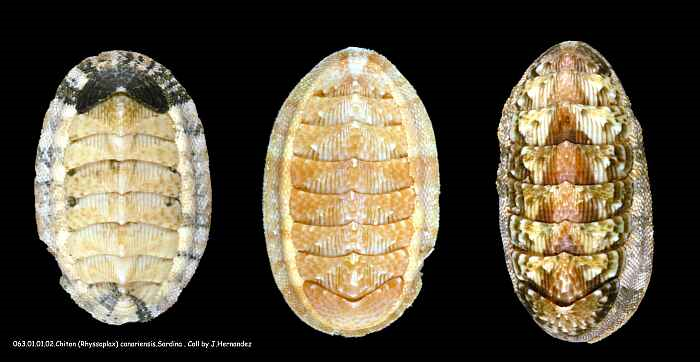
Rhyssoplax canariensis (Image: Jose Maria Hernandez Otero, BioLib)
“Many chiton species possess hundreds of small, mineralized scales arrayed on the soft girdle that surrounds their overlapping shell plates,” the abstract states. “Ensuring both flexibility for locomotion and protection of the underlying soft body, the scaled girdle is an excellent model for multifunctional armor design.”
Because many biological armors are based on hard and rigid armor plates, flexibility is tough to pair with it. Scale-like armors with many small, repeating elements, like that of chiton, can help maximize the combination of flexibility and protection. The team completed a study of the 3D geometry, interspecific structural diversity, material composition, and nanomechanical properties of chiton girdle scales, focusing on the chiton Rhyssoplax canariensis (Chitonidae: Chitoninae). This species is covered by a total of eight “bilaterally symmetrical overlapping mineralized shell plates,” in addition to the protective scaled girdle.
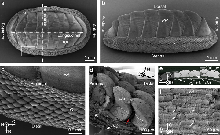
Figure 1. Biological flexible scaled armor in the girdle of the chiton Rhyssoplax canariensis. a, b Wide-field SEM images of the chiton R. canariensis, which show the dorsal and side view of the primary plates (PP) and peripheral scale-covered girdle (G), respectively. c Enlarged view of the girdle covered with dorsal scales. The image was acquired from the region indicated by the rectangular box in a, highlighting individual overlapping dorsal girdle scales, a fully covered protective armor. d Fractured cross-section of the girdle scaled armor, which consists of three components arranged from dorsal to ventral: (1) dorsal scales (DS); (2) fibrous layer (FL); and (3) ventral scales (VS). The white dashed lines indicate the height of the inter-scale organic matrix, and the red arrow indicates gaps between adjacent scales. e Cross-sectional view of scaled armor based on micro-computed tomography (μ-CT) data. Note the distance between the dorsal and ventral scale layers, which is occupied by the fibrous layer. f SEM image of the rod-shaped ventral scales, where the white arrows indicate small cracks.
“In contrast to most shelled mollusks where mobility is limited, as in the single shelled mollusks (gastropods, including snails, scaphopods or tusk shells, and some cephalopods such as Nautilus) or hinge-shelled bivalves (mussels, clams, scallops, etc.), most polyplacophorans (chitons) are characterized by eight overlapping, hard shell plates (Fig. 1a, b), which collectively accommodate a wide range of motion,” the researchers explained. “In addition to the eight overlapping shell plates (which are functionally analogous to the segmented plate-like exoskeleton of many crustaceans), additional protection is provided by a thick leathery girdle that skirts the animal’s periphery.”
Even though the girdle scales are nearly pure mineral and very rigid, they are also very flexible and able to conform to rough surfaces. Chiton scales are also more uniform in composition, with no porosity, sub-layering, or material heterogeneity.
“This observation underlines the suitability of chiton scales as a model for bioinspiration, as the mechanical performance of their armor can be ascribed primarily to geometric considerations, rather than fine scale material variation,” the team noted.
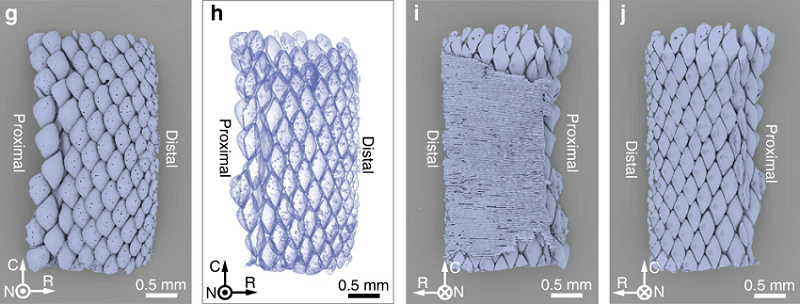
Figure 1. g–i: μ-CT 3D rendering of the chiton R. canariensis girdles in different viewing orientations and modes: g top view of girdle scales, h transparency mode showing the overlapping characteristics among adjacent scales, bottom view i with and j without ventral scales. Local coordinates: N (normal), from ventral to dorsal; R (radial), from proximal to distal; C (circumferential).
The team used many experimental and modeling approaches, such as mechanical testing, finite element modeling, electron microscopy, synchrotron X-ray micro-computed tomography, and instrumented nanoindentation, to investigate chitons, and the use of chiton-like scales in 3D printed flexible armor.
“Incorporating the physical and functional properties of chiton girdle scales characterized in these investigations, we design a bio-inspired flexible armor system, integrating parametric geometrical modeling and multi-material 3D printing,” the researchers wrote. “We explore the functional trade-offs between protection and flexibility in this model scaled armor system and its potential for informing the design of additional functional prototypes.”
A Connex 500 multi-material 3D printer from Stratasys was used to create prototypes out of both flexible and rigid photopolymers in different colors.
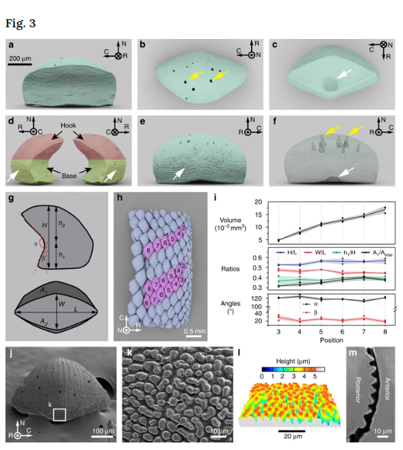
3D geometry and surface morphology of individual dorsal scales of the chiton R. canariensis. a–f: μ-CT data-based 3D rendering of individual girdle scales in different view angles and modes: a front view, b top view (yellow arrows indicate pore openings), c bottom view (white arrow shows a depression at the base of the scale), d two side-views (white arrows shows the surface roughness at the lower surface of backside), e back view, and f transparent mode (the yellow arrows show holes in the dorsal surface of scales and the white arrow indicates depression in base). g Projection contours along two orientations (transverse and bottom) are used to describe the geometries of chiton scales. h Top view of a μ-CT data-based reconstruction of the girdle scale assembly of R. canariensis. Three columns of scales used in the geometrical measurement are highlighted in pink color and their positions are indicated. i Variations of geometrical parameters as a function of scale position. The solid line represents the average and the shaded area shows the standard deviation. j SEM image of a scale’s back surface. k Magnified-view of scale surface with microscopic bumps at the underside of the back surfaces of chiton scales, as indicated by the white box in j. l SEM-derived stereographic reconstruction of microscopic bumps in a similar region shown in k. m Backscattered SEM image of a polished cross-section in the region of microscopic bumps of a scale, highlighting the difference in morphology between the front and back surfaces of the dorsal scales.
“In order to successfully mimic scale morphology for the production of a 3D-printed structural analogue…quantitative measurements of the scale geometry were conducted by defining several morphometric parameters,” the researchers stated.
The team also took “3D morphometric measurements” of the dorsal girdle scales from chiton species in the Ischnochitonidae and Chitonidae families. In order to reproduce the morphometrics for further modeling of scaled arrays, they created a parametric geometrical model.
“The successful 3D modeling of individual scales allowed us to design a composite scale armor assembly similar to that of chitons,” the team explained. “The bio-inspired armor system included rigid scales embedded in an underlying soft substrate.”
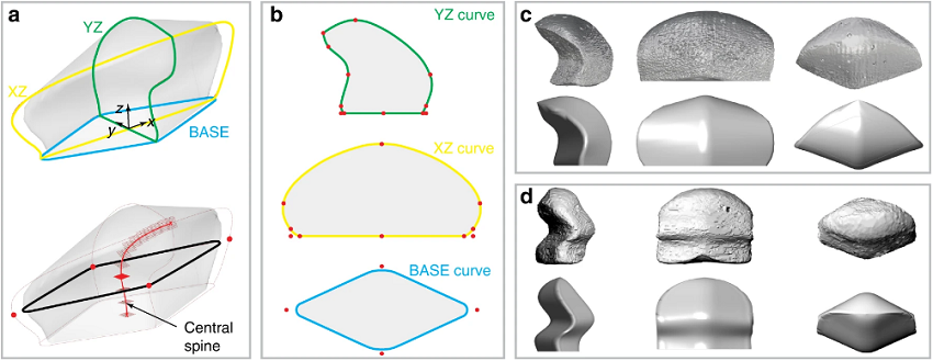
Figure 5. 3D parametric modeling of chiton scale geometries. a Top, the 3D scale model with three principal scaffolding curves. Bottom, 3D scale model highlighted with the central spine for generating the surface meshes. b Three principal curves with geometrical landmarks indicated. c, d Comparison of original chiton scales with corresponding mimicked scale models for two species: c, a single-curved scale from Rhyssoplax canariensis and d, a double-curved scale from Lepidozona mertensii.
They used materials with moduli of ca. 2 GPa and ca. 0.7 MPa, respectively, to 3D print the scales and matrix, in order to properly replicate how the scales would interact with soft girdle tissue. The scale assembly was very flexible, with a similar range of motion to real chiton scales, and the team was able to efficiently explore a variety of arrangements with the scales due to the “parametric nature of our model.”

Design and fabrication of bio-inspired flexible scaled armor. a Schematic diagram showing basic components of the armor; (1) scales, (2) matrix, and (3) soft underlying layer. b Side and c bottom view of the armor. d, inter-scale spacing. d Flat panel with uniform scales fabricated through additive manufacturing. e A bent panel showing excellent flexibility. f, g Design of scale pattern with size gradients. h Scale assembly in flat and curved substrate. i Scale assembly on double-curved surfaces. j X-ray projection images of a kneepad based on the bio-inspired scaled protective panel in j extended and k bent positions, demonstrating its conformability and flexibility. l Demonstration of the protection capability of the chiton scale-inspired kneepad on broken glass.
They also studied the mechanical performance of their multimaterial 3D printed prototypes, and even 3D printed a scaled kneepad prototype in order to demonstrate the usefulness of the chiton-inspired system for both protective and flexible applications.
“Current kneepad designs often fall in one of two extremes: hard and rigid plates that create heavy protection but limit flexibility, or elastomeric rubbers/foams that provide high flexibility but limited protection (especially against sharp objects). The chiton scale-inspired knee protection pad offers a unique solution to this dilemma,” the researchers noted.
The 3D printed scale assemblies had much higher puncture resistance than typical kneepads with rubber- or foam-based inserts, and also featured good shape-conforming capabilities in extended and bent configurations.
Co-authors of the paper are Matthew Connors, Ting Yang, Ahmed Hosny, Zhifei Deng, Fatemeh Yazdandoost, Hajar Massaadi, Douglas Eernisse, Reza Mirzaeifar, Mason N. Dean, James C. Weaver, Christine Ortiz, and Ling Li.
Discuss this and other 3D printing topics at 3DPrintBoard.com or share your thoughts below.
The post Collaborative Research Team Creates 3D Printed Armor Inspired by Chiton Scales appeared first on 3DPrint.com | The Voice of 3D Printing / Additive Manufacturing.
Defining a new paradigm: Harvard researchers invent multimaterial multinozzle 3D printing
Researchers at Harvard University have developed a new method of 3D printing. Termed multimaterial multinozzle 3D printing, or MM3D, the method seeks to help define the emerging paradigm of voxel by voxel fabrication. In this paradigm, 3D printed parts are composed of minute building blocks. Each of these blocks has a different material composition, and are […]
Harvard researchers 4D print the face of a new class of shape-shifting materials
4D printed, shape-shifting structures hold significant potential for the future of engineering. Be it self-assembling trusses, or medical devices that help inform cell regeneration, the field still has many challenges to overcome before its possibilities become practical. Up to now, one of the greatest challenges to 4D printing has been the ability to create complex, smoothly-curved shapes. […]
SWIFT: Uzel and Skylar-Scott are Paving the Way for the Future of Bioprinting
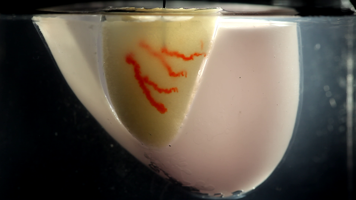
A few weeks ago Mark Skylar-Scott and Sébastien Uzel, researchers working in Jennifer Lewis’ Lab at Harvard´s Wyss Institute for Biologically Inspired Engineering and John A. Paulson School of Engineering and Applied Sciences (SEAS), came up with a breakthrough new technique that could one day provide organ tissues for therapeutic use. The method, called SWIFT (sacrificial writing into functional tissue), allows 3D printing to focus on creating the vessels necessary to support a living tissue construct.
![]()

All organs need blood vessels to supply their cells with nutrients, but most lab-grown organoids lack a supportive vasculature. This is were the SWIFT method comes into play, 3D printing vascular channels into living tissues. Two weeks ago, 3DPrint.com went into some of the main details of the research, but now we have gone straight to the source and spoken with two of the co-first authors of the paper which came out on September 6 in Science Advances, to understand the process behind the method, as well as the collaborative work shaping the future of Harvard’s bioengineering aspirations.
“Inspired by the 3D bioprinting techniques emerging from the Lewis lab and the community in general, Mark [Skylar-Scott] and I decided that is was time to tackle, head-on, the challenge of cell function and density, and tissue volume, which were keeping us from reaching organ manufacturing at therapeutic scale,” revealed Uzel. “Using patient-derived organoids or 3D cell spheroids as our building blocks appeared like a natural choice. They are cellularly dense and exhibit great functional and architectural similarities with the organs they are meant to mimic.”
A branching network of channels of red, gelatin-based “ink” is 3D printed into a living cardiac tissue construct composed of millions of cells (yellow) using a thin nozzle to mimic organ vasculature.
Uzel went on to explain that “the idea of this SWIFT printing process really took shape when we speculated that once jammed into a dense slurry, those organoids would behave as predicted by the science of colloid suspensions and therefore could serve as a supporting living matrix for the free form templating of perfusable vessels. The rest was many months of testing and optimization!”
Both researchers and their colleagues found a way to pack living cells tightly enough together to replicate the density of the human body. Actually they assembled hundreds of thousands of organ building blocks (OBBs) composed of patient-specific-induced pluripotent stem cell-derived organoids, which offer a pathway to achieving tissues with the requisite cellular density, microarchitecture, and function required. At the same time, they introduced vascular tunnels via embedded 3D bioprinting in between the OBBs to mimic blood vessels that are needed to deliver fluids, like nutrients and oxygen, that are vital to survival.
As an example, the group of researchers created a perfusable cardiac tissue that fuses and beats synchronously over a seven-day period. The SWIFT biomanufacturing method enables the rapid assembly of perfusable patient and organ-specific tissues at therapeutic scales. What is so novel about the new lab-grown heart tissue is that it beats, just like a normal human heart, and has an embedded network of the blood vessels that would be needed to survive if it was ever transplanted into a patient. It still needs to be tested before it can be used in humans, and their channels aren’t yet truly blood vessels, but if the innovation works for heart tissue, the experts expect SWIFT
could also be used for other organs.
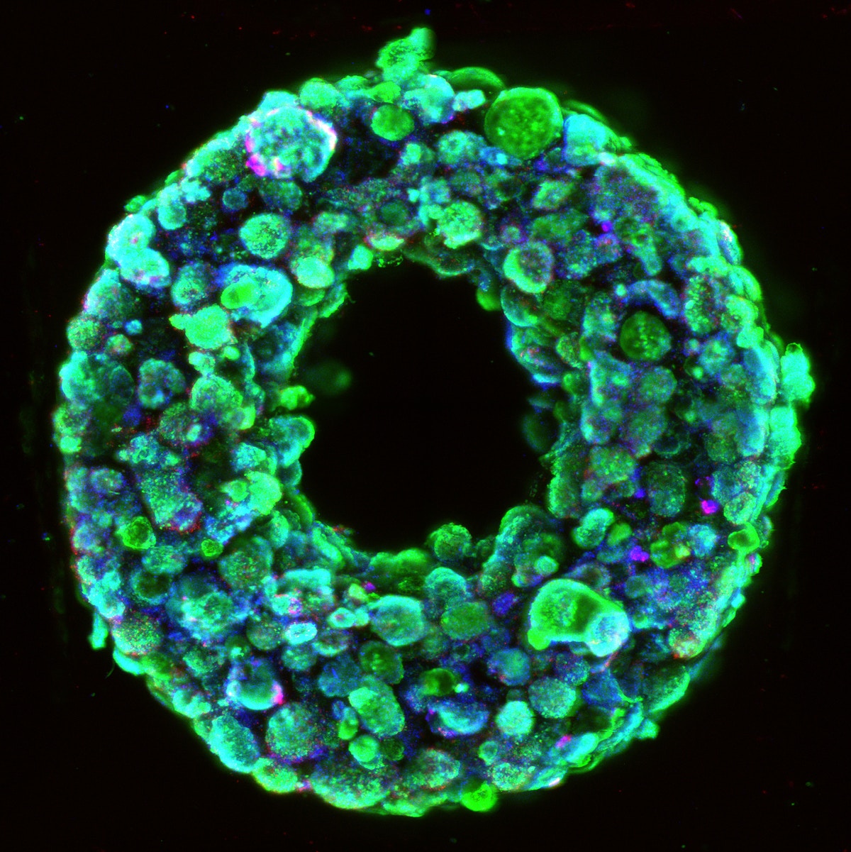
Living embryoid bodies surround a hollow vascular channel printed using the SWIFT method.
“We believe that this new technique addresses the technical roadblocks of cell density and manufacturing scalability. From a biology standpoint, making each building block more functional and performant, meaning being able to contract stronger in the context of cardiac tissues, for instance, is among the challenges that need to be overcome and will require gaining even more insights in pluripotent cell differentiation and how it can be recapitulated in vitro. We will also need to better emulate the multicellular and hierarchical complexity of the vessels as found in the human body,” proposed Uzel.
The researchers consider that on the manufacturing side of the process, the cost of reagents for scaling up cell culture and differentiation will have to be drastically reduced for de novo organ manufacturing to be a viable option looking into the long term.
When it comes to considering SWIFT as one of the main advances in the last few years towards bioprinting organs, Skylar-Scott claims “it would be presumptuous to say that SWIFT came out of a vacuum”.
“There have been many great works in this decade that have applied 3D printing to generate perfusable tissues, and our work builds on these efforts. What really does get us excited about SWIFT is how we have brought the matrix for embedded printing ‘to life’, and, by using organoids, we hope that SWIFT may serve as a bridge between the bottom-up self-assembly of developmental biology, and the top-down directed assembly of 3D printing,” Skylar-Scott asserted. “We can say, with reasonable certainty, that any successful engineering of a complex organ from scratch will require a combination of these two approaches.”
“The recent progress in the field of bioprinting has brought us a lot closer to the eventuality of 3D printed organs. The field is moving faster than we expected. Just five years ago, we were afraid to use “the big O word” [organs], but we are now, as a field, beginning to tentatively see a path forward,” he continued.
SWIFT is one of the projects at Harvard that could ultimately be used therapeutically to repair and replace human organs with lab-grown versions containing patients’ own cells. There is actually so much research at Wyss and SEAS, from scaling up tissue engineering to engineering miniature kidneys, it’s even one of the first places where researchers entirely 3D-printed an organ-on-a-chip with integrated sensing. Moreover, the creation of highly-organized multicellular biological tissues and organoids is structurally diverse and complex, so tissue manufacturing techniques require extreme precision, making us wonder what type of bioprinter the researchers are using. According to Skylar-Scott, they “exclusively use custom made printers and extruders” in the lab, that “for the purposes of wacky experimentation, they offer the most versatility by far.” He also suggests that these printers are large and expensive, “but, for many processes, including SWIFT, we’re confident that it can be replicated with commercially available or open-source alternatives.”
As part of the SWIFT project evolution, collaborations are underway with Wyss Institute faculty members Christopher Chen, Professor of Biomedical Engineering and director of the Tissue Microfabrication Laboratory at Boston University and Sangeeta Bhatia, Professor at MIT’s Institute for Medical Engineering & Science (IMES) and Electrical Engineering & Computer Science (EECS), to implant these organ-specific tissues created by SWIFT into animal models and explore their host integration, as part of the 3D Organ Engineering Initiative, co-led by 3D printing pioneer and Wyss core faculty member, Jennifer Lewis, and Chen.
“We are currently working on rodent models for our initial in vivo phase. Along with perfecting our technique and improving the performance of printed tissues, we are investigating how small vascularized SWIFT-printed cardiac constructs integrate within the animal and connect to the existing blood stream. Once confident that the SWIFT tissues behave appropriately in small animals, the hope is to move to larger chunks of tissue to be tested on larger animals, in preparation for tests in humans in the long run,” revealed Uzel.
The collaborative work to make SWIFT a reality is a great example of integrating various disciplines and professionals into bioprinting projects.
“A process like SWIFT combines various expertise, from developmental biology to materials science or mechanical engineering. The strength of the lab is that it is built around great talents in all those disciplines. The Lewis lab is roughly divided into bioprinting and non-bioprinting work, but the two groups share technologies, techniques, and printing inks very frequently,” said Scott.
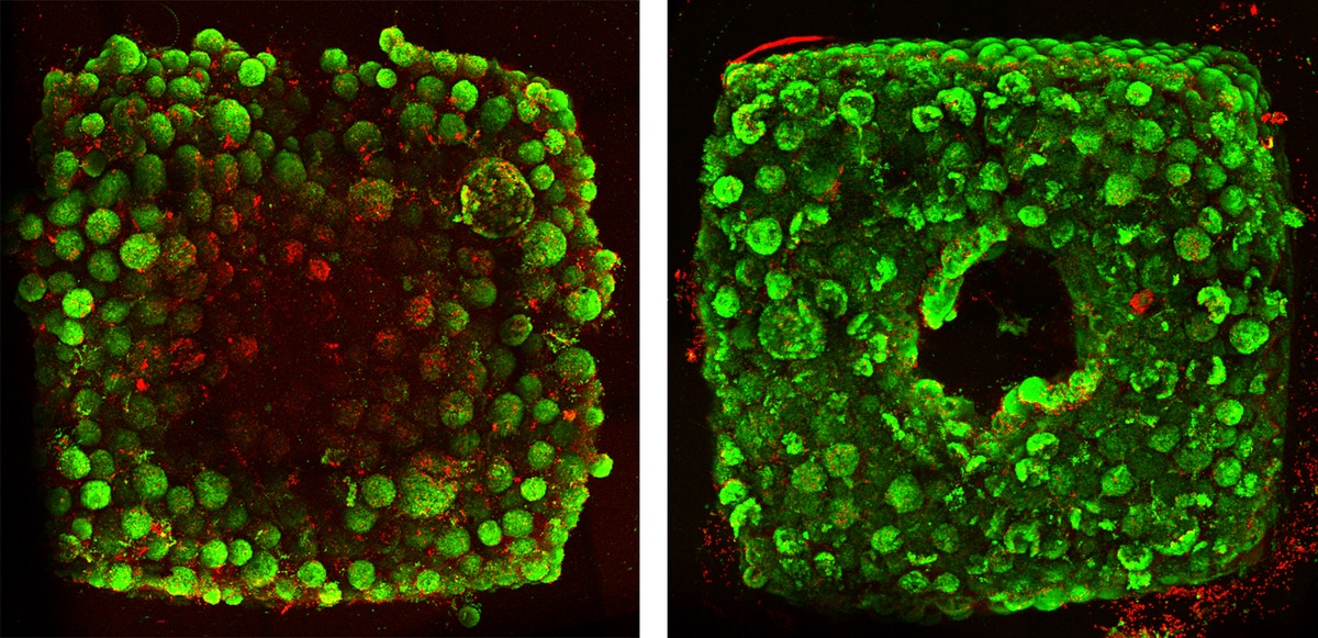
Tissues created without SWIFT-printed channels display cell death (red) in their cores after 12 hours of culture (left), while tissues with channels (right) have healthy cells.
He went on to explain that “it is unlikely that 3D printing can print all length-scales of an organ – from centimeter-scale ventricles to micrometer scale capillaries. So, we specifically designed the SWIFT process so that it can work with ‘organoids’ being built by the stem cell and developmental biology communities. By bridging the 3D printing and organoid fields, we believe there is a great potential for collaboration, and have already heard from researchers interested in using SWIFT to test scaling up their organoid systems. This interest has come from all sorts of specialists in different organs, including kidney, liver, heart, and brain.”
With so much going on, a typical day at the lab for Uzel and Skylar-Scott is not so typical. Although most of the daily tasks involve a combination of cell culture, printing ink formulation and characterization, CAD design and fabrication of printing and perfusion systems, tissue maintenance, imaging, and analysis. At busy times, Skylar-Scott says they could have upwards of four hours of work per day just to keep their cells fed, which has led to many long nights and weekends in the lab.
Similar to most academic labs, graduate students and postdocs all have two or three projects running in parallel. “For SWIFT, we had to culture so many cells for a single print, that we were only running about one print per week. Since staring at cells doesn’t make them grow faster, it is often helpful to have a second project to focus on,” joked Skylar-Scott. For example, they are currently working on new 3D printer hardware technology and focused on testing the SWIFT printed tissues in vivo so they can begin to test for additional function. All in a days work.
[Image Credit: Wyss Institute at Harvard University, John A. Paulson School of Engineering, Mark Skylar-Scott and Sébastien Uzel]
The post SWIFT: Uzel and Skylar-Scott are Paving the Way for the Future of Bioprinting appeared first on 3DPrint.com | The Voice of 3D Printing / Additive Manufacturing.
Harvard researchers develop SWIFT method to 3D print organ building blocks
Researchers from Harvard University’s Wyss Institute have developed a novel sacrificial ink-writing technique called SWIFT (sacrificial writing into functional tissue) to 3D print large, vascularized human organ building blocks (OBBs). Demonstrating its method, the team has created cardiac tissue that fuses and beats synchronously over a 7-day period. This enables the rapid assembly of perfusable […]
Harvard and Caltech 3D prints self-folding soft robots inspired by origami
Researchers from Harvard University and the California Institute of Technology have developed origami-inspired soft robotic systems that adopts task-specific configurations on demand. Of the systems fabricated is a 3D printed “Rollbot” that assembles into a pentagonal prism and self-rolls in programmed responses to thermal stimuli. Another 3D printed device is a self-twisting origami polyhedron that exhibits […]