3DHEALS2020: A Not So Lonely Planet

Only a few weeks away from 3DHEALS2020, and I just got off the phone with one of our speakers, Dr. Ho, from NAMIC Singapore. Our brief interview reminded me just how much I enjoyed Singapore—its start-up like government, incredible universities, and its beautiful modern architecture, chili crabs, and unpredictable rainstorms. Now, I’m on my way to some of the best meals in my life with another 3DHEALS community event in a foreign city. Looking back, there are many stories like that: in Detroit, Vigo, Paris, Shanghai, or Boston, my work with 3DHEALS communities has been a journey of adventures and friendships. 3DHEALS2020 is really a way to summarize my travels from the last two years. It is my version of Lonely Planet—the healthcare 3D printing version.

I really felt more alive when people have welcomed me into their cities; when they have showed off their latest innovations; when they have bantered enthusiastically with one another in a local pub till midnight after 3DHEALS events. And they felt the same way.

Sadly, however, this pandemic is putting old methods of human connection into question. Perhaps, a virtual summit is a stopgap solution for conferences, but, more likely, it is time for us to explore alternative and better ways to stay connected and informed.

The virtual 3DHEALS2020 summit will be a good start.

While we can’t serve you delicious San Francisco Blue Bottle coffee, there are three things we aim to do right with this conference:

1. Awesome live content

One upside about the virtual summit is that people who could not be available due to logistic barriers are now more available. We added 20+ speakers since the pandemic began and are still adding more parallel workshops to the existing program. Some of highlights include:

A. Biofabrication/Bioprinting Panels and Workshops:

Welcome to the holy grail of healthcare 3D printing applications!

These panels and workshops collect some of the brightest minds in the world of tissue engineering, biofabrication, and bioprinting. It includes the newest generation of startup founders. Names such as Stephanie WillerthAdam FeinbergJordan Miller are already well-established and loved in the scientific communities and just founded their own startups within the last 12 months. More established startup founders whose companies are also critical to the eventual success of biofabrication, tissue engineering, and cell therapy at large will also join us live, including Melanie Mathieu from Prellis Biologics, Jon Rawley from Roosterbio, John O’Reily from Xylyx, Taciana Pereira from Allevi, and Kevin Caldwell from Ossium Health. Qrquidea Garcia (“Orchid”) from JNJ Innovation will also join us on this panel, discussing how an industry leader can work with innovators and startups in this exciting, burgeoning field.

B. Regulatory and Legal Landscape of Healthcare 3D Printing

For those who put their skin in the game, this is probably one of the most must-attend sessions. 3D printing in healthcare is a super new field, and legal experts in this field who have established track records and legitimacy are only a handful. This session will include the most comprehensive list of legal and regulatory concerns specifically for healthcare 3D printing, including intellectual property/patent issues, product liability, FDA pathways, manufacturing standards, and more. Steven Bauer, from FDA CBER, just joined the panel to directly address concerns related to cell therapy from the biofabrication and stem cell communities. The speakers are not just well-versed on how to interpret the law and policies, but also how to interact with scientists, policy makers, organizations, and standards bodies at this early stage of the industry, with practical, real-life examples.

C. Global Perspectives

One lesson from this pandemic is that globalization has consequences. Having a well-rounded worldview of the global healthcare 3D printing ecosystem is a requirement for future success. Our early morning sessions are reserved for international speakers all over the world to meet the audience and share their unique perspectives, needs, and hopes. Both America Makes director John Wilczynski and NAMIC director Dr. Chaw Sing Ho, along with experts from Turkey, India, and Taiwan, will share how healthcare innovations can thrive in both local and global environments. On day two (June 6th), the audience will learn about how different countries are implementing the concept of 3D printing for Point of Care, which cannot be taken out of context of different healthcare systems and cultures. The audience will meet and learn from the leaders at UCSFStanfordGermany (Kumovis), India (Anatomiz3D), and developing countries.

2. Pre- and post-event networking opportunities

The attendees will have the opportunity to meet other attendees, speakers, and conference organizers as soon as they sign up the event using a dedicated conference app. They can send direct messages, post threads, share photos, host their own virtual events days before the conference. The app will be available to registered attendees for six months after the conference ends.

3. Entrepreneurship

One of the most exciting aspects of 3DHEALS2020 is its focus on entrepreneurship. Pitch3D has been a quarterly free and online pitch platform to selected early-stage startups in healthcare 3D printing and bioprinting spaces for the last two years, introducing 30+ startups from all over the world to institutional investors. 3DHEALS2020 also gathered some of the most experienced VCs and entrepreneurs in the space to share their stories, perspectives, and directly engage with the startups and the 3DHEALS2020 attendees directly during both pitch sessions and investor panels. There will be ten startups pitching each day at 5-6 PM PST. Interested startups can apply here.

This is the time of uncertainty and change.

Join us at 3DHEALS2020, connect with the world, and take control of your future. This is a Not So Lonely Planet.

The post 3DHEALS2020: A Not So Lonely Planet appeared first on 3DPrint.com | The Voice of 3D Printing / Additive Manufacturing.

Marco Valenzuela of Additive Design Studio Makes an Innovative 3D Printed Pipette

Marco Valenzuela is a designer who specializes in crafting innovative and new 3D printed products. Originally coming from the gaming world, his Additive Design Studio now is focused on using Additive Manufacturing and 3D printing exclusively in product design. The team works with Design for Additive Manufacturing methods and also works with services to deliver parts to customers in FDM, SLA, Polyjet, MFJ, and metals. Valenzuela made a pipette for a customer and this new design for a tried and true lab stalwart really interested us. We really believe that a wave of innovation will come to medical devices, medical supplies and even things like disposables through 3D Printing the right solution in medicine, and spoke with Valenzuela about his creation.
Why did you print it? 
The dual pipette was designed and 3D printed to fulfill specific needs in the fluid piping process. These needs were previously met. However the process was time consuming. The combining of the two pipettes into one provides for a speedy and more efficient workflow when processing large amounts of medicine.
The need was for two different functions:
1.The ability to suction a single fluid mixture into separate reservoirs for individual ejection into separate containers.
2.The ability to suction two different fluid mixtures separately and eject them into a single container together.
How does it work?
The dual pipette utilizes the same principles of pulsion and suction as a common syringe. The plungers are pulled up to create a vacuum and draw fluid up the spout and pressed down to eject fluid from the spout. The 3D printed plungers are fitted with normal rubber plunger tips to ensure an air-tight seal.
What is different about it?
The pipette is a 3d printable, simplistic design. The primary difference is the ability to 3D print this pipette quickly.  This means that we’re producing copies without the need for large-scale manufacturing. Reducing production time and availability to technicians by weeks.
What materials and processes were used to print it?
A variety of materials have been experimented with. The most suitable 3D printed material for the device will be EnvisionTEC’s E-Shell 200. A liquid photopolymer designed for DLP 3D printers that produces strong, tough, water-resistant ABS like parts with high detail that are Class IIa biocompatible according to ISO 10993/Medical Product Law and are CE certified for use as hearing aid products, otoplastics, and medical devices.
What software did you use?
I utilized a variety of 3d CAD design software in the development process. The final design was created in Lightwave 3D. I find a mix of traditional CAD and Polygon based modeling software helps me produce better more ascetic product designs.
Why is it a good design?
I enjoyed the creation of the dual pipette and consider it a good design because it has succeeded in fulfilling a specific need without otherwise costly measures. The dual pipette design aids in the development of medicines related to many medical treatments including Cerebral palsy.
We think that there is a bright future for 3D printed medical devices and medical supplies. Yes, this is a high touch regulatory environment so operating in it will never be simple. Medical supplies and devices have a lot of niche products however and a lot of comparatively low volume high priced goods. On the whole it will be exciting to see real low-cost innovation come to the medical world via 3D Printing.

3D Printing Pioneer Interview With Lars Brouwers on 3D Printing for Complex Fractures and Helping in Sierra Leone

Lars Brouwers is a surgeon in training and a researcher at the Radboudumc and the Elisabeth-Tweesteden hospital. He investigates the value of 3D printing and Virtual Reality in the treatment of complex fractures. With his research he received the AOTrauma Europe Research Grant and has won the Technical Innovation Award from the European Society for Surgical Research (ESSR), the international award of the Orthopedic Trauma Association (OTA), the Canadian and American sister association of the Dutch Nederlandse Vereniging voor Traumachirurgie (NVT) and the national IT award for “best IT project in healthcare” (Computable awards). In December 2017 he drove with an old car to Sierra Leone, equipped with a 3D printer (Ultimaker 2+), to develop a sustainable way in improving prosthetic arms and legs for handicapped people and making spare parts for a hospital there. One-and-a-half years later, a medical 3D printed lab is up and running in the Masanga Hospital. Now for the second time, a technical medicine student from the Netherlands is investigating 3D printed prostheses there. For this work, he received the Dutch Albert Schweitzer fund (NASF) grant. Lars is a really inspirational person really making more happen with 3D printing in medicine so we decided to interview him.

Who are you?

I’m Lars Brouwers, live in a small town near ‘s-Hertogenbosch in the southern part of the Netherlands, 30 years old and work in two major trauma centers (Elisabeth-Tweesteden Hospital, Tilburg and Radboudumc, Nijmegen) as a surgeon in training.

 What do you hope to achieve?

Besides being trained to be a surgeon in two top-clinical healthcare centers, I investigate the role of 3D printing in the medical world. I believe 3D printing is the next important invention after the internet. With 3D printing, we could provide patients, residents, nurses and physicians the best medical care there is.

 How do you use 3D printing?

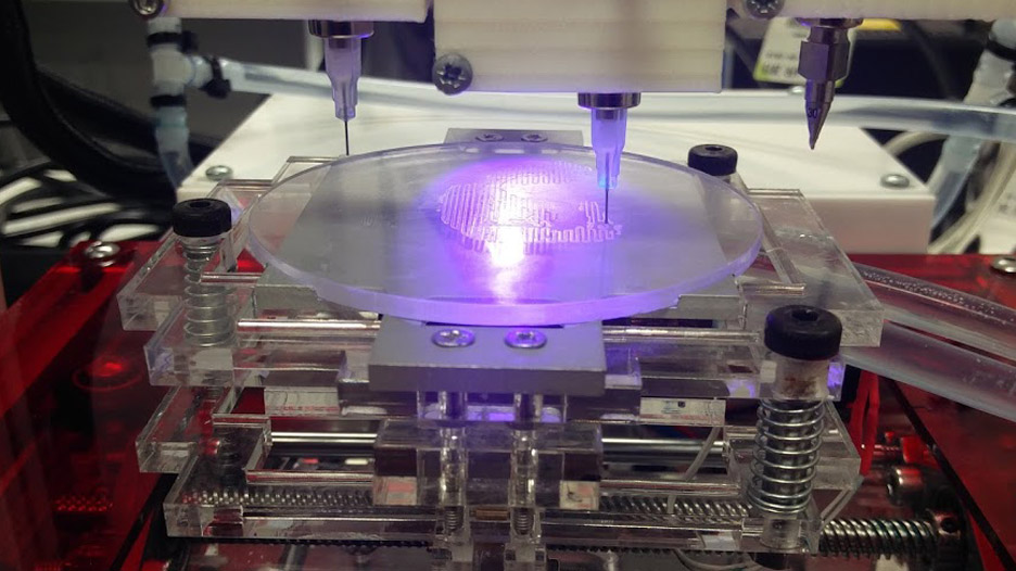
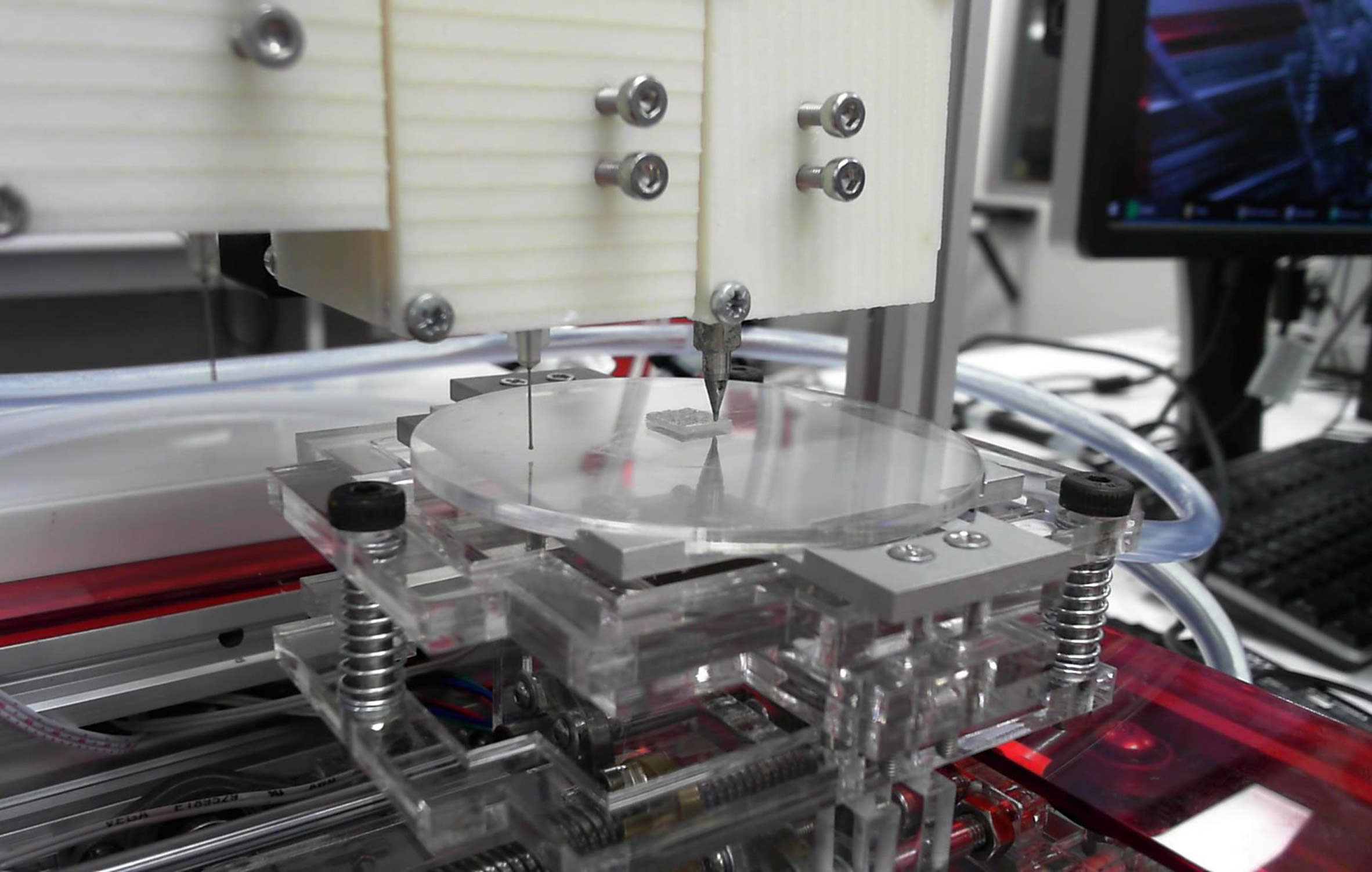
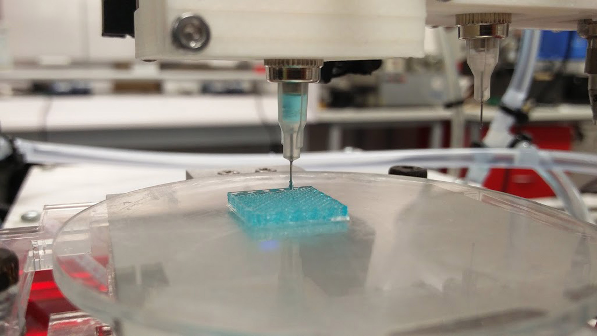
We use 3D printing for building life-size anatomical models and molds for surgical procedures. We set up several in-hospital medical 3D labs to ensure a 24/7 workflow without the need for technicians. 3D printing for doctors, by doctors. Low costs, and with the use of easy to use software systems.

 How does it help doctors and what research do you need to do in order to use more 3D printing in your work?

Dr. Mike Bemelman, traumasurgeon, Dr. Jan Heyligers, vascular surgeon and I set up our medical 3D lab in 2016 in the Elisabeth-Tweesteden hospital. We have expanded our 3D lab to a fully up and running 3D print lab in which we can 3D print anatomical models for surgeons, residents, nurses and patients 24/7. Our 3D print lab is now an example in the Netherlands of how a non-profit lab could and should work.

We think 3D printing, is the next invention after Xray and CT. Using 3D printed models, surgeons are able to expand their surgical preparations. 3D printed models provide a 3D view, while using conventional diagnostics brains are needed to form “virtual 3D models” in the head of the surgeon. Some people say, 3DCT is another solution. However, last year we demonstrated the added value of 3D printing in classifying complex fractures fractures (Brouwers, Lars, et al. “The Value of 3D Printed Models in Understanding Acetabular Fractures.” 3D Printing and Additive Manufacturing 5.1 (2018): 37-46.). Furthermore, we have showed for the first time that a 3D printed model is exactly the same as a human cadaver (Brouwers, Lars, et al. “Validation study of 3D-printed anatomical models using 2 PLA printers for preoperative planning in trauma surgery, a human cadaver study.” European Journal of Trauma and Emergency Surgery (2018): 1-8.)

At this moment we are investigating the role of 3D printing in explaining fractures and aneurysms to patients. Is it possible that patients are more satisfied or have a better understanding of their injury when 3D printed models of their own body are using during the explanation.

The 3D printing industry sometimes sees 3D printing as the holy grail. However, we have to be critical. If you treated one case with a 3D printed model and you are satisfied with the result, is it because of the 3D printed model or something else? The supposed advantages of 3D printing can only be proven by doing a lot of research. Some people say, that because of 3D printing, the operation time is shorter. However, is 10 minutes shorter really important? And they say that less blood loss occurs and operations will get cheaper. However, the preoperative workup gets more expensive. So, in what way is it really true that 3D printing is cost-effective?! A lot of research is needed and not case reports about how fantastic 3D printing is, based on 1 or 3 cases.

 For what kind of cases do you use it?

We use 3D printing mainly for traumasurgery cases. Patients with broken bones due to a fall, car accident or shooting, stabbing. We also 3D print aneurysms of the aorta to visualize the abdominal aneurysms for the patients preoperatively and postoperatively. At the moment we are testing with 3D printing soft tissue, such as esophagus cancer or anal fistulas. However, this is much more complex because the density of the tissue looks more like the surrounding tissue. An automatic threshold is in these cases not possible and we have to extract all the specific tissue by hand.

 What printers and materials do you use?

We use several 3D printers. In Sierra Leone an Ultimaker 2, in Tilburg an Ultimaker 3+, Makerbot Replicator Z18 and a Formlabs 2 and in Nijmegen two Ultimaker S5’s. We print with PLA, PVA and TPU 95A.

 How is 3D printing limited for you?

I do not see any limited subject regarding 3D printing at the moment. How I see it, is that we are working with an ”iPhone 1.0”. The technique is here, however, it can get faster and better. But we are satisfied with what we have accomplished so far.

 What partnerships do you seek?

We cooperate with both 3D printing companies such as Ultimaker and other hospitals to gain more knowledge about medical 3D printing. We have helped set up non-profit low-budget medical 3D labs in the Hospitals of Assen, Enschede, Nijmegen, Roosendaal and Rotterdam. Every hospital has his own radiology department with their own software. So converting CT files (DICOM) into STL files is in every hospital different. Some hospitals have own 3D software (Philips, Siemens) and other buy 3D software from 3D software companies (Mimics for example.). Therefore, we are cooperating with the largest medical 3D lab of Europe: 3D lab Radboudumc, head of department is Prof. dr. Thomas Maal. They are investing the role of artificial intelligence on the operation room and the role of augmented reality. Furthermore, his team takes care of several preoperative evaluations for all kinds of operations using molds and guides. Together we are investigating non-profit 3D software in order to set up 3D labs worldwide.

Last, we cooperate with the technical University of Twente. Technical medicine students are the connection between doctors and medical technique and are becoming more and more important. Together with their students, we can expand our 3D labs and ensure a proper 3D understanding for doctors and patients. For example, if we have a case with a malunion of the wrist, a technical medical engineer and treating physician are sitting together to prepare the operation and design guides or molds in order to correct the malunion in the best way there is. The engineer is also present during the operation. So they think, act and discuss together. A new way in improving patient outcomes!

 Why Sierra Leone?

A friend of mine, Wouter Nolet, is working as a doctor in the Masanga hospital, in the heart of Sierra Leone. Furthermore, he runs the Capacare programma in which local physicians are being trained in order to perform the most common surgical procedures. After graduation, they will work in several small remote hospitals of the country. A beautiful sustainable project! Together we decided to drive to Sierra Leone using his old Swedish car, a Saab 9.5 Turbo from the year 2001. I already thought for a while about the idea of implementing 3D printing in third world countries. This was an excellent opportunity to investigate the role of 3D printing in a local hospital. We put an Ultimaker 2+ in de back of the car and drove in 3 weeks time from the Netherlands, through Belgium, France, Spain, Morocco, Mauritania, Senegal, Guinea to Sierra Leone.

 What have you accomplished there?

In cooperation with the 3D lab of the Radboudumc, we set up a backup team for soft- and hardware problems. On-site we discussed with local people and tropical doctors to check the need of 3D printing. Based on this fieldwork, we found out that 3D printing could be of importance for making prostheses, spare parts and anatomical models for physicians. Many patients were living in camps nearby and we found out that the conventional prostheses were expensive to make. First we tried to help a woman with a burned hand. She wanted to write again. We took photos, used whatsapp to send the photo’s to the 3D lab in the Netherlands, designed a functional prosthesis and 3D printed this in the hospital. Due to this, she could write again!

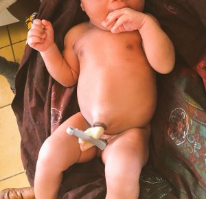
Furthermore, the umbilical cord clamps appeared to be finished in the hospital, so we 3D printed them as well. However, we were not satisfied with the workflow and added the head of the department of Utwente, Prof. Slump and prof. Grobusch, professor of tropical medicine, of the University of Amsterdam. Together with Jonathan Vas Nunes, the tropical Doctor, and director of the Masanga Hospital and Arico Verhulst, medical technician, we improved our concept and send for the first time a Technical medicine student, Merel van der Stelt, for 3 months to Sierra Leone, to investigate the role of 3D printing. She soon found out that patients found it more important to wear aesthetic prostheses when compared with functional prostheses. She used a 3D handscanner to scan the contralateral arm and mirrored this model. We are now able to 3D print prosthestic arms in less than 24 hours. Merel is investigating the lower extremity 3D printed prosthesis at the moment (even more challenging!) and a new technical medicine student, Andrea Sterkenburg, will soon fly to Sierra Leone to test different 3D printed prosthesis and investigate the sustainability. We continue with the follow-up of our patients to investigate the short-term and long-term quality of life and satisfaction about the prostheses. So far, it looks like that patients are satisfied about the strength and design. Even though the prostheses were aesthetic, they also seem to exhibit some degree of functionality. Even more important, ethical approval for this study has been obtained by the Sierra Leone Ethics and Scientific Review committee.

 What advice do you have for people who wish to help developing countries with 3D printing?

Helping people in third world countries is all about sustainability. With all respect to the many international teams who are going to African countries to help for 2-3 weeks, “giving” people prostheses for free is not sustainable. If you give local people everything for free, they do not understand that is normal to pay for a product, although it is a small amount of money. I will give you an example. In Africa, several hospitals are being run by local doctors. Local people have to pay a small amount of money for being treated, like in the western world. Only in this way, employees can be paid and the hospital can survive of course. It is not sustainable to live and survive on grants from western countries. However, every now and then a small (surgical) team from a western country arrives and announces that they treat as many as people they can treat in 2-3 weeks for free. Besides the lack of aftercare, it is confusing for local people when to pay or not? And people then refuse to pay the local doctors for treatment after this period. Therefore, our concept is to build a sustainable 3D workplace, in cooperation with local prostheses shops and in cooperation with local hospitals. We examine at the moment, how much local people can afford to pay for a 3D printed prosthesis and are making contracts with local shops in order to get sustainable.

 What advice do you have for physicians who wish to use 3D printing more?

Trial and error! Innovations and failing are related to each other. If there is no failing, anyone can innovate. Therefore it is important to keep on trying an believe in your own ideas! Be critical and do research in order to prove 3D printing is the technique of the future. Proper understanding and research is needed to draw conclusions!
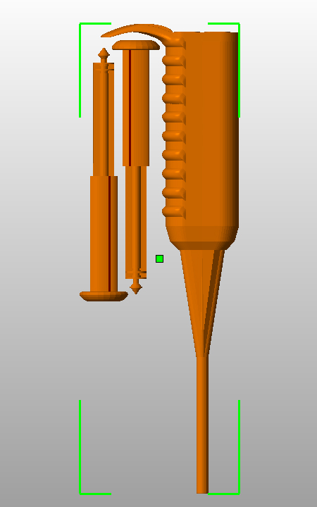
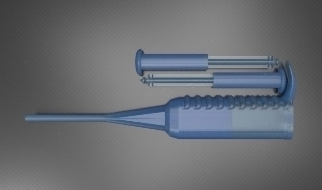
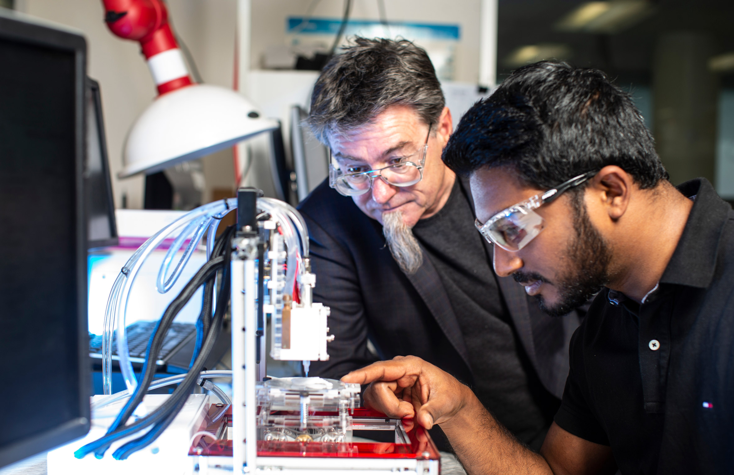
3D Alek: The Bioprinter That Replicates Human Ears for Patients with Microtia

There is no doubt that 3D printing offers more and more promise to the medical industry, and that has helped to save and improve lives in many ways.

Image via Dezeen

A team from the University of Wollongong (UOW), Australia, who are working at the ARC Centre of Excellence for Electromaterials Science (ACES) in collaboration with Payal Mukherjee, a nose and throat surgeon, and Associate Professor at the University of Sydney School of Medicine, have developed a 3D bioprinter that replicates human ears. These replicas can be used in a reconstructive surgery. Their 3D bioprinter called “3D Alek”, is a multi-materials 3D printer customized for biofabrication.

Bioprinters use a pipette, which is computer-guided. Instead of using metal or plastic, the pipette layers living cells, which are referred to as “bio-ink”. Bio-ink was developed by researchers at University of Wollongong and the Australian National Fabrication Facility and it uses stem cells to print tissue constructs or oragnoids for medical research. In the case of 3D Alek, it prints human ear cartilage as to create a “living ear” to treat congenital ear deformities.

Image via Dezeen

This new technology, as researchers say, brings them closer to revolutionizing a complex medical procedure for children with microtia. To understand this congenital condition better: microtia is the deformity of the outer ear where the ear doesn’t develop fully during the first trimester of pregnancy. Mukherjee, claims that this particular ear deformity treatment is demanding: the outer ear 3D shape is extremely complex because of its length, breadth, height, and projection from the skull.

Mukherjee believes that bioprinting is extremely exciting since “it allows an ear graft to be designed and customized according to the patient’s face using their natural tissue”. For her, it would result in a reduced operating time and an improved cosmetic outcome, as well as avoiding the complexity of finding a cartilage donor site.

Image via Dezeen

3D Alek was recently installed at the Royal Prince Alfred Hospital (RPA) in Sydney, and has become the world’s first hospital in New South Wales to have a 3D bioprinter. However, researchers hope to improve their bio-ink for it to match the patient’s anatomy by using the patient’s own stem cells to grow the ear cartilage. The ear abnormality is specific on every patient and it depends on their facial features, thus the idea is to print a customized ear perfect for each patient.

Professor Gordon Wallace, ACES Director, said: “The 3D Alek collaboration had benefited from drawing upon the diverse skills of a range of scientists, engineers and clinicians to bring about real advances to tackle this significant medical challenge.”

Image via Dezeen

For Wallace, the project illustrates their ability to manage a successful pipeline to turn fundamental research into a strategic application to create a new health solution to improve people’s lives.

“We have been responsible for the primary sourcing of materials; the formulation of bio-inks and the design and fabrication of a customized printer; the design of required optimal protocols for cell biology; through to the final clinical application. With one 3D Alek now established in a clinical environment at RPA and a replica in our lab at TRICEP (The Translational Research Initiative for Cell Engineering and Printing) , our new 3D bioprinting initiative, we will be able to fast-track the next stages of our research to deliver a practical solution to solve this clinical challenge,” Wallace added.

Sources: [Dezeen, University of Wollongong, Verdict Medical Devices]

Printing Lungs Using 3D, Bioprinting, and Stem Cells

via MIT Technology Review

Last month I had the chance to hold a replica of the upper part of a human airway—the windpipe plus the first two bronchi. It had been made from collagen, the biological cement that holds our bodies together. It was slippery and hollow, with the consistency of undercooked pasta.

The structure had emerged from a refrigerator-size 3-D printer in Manchester, New Hampshire, at an outpost of United Therapeutics, a company that earns more than a billion dollars a year selling drugs to treat lung ailments.

One day, the company says, it plans to use a printer like this one to manufacture human lungs in “unlimited quantities” and overcome the severe shortage of donor organs.

Bioprinting tissue isn’t a new idea. 3-D printers can make human skin, even retinas. Yet the method, so far, has been limited to tissues that are very small or very thin and lack blood vessels.

United instead is developing a printer that it believes will be able, within a few years, to manufacture a solid, rubbery outline of a lung in exquisite detail, including all 23 descending branches of the airway, the gas-exchanging alveoli, and a delicate network of capillaries.

A lung made from collagen won’t help anyone: it’s to a real lung what a rubber chicken is to an actual hen. So United is also developing ways to impregnate the matrix with human cells so they’ll attach and burrow into it, bringing it alive.

“We are trying to build the little stick houses for cells to live in,” says Derek Morris, a project leader in United’s organ manufacturing group.

Read more!

Using Patient-Specific 3D Printed Surgical Guides for Total Knee Replacement

While surgery has always, ultimately, been about the patient, it hasn’t always been patient-centered. Historically, patients have not had an easy time of understanding exactly what their surgery entails and have often been treated as if they were ancillary to the surgical problem presented. This can’t all be blamed on uncaring medical staff, as most people involved in medicine do care and care profoundly. Instead, it has largely been a result of resources and standing custom. Surgical procedures are complicated and difficult to understand, hence the reason why experts are the ones who address them, and the pressure and stress involved in going into a procedure largely blind has made it difficult for surgeons to relax and broaden their focus to include the patient beyond the problem.

3D technology is making great contributions to medicine, from aiding in research to assisting in the preparation of students to practice medicine to producing the tools necessary to perform better operations. It is being integrated into the surgical theater and changing the face of surgery as we know it. One of the ways it is doing this is through the provision of a greatly improved ability to plan for the procedure. 3D technology not only allows the medical team a sneak preview, 3D printing can create models of the particular areas to be addressed and allow surgeons to study them in advance. This helps minimize surprises and therefore reduces the stress on both the patient and the medical staff.

The staff at Orthoparc in the Netherlands has figured out another way to help create and deliver the best in patient centered care. Using 3D technology, they have developed a method of patient-centered total knee replacement that allows a patient to walk in, in the morning, and walk out that same day. Such a possibility requires a highly interconnected team of specialists working together to ensure that not only does the patient get the knee replacement they need, but their psychological, nutritional, and whole health needs are met as well.

Dr. Saskia Boekhorst

One component of this is the integration of 3D printed, patient-specific surgical guides that take the uncertainty out of the procedure itself. These surgical guides are produced using data gathered about an individual patient’s knee and are fabricated in-house on a 3D printer. When placed upon the patient during surgery, they guide the surgeon to exactly where cuts need to be made in relationship to where the knee is resting. Dr. Saskia Boekhorst is an orthopedic surgeon at Orthoparc, and she described the impact these guides have had in her experience:

“I’m a big fan of the patient-specific knee guides because this technology allows me to place the components of the knee arthroplasty exactly in the right axis of the leg in all dimensions. The first one hundred cases I’ve double checked by manual measurements, because it was a different approach and I am very careful. But after seeing very nice and consistent results, I became convinced.”

This increase in precision means that the surgery is more exactly what the patient needs and reduces the risk of unnecessary or erroneous incisions. The surgical guides also make the surgery less invasive, removing the need to drill into the femur canal as in traditional knee replacement surgery procedures. They also help decrease the amount of time the patient has to spend in surgery, as Dr. Boekhorst explained:

“The positioning and alignment are already done by me within an interactive planning software in which I can rotate the knee in all directions and see how the prosthesis could be placed for a particular patient. You will never be able to see this in all these dimensions and directions with a real patient because of soft tissue.”

All of this adds up to mean that there is less anesthesia necessary and less powerful post surgery painkillers required. Not only does that reduce the risks associated with such powerful medicines but allows the patient to be more fully cognizant and mentally focused on an increased timeline, something which is necessary for the implementation of a physical therapy regimen.

It’s not just good for the patients, it also makes sense for the surgical team in terms of reducing stress and ensuring they are better prepared for the operation. In addition, using these guides means that fewer surgical tools need to be prepped and sterilized as the knee guides come in a comprehensive ‘knee in the box’ package that includes all the necessary instrumentation and two sizes of preselected implants to treat a single patient. This means less overhead and a reduction in logistics cost, something which allows the medical practice to reinvest its resources elsewhere, such as in its staff and patients.

What do you think of this news? Let us know your thoughts; join the discussion of this and other 3D printing topics at 3DPrintBoard.com.

[Source/Images: Materialise]


Indiana University and Lung Biotechnology Partner to Advance 3D Printing of Organs

One of the reasons why I really enjoy writing about 3D printing is because it is a fabulous mix of the fun and the important. The stories we cover can be anything from the enjoyable experience of standing before a giant 3D printed skeleton to the very serious nature of fabricating necessary objects on a 3D printer onboard the International Space Station. The work done by these machines and the people that operate them has, time and again, proven to offer a helping hand (sometimes literally as in the case of prosthetics) to boosting the quality of life of a growing number of individuals.

From left to right: Lester Smith, PhD, Burcin Ekser, MD, PhD and Ping Li, PhD [Image:Eric Schoch, IUSM]

One area in which 3D printing is making particularly important contributions is in the field of medicine. Over the past several years, we have seen stories about students getting hands-on experience through 3D printed models and of improved patient outcomes as a result of preparation and the fabrication of custom surgical equipment for the medical team. With the introduction of bioprinting, the ultimate dream in medicine has been to advance to the point of being able to 3D print whole organs that could be used to replace those that are failing in patients. Thus far, that is still a dream for the future, but important advances are being made in that direction, sometimes great strides, other times only baby steps.

One of the most recent steps forward has come in the form of an agreement between faculty at Indiana University (IU) School of Medicine and the Maryland-based company Lung Biotechnology PBC, that is focused on organ transplantation technologies. The hope is that the $9 million project will result in the knowledge necessary to make the dream of 3D printing organs into a reality. They won’t be starting from scratch; the IU team is already able to generate tissues, but they will use the funding provided through this partnership to analyze the tissues and their structures in order to possibly unlock the key to more advanced organ creation. Dr. Lester Smith, an Assistant Professor of Radiology and Imaging Sciences at IU School of Medicine and the head of the research team, explained the prolonged nature of any such investigation:

“[I]f someone has a skin burn, maybe we can replace skin. Or if someone has a bad liver then we can replace the liver entirely. But this is way down the road. Most of our tissues which make up our organs have a lot of different cell types. They are also vascularized, which means they have a lot of blood vessels that are basically channeling through them. When we get there that’s when I can tell you how long it took. That’s because the body is so complex and there’re so many different parts and so many responses. I couldn’t tell you how long it would take but we’re on the road to that destination.”

Luckily, Indiana University and Lung Biotechnology don’t have to make all the headway by themselves; there are a large number of organizations, from large to small and public to private, pursuing the dream of fabricating organs. This is more than just an effort to do something to see if it can be done; there are people dying every year because they cannot get access to the organs that they need, and further deaths and astronomical medical expenses to deal with for those whose bodies strongly reject the foreign organs. Should it become possible to create a custom organ for someone using their own cells, the entire process from the surgery to simple day to day functionality would be vastly improved, and this partnership should help push that research closer to the gold standard.

What do you think of this news? Let us know your thoughts; join the discussion of this and other 3D printing topics at 3DPrintBoard.com or share your thoughts below.

[Source: Indiana Public Media]