Improving Polymers: 3D Printing Polycaprolactone with Gum Rosin and Beeswax Additives

Researchers from Spain and Ecuador are focused on nature-driven materials for digital fabrication, outlining their findings in the recently published ‘New Materials for 3D-Printing Based on Polycaprolactone with Gum Rosin and Beeswax as Additives.’

Nature is often the inspiration for scientific findings and innovations, and the world of 3D printing is no exception, from the intense study of fish to seashell material to the ever-changing color of the chameleon’s skin, and more. In this study, the researchers experiment with the potential of gum rosin and beeswax as additives, analyzing mechanical, thermal, and structural properties.

Reminding us that polymers are indeed useful in manufacturing and many applications, some do present a hazard to the environment regarding the build-up of waste on the planet. With no desire to add to that problem, the authors sought alternative materials such as biopolymers.

While there are many benefits to avoiding the use of conventional plastics, affordability has typically been an issue, along with finding materials that have suitable mechanical properties. Blends, fillers, and composites are often the key, however, for scientists and innovators when it comes to materials like polycaprolactone (PCL) that require some refining—despite offering benefits such as biocompatibility, biodegradability, and non-toxicity.

The researchers intended to find out whether gum rosin, beeswax, and PCL would offer the ‘synergistic’ effect expounded on by other scientists as it is expected that the mixture not only will support initial benefits of all the materials but also ‘enhance the antimicrobial properties.’ Beeswax has also been known to complement polymers being used in biomedical applications like drug delivery systems. Both GR and BW are known to offer improvements to other materials in terms of adhesion (often an issue in 3D printing), toughness, and behavior of plastic overall.

The researchers used a BCN3D 3D printer with a 0.6 mm diameter nozzle to print samples that could then be compared to standard test specimens. A bed temperature of 40 °C was set for the printing of all materials, but nozzle temperatures varied among the samples, from 90 °C and 150 °C, ‘depending on the easiness of traction of the materials in the printer.’

“These differences aim to achieve and adequate printability,” explained the researchers, noting that just an ‘increment’ in the nozzle temperature could offer increased mechanical strength.

Temperatures of 110 °C for PCL-GR and 150 °C for PCL-BW were chosen as the printing temperatures.

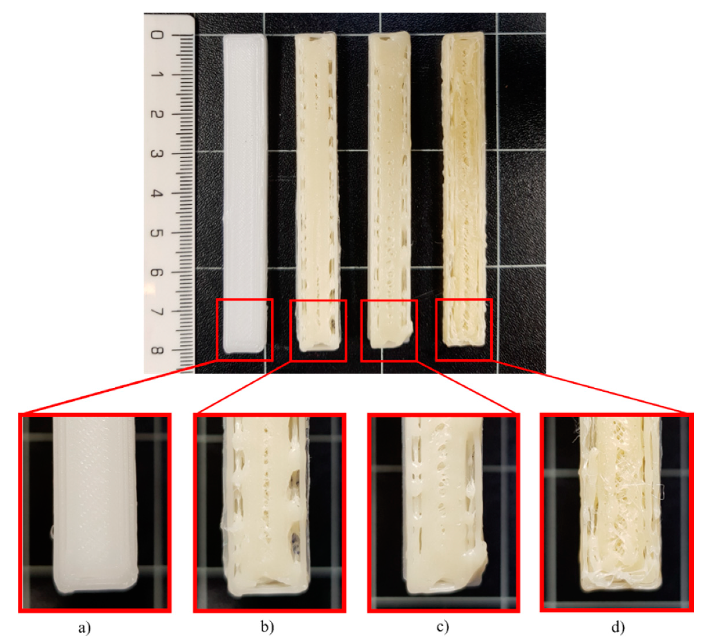
Standard test specimens (STS)surface obtained in the printing test at 80 °C for (a) PCL, (b) PCL-GR, (c) PCL-GR-BW and (d) PCL-BW.


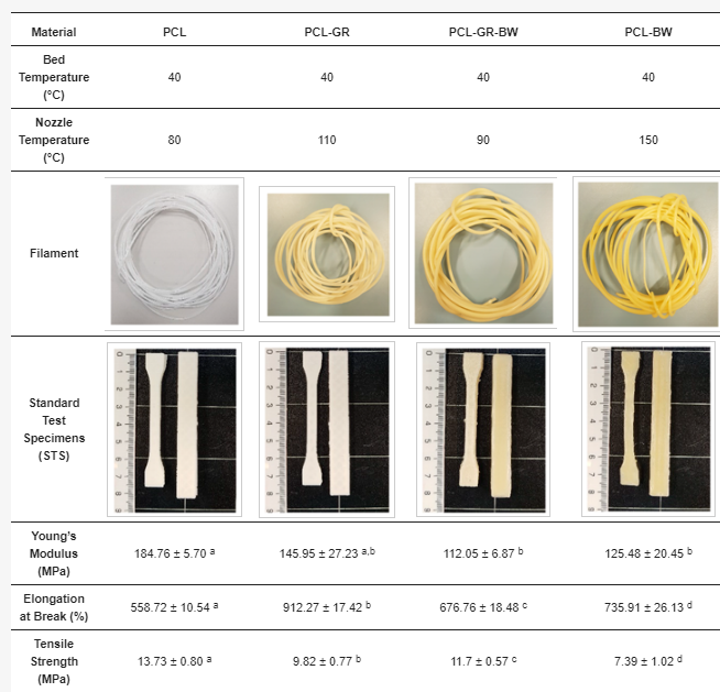
Three-dimensional (3D)-printing parameters and tensile mechanical properties of filaments of neat polycaprolactone (PCL) and the formulations with gum rosin (GR) and beeswax (BW).

Thermal characterization showed ‘good miscibility’ in the PCL matrix, upon examination of GR and BW, with the added note that GR did increase the thermal stability of PCL.

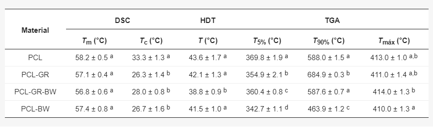
Thermal properties of neat PCL and its formulations with GR and BW.

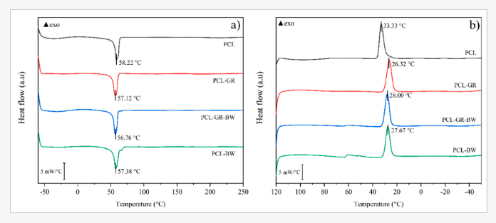
(a) DSC second heating curve and (b) DSC cooling curve of neat PCL and the formulations with GR and BW.

(a) TGA curves and (b) DTG curves with an expanded area for temperatures between 395 °C and 430 °C for the neat PCL and the formulations with GR and BW.

With GR being used as an additive, the authors noted that the material was then limited to just one phase—while with BW, two phases existed, causing low miscibility and lowered mechanical properties.

“Color measurements showed that the intrinsic coloration of natural additives has a significant effect on the color of the final materials. With respect to wettability, the addition of GR and BW increased the hydrophobic behavior of neat PCL,” said the researchers.

“Finally, it was concluded that the PCL-GR-BW formulation is the most suitable material for a 3D-printing process as it behaves better in the traction mechanism of the printer. Further, it exhibits the thermal and mechanical properties closer to neat PCL.”

What do you think of this news? Let us know your thoughts; join the discussion of this and other 3D printing topics at 3DPrintBoard.com.

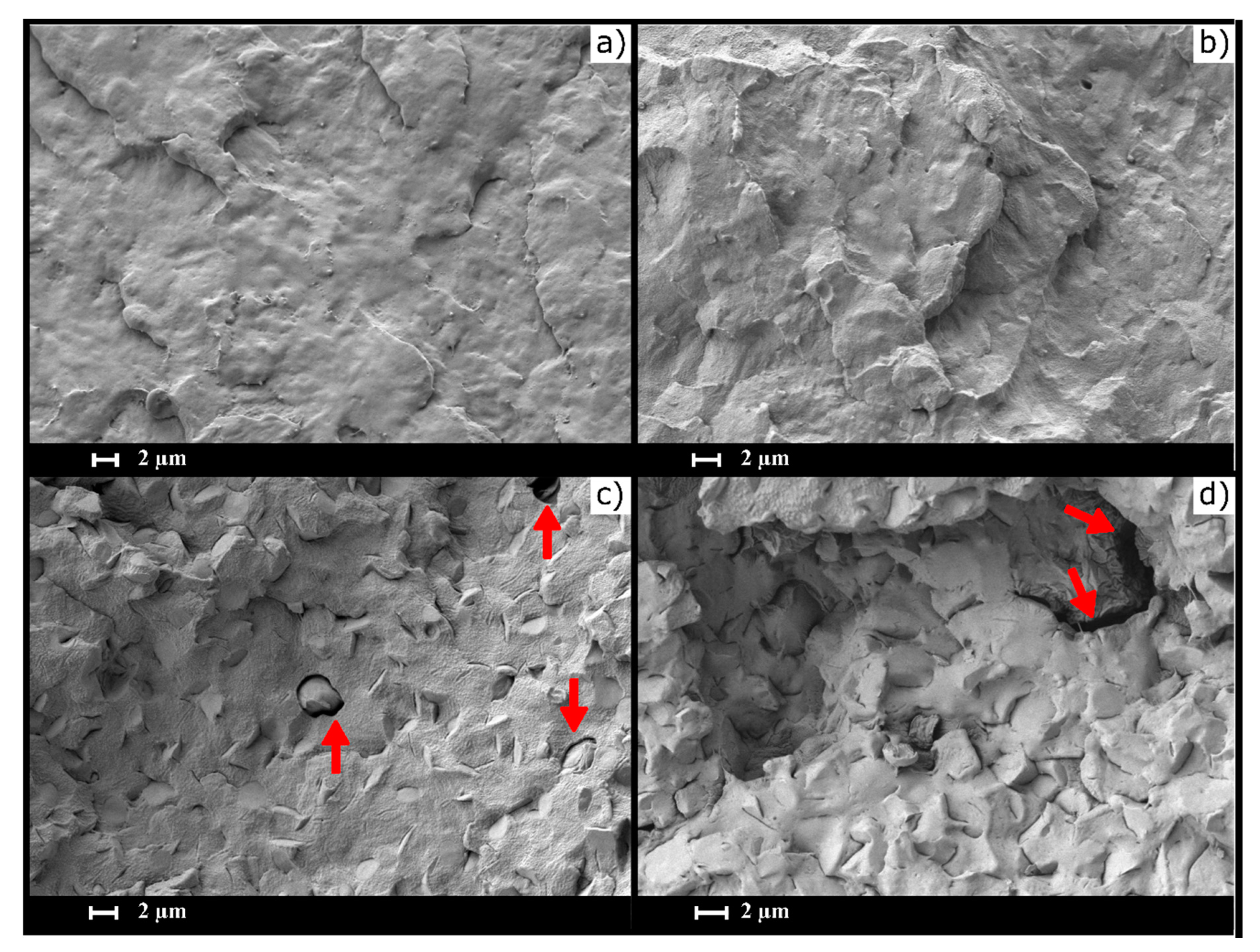
Scanning electron microscopy (SEM) images of (a) PCL, (b) PCL-GR, (c) PCL-GR-BW, and (d) PCL-BW, red arrows show holes and discontinuities in the material surface.

[Source / Images: ‘New Materials for 3D-Printing Based on Polycaprolactone with Gum Rosin and Beeswax as Additives’]

The post Improving Polymers: 3D Printing Polycaprolactone with Gum Rosin and Beeswax Additives appeared first on 3DPrint.com | The Voice of 3D Printing / Additive Manufacturing.

Beijing University of Chemical Technology: 3D Printed HA/PCL Tissue Engineering Scaffolds

3D printed bone scaffolds used for tissue engineering purposes need to have a good amount of mechanical strength, since the scaffold needs to be able to provide support for the tissue. As bone scaffolds also require the correct pore structure to help provide a good environment for the differentiation, proliferation, and repairing of damaged tissue cells, bioactive materials, such as polycaprolactone (PCL) and hydroxyapatite (HA), are needed.

Researchers Zhiwei Jiao, Bin Luo, Shengyi Xiang, Haopeng Ma, Yuan Yu, and Weimin Yang, from the Beijing University of Chemical Technology (BUCT), published a paper, titled “3D printing of HA / PCL composite tissue engineering scaffolds,” about their work constructing nano-HA/PCL and micro-HA/PCL tissue engineering scaffolds using the melt differential FDM 3D printer they developed.

The abstract reads, “Here, the internal structure and mechanical properties of the hydroxyapatite/polycaprolactone scaffolds, prepared by fused deposition modeling (FDM) technique, were explored. Using hydroxyapatite (HA) and polycaprolactone (PCL) as raw materials, nano-HA/PCL and micro-HA/PCL that composite with 20 wt% HA were prepared by melt blending technology, and HA/PCL composite tissue engineering scaffolds were prepared by self-developed melt differential FDM 3D printer. From the observation under microscope, it was found that the prepared nano-HA/PCL and micro-HA/PCL tissue engineering scaffolds have uniformly distributed and interconnected nearly rectangular pores. By observing the cross-sectional view of the nano-HA/PCL scaffold and the micro-HA/PCL scaffold, it is known that the HA particles in the nano-HA/PCL scaffold are evenly distributed and the HA particles in the micro-HA/PCL scaffold are agglomerated, which attribute nano-HA/PCL scaffolds with higher tensile strength and flexural strength than the micro-HA/PCL scaffolds. The tensile strength and flexural strength of the nano-HA/PCL specimens were 23.29 MPa and 21.39 MPa, respectively, which were 26.0% and 33.1% higher than those of the pure PCL specimens. Therefore, the bioactive nano-HA/PCL composite scaffolds prepared by melt differential FDM 3D printers should have broader application prospects in bone tissue engineering.”

Melt differential 3D printer.

PCL is biocompatible, biodegradable, and has shape retention properties, which is why it’s often used to fabricate stents. But on the other hand, due to an insufficient amount of bioactivity, the material is not great for use in bone tissue engineering. HA, which has been used successfully as a bone substitute material, has plenty of bioactivity, which is why combining it with PCL can work for bone tissue engineering scaffolds.

“On the whole, the existing tissue engineering scaffolds preparation process have problems of low HA content, easy agglomeration, low stent strength, and single printing material,” the researchers explained.

“The HA/PCL composite particles are used as printing materials, and the mechanical properties and structural characteristics of the two tissue engineering scaffolds are compared and analyzed. The raw material of the melt differential 3D printer is pellets, which eliminates the step of drawing compared to a conventional FDM type 3D printer. The 3D printer is melt-extruded with a screw, and a micro-screw is used for conveying and building pressure. At the same time, precise measurement is performed by a valve control system. This printing method shows advantages in simple preparation process of the composite material, higher degree of freedom in material selection, simple printing process, and shorter preparation cycle of tissue engineering scaffolds.”

The team mixed PCL particles and HA powder together to make the scaffolds. Their melt differential 3D printer uses pellets, and features a fixed nozzle with a platform that moves in three directions. A twin-screw extrusion granulator was used to prepare the PCL material, and the melt differential 3D printer fabricated the tissue engineering scaffolds out of the nano-HA/PCL and micro-HA/PCL composite particles.

The working principle diagram of the polymer melt differential 3D printer.

A microcomputer-controlled electronic universal testing machine was used to test the scaffolds’ bending and tensile properties. A scanning electron microscope was used to observe the micro-HA particle size, as well as the scaffolds’ cross section, while an optical microscope was used to observe their surface structure and a transmission microscope was used to look at the nano-HA particles’ particle diameter and morphology. The scaffold material’s crystallization properties were analyzed using a differential thermal analyzer.

3D printing tissue engineering scaffolds.

Testing showed that the micro-HA was spherical, with a 5–40 μm diameter, and contained some irregularly-shaped debris. The nano-HA was rod-shaped, with a 20–150 nm length.

The crystallization peak temperature of the HA/PCL composites was higher than pure PCL material, because adding HA caused its molecular chain to form a nucleate after absorbing on the HA’s surface. Additionally, adding HA to pure PCL increased the material’s melting temperature, as the latter material had crystals “of varying degrees of perfection.”

The nano-HA/PCL and micro-HA/PCL tissue engineering scaffolds “could form a pre-designed pore structure and the pores were connected to each other,” which is seen in the image below.

“…the micro-HA/PCL and the nano-HA/PCL composite tissue engineering scaffolds can form a three-dimensional pore structure with uniform distribution and approximately rectangular shape.”

External views of micro-HA/PCL and nano-HA/PCL composite tissue engineering scaffolds.

These rectangular pores, with a 100-500 μm length and width, are good news for cell adhesion and proliferation, and the fact that they’re interconnected is positive for nutrient supply.

As for mechanical properties, the nano-HA/PCL specimens had the highest tensile and bending strengths – between 25 and 35% higher than the pure PCL. The micro-HA/PCL specimens had higher tensile and flexural strengths than the PCL, but the nano-HA/PCL was stronger than the micro-HA/PCL, because the HA’s modulus is higher than the PCL’s.

“In addition, nano-HA was more evenly distributed in the composite, while micro-HA had obvious agglomeration in the composite, so the tensile strength and flexural strength of nano-HA/PCL specimens were higher than that of micro-HA/PCL specimens,” the researchers wrote.

Finally, the pore structure of the nano-HA/PCL and micro-HA/PCL tissue engineering scaffolds offered a favorable environment for the discharge of cellular metabolic waste, in addition to facilitating nutrient transport and blood vessel growth. The researchers concluded that their 3D printed composite scaffolds had more potential applications in bone tissue engineering.

Discuss this and other 3D printing topics at 3DPrintBoard.com or share your thoughts below.

The post Beijing University of Chemical Technology: 3D Printed HA/PCL Tissue Engineering Scaffolds appeared first on 3DPrint.com | The Voice of 3D Printing / Additive Manufacturing.

Bio-printing 101: How to Bioprint at Home

<em>Picture 2: </em>Hydrogel fabricated by PSL 3D printing technique” width=”565″ height=”314″></p>
<p style=Bioprinted Hydrogels

Bioprinting is an exciting area to follow as it invigorates the ideas of Frankenstein and a bunch of other sci-fi scenarios that make us slightly paranoid. So how does someone create their own monster within their garage? Well, that is pretty far fetched at the moment, but there are ways to get involved in this field in small ways.

Consistently, within bioprinting, there does not seem to be a significant presence within the general maker community. The healthcare industry as a whole is typically private sector driven. So how does someone get the chance to work on 3D bioprinting when they do not even know the resources they need, or how to start?

An essential consideration for 3D bioprinting is the material used for prototyping. Typically 3D printers use different materials to create products such as PLA, ABS, Wood Fiber, PET, PVA, Nylon, and TPU. The issue of creating bioprinted materials is not within the actual structure of the model and design. The problem lies in creating objects that also follow the rules of biology. This limitation forces a material to have specific heating and cooling properties in relation to where it is within the body. Specific heat and tolerance to different temperature ranges are vital in a material used for bioprinting. Even with the creation of 3D structures, there is still a difficulty in replicating the intricate vascular structures of different organs within our bodies. This makes for a variety of problems that need to be worked on within the field of 3D bioprinting in general.

So what are some first steps within 3D bioprinting? Let us focus on some materials that would be ideal to focus on as they are good candidates for current and future use. Here is a list of some current materials used in bioprinting:

  • Bioink
  • Hydrogels
  • Alginate
  • PEG (polyethylene glycol)
  • PCL (Polycaprolactone)
  • PGA (Polyglycolic Acid)
  • Pluronics

In later articles, we will discuss each material above in more depth. For now we will address them briefly.

Bioinks are substances made of living cells that can be used for 3D printing of complex tissue models. Bioinks are materials that mimic an extracellular matrix environment to support the adhesion, proliferation, and differentiation of living cells.

A hydrogel is a network of polymer chains that are hydrophilic, sometimes found as a colloidal gel in which water is the dispersion medium. A three-dimensional solid results from the hydrophilic polymer chains being held together by cross-links. Alginate is a polysaccharide distributed widely in the cell walls of brown algae, where through binding with water it forms a viscous gum.

Alginate is made from brown seaweed.

Alginate is a polysaccharide distributed widely in the cell walls of brown algae, where through binding with water it forms a viscous gum.

PEG is marketed as a laxative but is also a stabilizing agent in toothpaste.

Polyethylene glycol (PEG) is a polyether compound with many applications, from industrial manufacturing to medicine and is often used in making hydrogels for 3D printing.

Polycaprolactone (PCL) is a bioabsorbable polyester with a low melting point of around 60 °C and a glass transition temperature of about −60 °C.

Polyglycolic acid (PGA) is a biodegradable, thermoplastic polymer and the simplest linear, aliphatic polyester. PGA is used for scaffolds and as a support material.

Pluronics are nonionic triblock copolymers composed of a central hydrophobic chain of polyoxypropylene (poly(propylene oxide)) flanked by two hydrophilic chains of polyoxyethylene (poly(ethylene oxide)).

Now that we understand a little bit about the materials that we can use, it becomes a question of what type of printer to use. Most industrial bioprinters are far from a viable price for consumer purchase.  Communities of makers have few options for buying a 3D bioprinter. There also seems to be a lack of internet resources to instruct people on how to bioprint. To help people at home, we will try to build a 3D bioprinting setup with a guide for all those who are interested in bioprinting. As a follow up on this article, be sure to look out for information on the biomaterials mentioned above and separate articles on each of them and how they relate to 3D bioprinting. Stay tuned for more DIY bioprinting tips and tricks.

3D printed tracheal splints used in pediatric surgery

Children’s Healthcare of Atlanta (CHoA) and the Georgia Institute of Technology (Georgia Tech) have successfully used 3D printed tracheal splints in the pediatric surgery of a 7-month old patient with a life-threatening airway obstruction. The surgeons of CHoA used three custom-made splints, which were made by biomedical engineers of Georgia Tech. Donna Hyland, President and […]