

(a) Bright-field transmission electron microscopy (TEM) image of the cells oriented perpendicular to the observation direction; (b) Bright-field TEM image of the cells oriented parallel to the observation direction, diffraction pattern illustrates the <100> fcc zone axis orientation of the cells; (c) nanoparticles observed in the microstructure of LPBF 316 L steel marked by arrows; and (d) TEM energy dispersive X-ray (EDX) spectra taken from one of the particle illustrated in (c).
A great deal of work is involved in optimizing materials for additive manufacturing. Porosity is a consistent problem in metal 3D printing, and scientists spend a lot of time studying each metal material to try to minimize or eliminate flaws. In a paper entitled “Microstructure, Solidification Texture, and Thermal Stability of 316 L Stainless Steel Manufactured by Laser Powder Bed Fusion,” a team of researchers examines 316 L stainless steel using techniques including scanning and transition electron microscopy, diffraction methods and atom probe tomography.
Porosity can be eliminated by controlling the laser power and laser scanning speed during the 3D printing process, the researchers point out.
“The final properties are governed by the microstructure of the material,” they continue. “The microstructure of the LPBF material is formed under the conditions of high temperature gradients and solidification rates, far from the ones of conventional materials. This results in the formation of a nonequilibrium microstructure with a unique set of properties. Epitaxial nucleation of cellular colonies has commonly been observed, which results in the solidification texture and anisotropic mechanical properties of LPBF materials.”
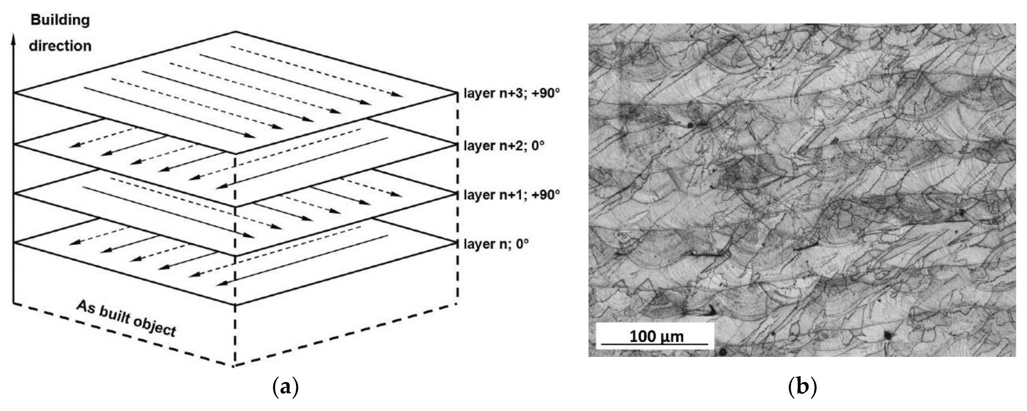
(a) Scanning strategy used to manufacture laser powder bed fusion (LPBF) 316 L; (b) microstructure of the LPBF 316 L steel, optical micrograph.
The study, which was conducted over several years, focuses on the metallurgical aspects of the material, as well as its microstructure. The formation of a cellular structure in a molten pool was discussed in relation to the thermal gradient and solidification rate. The correlation between the primary cell spacing and hardness was also discussed in relation to additive manufacturing process parameters and the presence of porosity.
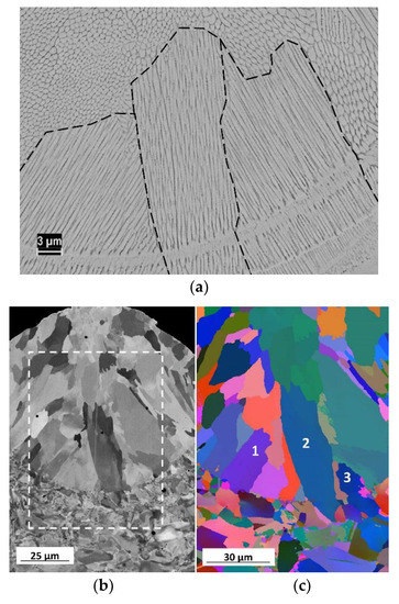
(a) Scanning electron microscopy (SEM) micrograph of the etched cross section of the LPBF 316 L. Colony boundaries are marked by a dashed line; (b) channeling contrast SEM image of a cross section of the LPBF 316 L single track; (c) an electron back-scattering diffraction (EBSD) orientation map of the marked in the (b) region; grains 1, 2, and 3 illustrate the epitaxial nucleation of colonies from the substrate.
Several experiments were carried out with the stainless steel material. Specimens were additively manufactured using a Phenix Systems PM 100 machine. For the microstructural analysis, parameters of 50 W laser power and a 120 mm/s laser scanning speed were used because they provided the lowest porosity. Microstructural analysis was performed using optical and electron microscopy methods.
Several conclusions were reached. The as-built microstructure of the stainless steel consists of colonies of cells, and the boundaries between the cells are not regular high-angle grain boundaries, but rather dislocation structures of 100-300 nm in thickness. The size of the cells in the colonies depends on the manufacturing conditions, and may vary within a single track.
“The segregation of elements on the cell boundaries is presumably a function of the solidification conditions, and it may vary in AM 316 L manufactured at different laser powers and scanning speeds,” the researchers state. “Primary cell spacing is the key parameter that controls strength, following the Hall–Petch relationship. In many cases, deviations from the Hall–Petch relationship can be explained by variations of the primary cell spacing through the LPBF material and porosity.”
Solidification texture was formed by colonies of cells that grew through several layers. The texture was controlled by the manufacturing strategy. Cells within colonies were stable up to 800-900°C, after which point they disappeared. The disappearance of the cells resulted in a decrease in hardness. Colony growth was not significant until 1050 °C.
“Nanoscale oxide particles probably form from surface oxide, or due to oxygen pick up during manufacturing,” the researchers continue. “They are stable and do not coalesce or change shape after heat treatment up to 1050 °C. The contribution of these nanoscale particles to hardness of LPBF 316 L material seems to be insignificant, since after heat treatment the hardness of LPBF 316 L steel approached values typical for conventional coarse-grained material.”
Authors of the paper include Pavel Krakhmalev, Gunnel Fredriksson, Krister Svensson, Igor Yadroitsev, Ina Yadroitsava, Mattias Thuvander, and Ru Peng.
Discuss this and other 3D printing topics at 3DPrintBoard.com or share your thoughts below.