T3D Announces New LCD-Based High-Speed 3D Printing System

Taiwan 3D Tech, also known as T3D, is a startup spin-off from the National Taiwan University of Science and Technology (NTUST). Headquartered in Taipei, the company was officially founded in 2017 by Jeng Ywan-Jeng, a Distinguished Professor of Mechanical Engineering and Director of the High Speed 3D Printing Research Center at the university, as well as the Founder of the 3D Printing Association in Taiwan.

Jeng had been working on a 3D printing system since 2012, and finally showed off his smartphone-based 3D printer to the world at Inside 3D Printing Shanghai 2015, launching a Kickstarter campaign for the small SLA system two years later. He told 3DPrint.com at formnext 2017 that T3D’s unique printer, which he had once referred to as “a cyber physic system (CPS) machine,” can cure a 100 micron layer in 15 seconds.

“The idea is to use only a smartphone, no PC; we use this light for its energy to do something. We have already proved it can be done,” Jeng told us at the event in Frankfurt.

The 3D printer uses light from the smartphone to cure specialty resin from a vat sitting on top of the phone to the print bed above, a concept we’ve seen before in the OLO smartphone-powered 3D printer. Both 3D printing systems had successful Kickstarter campaigns, but the difference between the two is that while there has been no news on the OLO, now the ONO, for roughly two years, T3D is actively getting its product to customers, while also continuing to innovate.

“T3D is the first mobile 3D printer in Taiwan,” the company states. “No complicated operation and no restrictions. Just print your lifestyle. We are a team of hardware, software, and chemical engineers aiming to disrupt the traditional 3D printing industry.”

Recently, the T3D team announced its newest product, the T3D LCD High-Speed 3D Printer, which will officially be launched at the Taiwan Innotech Expo event in Taipei this September.

According to T3D, its new High-Speed 3D Printer is able to speed up the 3D printing process by achieving fast print speeds of 10 cm per hour. In addition, thanks to the startup’s multiple colors of visible light curing photosensitive resin and “special fep film,” as a press release states, the system can also print continuously.

Just like with the original T3D smartphone-powered system, the T3D High-Speed 3D Printer also comes with an app that appears to make the process quick and easy. Users can search the Cloud Gallery for a variety of public models, and with one click can select their desired print. The T3D app works with many kinds of mobile phones, so you shouldn’t need to worry about corrupting any files, and you can also select your print settings in the app as well.

T3D, which aims to make 3D printing easier for consumers, states that the High-Speed 3D printer features “high productivity and accuracy,” which is definitely in line with this mission. Other competitive advantages the new T3D High-Speed 3D Printer features include 47 um precision and advanced software to ensure an easier workflow.

(Images courtesy of T3D)

The post T3D Announces New LCD-Based High-Speed 3D Printing System appeared first on 3DPrint.com | The Voice of 3D Printing / Additive Manufacturing.

Evaluating the Performance of 3D Printed Foot Orthoses for People with Flat Feet

People with conditions such as flat feet often turn to custom foot orthoses (FOs), which can be fabricated using 3D printing and scanning technologies at a reduced cost in less time. A team of researchers from Taiwan recently conducted a study, titled “Biomechanical Evaluation and Strength Test of 3D-Printed Foot Orthoses,” in order to evaluate the use of 3D printed FOs by people with flat feet.

“The purposes of this study were to fabricate FOs using low-cost 3D printing techniques and evaluate the mechanical properties and biomechanical effects of the 3D-printed FOs in individuals with flexible flatfoot,” the researchers wrote.

Figure 1: Fabrication of the 3D printed FOs. (c) Extraction of the FO shape from the foot model. (d) Solid FO model imported into Cura to be sliced and output as G-Code.

They 3D printed 18 FO samples, at orientations of 0°, 45°, and 90°, and subjected them to human motion analysis, with 12 flatfooted individuals, as well as mechanical testing to determine their maximum compressive load and stiffness.

The researchers 3D scanned the participants’ feet, and exported the result as an STL file, which was edited with Autodesk Meshmixer software and 3D printed out of PLA filament on an Infinity X1 FDM 3D printer. The build parameters of the FOs were defined using Ultimaker Cura 3.3 software.

“Because no standard tests for FOs exist, we designed a procedure to test the stiffness of the FOs,” the researchers explained. “A rectangular fixture measuring was placed on the lateral side of each FO.”

Then, six 3D printed FOs for each build orientation were put through dynamic compression, and the team collected displacement and reaction force data. An ANOVA, or one-way analysis of variance, test, and a post hoc Tukey’s test, were also completed in order to compare the maximum compressive load and stiffness of the FOs.

(e) FO 3D printed using an Infinity X1 3D printer. (f) Top view and (g) rear view of the 3D printed FO.

“The executed compressive tests revealed that the 45° and 90° build orientations engendered similar load and displacement behaviors in the FOs when the displacement was less than 5 mm,” they wrote. “The ANOVA revealed differences between groups. The Tukey test demonstrated that the maximum load in the FOs fabricated using the 45° build orientation ( N) was significantly greater than those in the FOs fabricated using the 90° ( N) and 0° ( N) build orientations.”

The participants were also subjected to a motion capture experiment, where both kinematic and kinetic data were collected by an eight-camera 3D Vicon motion analysis system. They had to “perform five trials of level walking at a self-selected speed” wearing standard shoes, and then the shoes embedded with 3D printed FOs.

The team again performed an ANOVA test to compare mechanical parameters of the FOs from each of the three build orientations; a paired-sample test was also conducted in order to compare biomechanical variables from the motion capture tests.

“The results indicated that the 45° build orientation produced the strongest FOs. In addition, the maximum ankle evertor and external rotator moments under the Shoe+FO condition were significantly reduced by 35% and 16%, respectively, but the maximum ankle plantar flexor moments increased by 3%, compared with the Shoe condition. No significant difference in ground reaction force was observed between the two conditions,” the researchers wrote. “This study demonstrated that 3D-printed FOs could alter the ankle joint moments during gait.

“We can conclude that the low-cost 3D printing technology has the capability of fabricating custom FOs with sufficient support to correct foot abnormalities. We provide evidence that such FOs engender biomechanical changes and positively influence individuals with flexible flatfoot.”

Co-authors of the paper were Kuang-Wei LinChia-Jung HuWen-Wen YangLi-Wei ChouShun-Hwa WeiChen-Sheng Chen, and Pi-Chang Sun.

What do you think about this news? Discuss this story and other 3D printing topics at 3DPrintBoard.com or share your thoughts in the Facebook comments below. 

The post Evaluating the Performance of 3D Printed Foot Orthoses for People with Flat Feet appeared first on 3DPrint.com | The Voice of 3D Printing / Additive Manufacturing.

Taiwan: Researchers Rely on 3D Printed Models & Surgical Guides for Pediatric Orthopedic Surgery

Medical researchers and orthopedic surgeons in Taiwan at Kaohsiung Veterans General Hospital continue to explore better ways to heal bones and manage defects, with their findings outlined in the recently published ‘Anatomic three-dimensional model-assisted surgical planning for treatment of pediatric hip dislocation due to osteomyelitis.’

While bone defects are already a challenge to manage, obviously the problem is compounded in children, with smaller bones being even more difficult to repair in surgery. Currently, there are few options for a good device meant for small bone repair during pediatric osteotomies—making it difficult for surgeons around the world to correct both subluxated hip joints and deformed femurs in children.

The authors (and surgeons) performed corrective surgery on a four-year-old boy with a post-osteomyelitis deformity. In preparing for the surgery, they relied on a 3D printed model of the bone for studying the condition, surgery and preparing the site for the appropriate implant. Because this type of surgery requires ‘meticulous planning,’ the doctors required both 2D and 3D assistance, in the respective forms of axial images and 3D virtual models of patient anatomies.

Radiographs taken before corrective surgery. (a) Triple film showing the proximal femur deformity with osseous recovery. Three-dimensional computed tomography image: (b) anteroposterior and (c) lateral views

As the surgeons examined the patient and reviewed the CT, they noticed a genu valgus deformity (more commonly known as a ‘knock-knee’ condition). Another corrective surgery was scheduled, with 3D CT imaging examined for bone tissue analysis. The surgeons realized, however, that the procedure would be more successful overall with a life-size 3D model. They were able to outline a patient-specific plan, also bringing in additional assistance from an orthopedic consulting firm focused around 3D orthopedics and ‘patient-specific instrumentation.’

Customized-to-patient three-dimensionally–printed guide. (a) The patient-specific guide for our patient. (b) Two resecting osteotomies can achieve optimal joint congruency and varus angle correction. (c) Correcting the femoral rotation would result in joint translation in both the coronal and axial planes

What was also very valuable to the surgery—and the outcome for the little boy involved—was that the surgeons could use the model to practice on, exercising ‘simulations of possible osteotomy options.’

“After a few osteotomy options had been analyzed, one osteotomy cut was made vertically to the femoral shaft on the subtrochanteric area, and another was made on the middle third of the femur to correct the bowing deformity of the midshaft,” stated the researchers. “Correction of femoral rotation can result in either joint translation in the coronal and axial planes or difficulty with fixation, both of which could be prevented with the help of the 3D model in the present case.”

The results of the surgeries were successful, with the patient able to stretch and begin other mobilization activity after four months.

Postoperative (a) anteroposterior and (b) lateral views. Fifteen-month postoperative (c) anteroposterior and (d) lateral views

“The result of our case suggests that the use of 3D printing models improves the postoperative performance as shown by both physical function and radiological evidence,” stated the authors in the concluding discussion.

“The use of a 3D-printed patient-specific guide is a safe, modern, affordable, and promising method that offers advantages including a shorter surgical time, optimally positioned implant placement, acceptable alignment, and a probable lower rate of complications. The utilization of 3D-printed models for skeletal deformity surgery, especially complex and difficult pediatric surgery, provides superior precision and foreseeably better outcomes. We strongly believe that with the promotion of 3D printing methodology, models for preoperative planning may soon become the gold standard for pediatric deformity correction surgery.”

3D printing continues to make impacts in the area of healing bones, regeneration and planning for complex surgeries with a range of medical devices and models. What do you think of this news? Let us know your thoughts! Join the discussion of this and other 3D printing topics at 3DPrintBoard.com.

Triple film at 2-year postoperative follow-up showing no significant leg length discrepancy (<0.5 cm)

[Source / Images: ‘Anatomic three-dimensional model-assisted surgical planning for treatment of pediatric hip dislocation due to osteomyelitis’]

The post Taiwan: Researchers Rely on 3D Printed Models & Surgical Guides for Pediatric Orthopedic Surgery appeared first on 3DPrint.com | The Voice of 3D Printing / Additive Manufacturing.

Researchers Use Aerosol Jet 3D Printing to Develop Neuronal Interface with More Anti-Inflammatory Ability

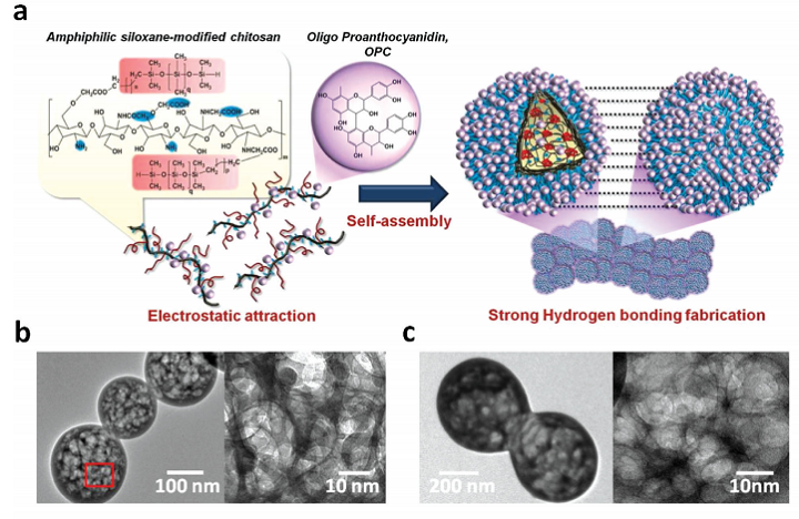
a) Schematic illustration of the mechanism for formation of nanogel-based membrane based on the self-assembly of OPC-incorporated amphiphilic polydimethylsiloxane-modified N, O-carboxylic chitosan (OPMSC), followed by hydrogel-bonding interaction of OPC. The TEM images display the network structure of b) PMSC and c) OPMSC spherical nanogels.

3D printing has been used in the past to help treat degenerative diseases, or at least make it easier to cope with them. In terms of neurodegenerative diseases, implanted prosthetic devices are often used, but adverse biological reactions in host tissues can result in signal failure. it’s important to create tissue that can mimic the mechanical and structural properties of neural implanted devices, and while flexible polymer-based implants have helped to alleviate some injuries, the mechanical stress doesn’t quite match brain tissue. That’s why a lot of research has been conducted about using conductive polymer (CP) composites or conductive hydrogels to coat the devices so the biocompatibility and electrochemical performance of neural electrodes can be improved.

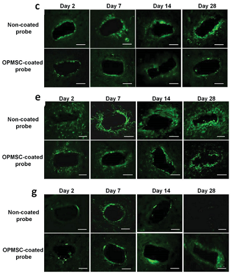
Representative fluorescent images demonstrate tissue responses around the tip of the non-coated probe and the OPMSC-coated probe at days 2, 7, 14, and 28 post-implantation. (c) ED1 staining; (e) GFAP staining; (g) NeuN staining.

But, a team of researchers from China and Taiwan say that it’s more important to design biocompatible coatings for implanted devices that mimic mechanical and structural properties of brain tissues, so tissue responses after long-term utilization can be reduced.

The researchers believe that 3D nanostructural coatings should be developed for the insulated regions, and not the implant electrode sites, so implants can interface with nearby brain tissues with more stability. They explained their findings in a recently published paper, titled “Multifunctional 3D Patternable Drug-Embedded Nanocarrier-Based Interfaces to Enhance Signal Recording and Reduce Neuron Degeneration in Neural Implantation.”

“Although the nanomaterial-based substrate coatings incorporated into drug delivery systems such as poly(lactic-co-glycolic acid) (PLGA) nanoparticles, pHEMA, or PLGA nanoparticles-embedded matrix have been developed, these systems lack stable physical and chemical properties for reducing tissue responses, including an appropriate nanostructural interface, mechanical properties, and biofouling ability,” the researchers wrote. “Multifunctional drug-embedded coatings must be developed and integrated into the nanostructural neural interfaces to allow sustained release of bioactive molecules (anti-inflammatory drugs) and simultaneous construction of a brain tissue-mimic but bioinert microenvironment for reducing both acute and chronic inflammation reactions during long-term implantation.”

The researchers used aerosol jet 3D printing to develop a neuronal interface with prolonged anti-inflammatory ability, structural and mechanical properties that mimicked brain tissue, and a sustained nonfouling property in order to inhibit tissue encapsulation.

Using aerosol jet printing, the OPMSC suspensions were directly patterned on a neural probe to create an anti-inflammatory neural interface.

“With the integration of nanomanufacturing technology and multifunctional nanomaterials into the neural implants, we can extensively reduce the reactive tissue responses, provide continuous protection of surviving neurons, and ensure long-term performance reliability of implants,” the researchers explained.

They created a new 3D nanocarrier-based neural interface that could possibly be used to support long-term neural implantation, as well as achieve better therapy for chronic and degenerative diseases. The researchers used a “novel combination of antioxidative zwitterionic nanocarriers and nanomanufacturing technology” to make the interface. The team developed a new type of anti-inflammatory nanogel, based on the amphiphilic polydimethylsiloxane-modified N, O-carboxylic chitosan (PMSC) incorporated with oligo-proanthocyanidin (OPC), called OPMSC.

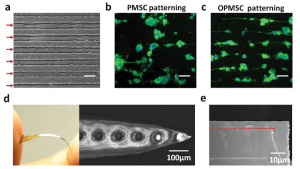
a) Optical microscopy image showing patterning morphology of PMSC and OPMSC arrays with a thickness of ≈30 µm obtained by aerosol jet printing. The red arrows indicate the patterned location. Comparison of PC12 cell patterning on b) PMSC and c) OPMSC arrays demonstrates that OPMSC can maintain structural stability in a biological microenvironment. d) An overview and SEM images of the flexible OPMSC-coated polyimide probe. e) SEM image showing a cross-sectional view of OPMSC-coated probe after washing with water.

“The natural OPC can be used as an anti-inflammatory drug due to its multipotent therapeutic effects on neurodegenerative diseases,” the researchers explained. “Furthermore, given the abundance of hydroxyl groups and the aromatic architecture, the semi-hydrophilic OPC can act as a structural stabilizer to help the self-adhesion of nanogels, making the structure evolve into a biostable 3D anti-inflammatory neural interface.”

The team directly fabricated OPMSC nanogels onto a membrane using aerosol jet printing technology, because it is a low-temperature technology. When developing neural implants, mechanical properties are the main concern, which is why the researchers conducted a tensile test, among other experiments, on their new 3D nanocarrier-based neural interface, which was also implanted into rodents.

“After short-term and long-term in vivo implantation, the OPMSC-coated neural probe displayed a relatively lower impedance value and much higher signal stability compared to noncoated probe,” the researchers concluded. “The ADC obtained by magnetic resonance imaging (MRI) demonstrated that the OPMCS-coated probe alleviated edema at the acute phase, and further reduced tissue trauma in the chronic phase. Immunostaining of anti-NeuN, anti-ED1, and anti-GFAP around the implanted site further demonstrated that the OPMSC-coated probe significantly reduced the population of activated microglia and astrocytes for all durations, resulting in increased survival 28 d after implantation. Such multifunctional nanostructured OPMSC-coated neural probes can provide a long-lasting functional neural interface for long-term neural implantation.”

Co-authors of the paper are Wei-Chen Huang, Hsin-Yi Lai, Li-Wei Kuo, Chia-Hsin Liao, Po-Hsieh Chang,Ta-Chung Liu, San-Yuan Chen, and You-Yin Chen.

Discuss this and other 3D printing topics at 3DPrintBoard.com or share your thoughts below.