3D Printing at University of Utah: Bioinspired Materials Managed with Ultrasound Directed Self-Assembly

In the recently published ‘Manufacturing bioinspired flexible materials using ultrasound directed self-assembly and 3D printing,’ researchers from the University of Utah are exploring a new process for engineering materials with innovative substructures that help tune mechanical properties.

Previous research has resulted in integrated microstructures brought on by nature’s designs for improved biological materials with improved properties overall. In the past, other research labs have used electrical properties with polymers, a variety of actuators, and more.

Hierarchical microstructures have been created via:

  • Stereolithography (SLA)
  • Traditional mold casting
  • Freeze casting

“Electric fields in combination with molding microwires from colloidal suspensions of metallic nanoparticles in water have also been used. These fabrication methods enable manufacturing of homogeneous composite materials and some have been combined with 3D printing, including electric field printing, shear force assisted 3D printing, and bioinspired 3D printing,” explain the researchers.

“However, these fabrication techniques place strict requirements on material choice, possible material geometry, and dimensional scalability, often requiring ultra-high field strengths. Thus, they may limit the potential properties and usefulness of the resulting engineered materials.”

In this study, the research team creates a new direct-writing process for 3D printing. Using a syringe, liquid feedstock is extruded, with ultrasound DW controlling the creation of the microstructure.

Ultrasound DW setup. Fibers are aligned by the acoustic radiation forcefield generated by the transducers before being extruded from the nozzle (shown in blue) and deposited on the print bed. P. Wadsworth et al. / Materials&Design 185 (2020) 1082432

The researchers used a Cartesian-style RepRap Guru 3D printer in this study, replacing the print head with a custom syringe designed in their lab, and meant for photopolymer resins. They created three syringes, all modified with custom nozzles.

“These custom nozzles were driven by a function generator and 45 dB RF amplifier (ENI 440LA, Rochester, NY, USA), to perform ultrasound directed self-assembly of microscale nickel-coated car-bon fibers (NCCFs) (100mm length, 10mm diameter, Conductive Composites, Heber City, UT, USA) dispersed in the photopolymer resin, into discontinuous line patterns,” explain the researchers.

In creating samples, feedstock was exposed to waves of:

  • MHz
  • 5 MHz
  • 0 MHz

Piezoelectric actuators with corresponding center frequencies were used with matching center frequencies. Along with evaluating mechanical properties and the effects of off-fiber loading, the research team created samples with 3 and 5 wt percent nickel-coated carbon fibers (NCCFs). The researchers noted that although 3D printing of multi-layer samples would have been possible, it was ‘beyond the scope’ of the research; however, six samples were printed for each ultrasound frequency, each at 1 wt percent NCCFs. Ultimately, there were 72 samples 3D printed.

(a) Custom fixture used to place needle probes during electrical conductivity measurements. (b) Diagram of a same line measurement for electrical conductivity.(c) Diagram of a neighboring line measurement for electrical conductivity.P. Wadsworth et al. / Materials&Design 185 (2020) 1082434

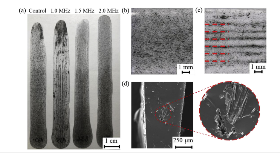
(a) Examples of the samples made with the ultrasound DW process. (b) Image of a printed control sample with randomly dispersed NCCFs throughout. (c) Image of a printed sample with linearly aligned NCCFs. (d) SEM image of a sample cross-section showing a linear feature concentration of NCCFs (in the expanded image).P. Wadsworth et al. / Materials&Design 185 (2020) 1082435

Single-layer materials were fabricated with aligned NCCFs after applying control via the ultrasound field, with the number of parallel lines for aligned NCCF’s figured by ultrasound frequency.

“No significant difference in any of the mechanical properties existed between samples with and without aligned NCCFs, including two separate moduli, ultimate tensile strength, and elongation at failure,” concluded the researchers. “The ultrasound DW technique created flexible single layer samples with integrated lines of aligned NCCFs inspired by biological and natural materials. Using electrically conductive fibers enabled sections of the engineered material to be electrically conductive and, therefore could potentially be used in shielding or other energy applications.”

The study of materials in 3D printing is growing to enormous proportions as researchers refine fabrication processes according to project needs, from exploring large-scale manufacturing solutions with composites to creating bioinks for tissue engineering, water-soluble filaments, and more.

What do you think of this news? Let us know your thoughts! Join the discussion of this and other 3D printing topics at 3DPrintBoard.com.

(a) Two typical conductivity test results (conductive or insulating). (b) Percentage of conductive test as a function of operating frequency and print speed.P. Wadsworth et al. / Materials&Design 185 (2020) 1082437

[Source / Images: ‘Manufacturing bioinspired flexible materials using ultrasound directed self-assembly and 3D printing’]

The post 3D Printing at University of Utah: Bioinspired Materials Managed with Ultrasound Directed Self-Assembly appeared first on 3DPrint.com | The Voice of 3D Printing / Additive Manufacturing.

New Study Discusses Unmet Clinical Needs Being Addressed by 3D Printing

3D printing continues to make a huge impact on the medical field – the evidence more than speaks for itself. But this important work is not done yet. A team of researchers based at the University of Utah recently published a review paper that, as Yong Lin Kong, PhD, an assistant professor in the university’s Department of Mechanical Engineering, told 3DPrint.com, “highlights the progress of 3D printing technologies in addressing unmet clinical needs.”

The paper, titled “Addressing Unmet Clinical Needs with 3D Printing Technologies” and published in Advanced Healthcare Materials, was written by Udayan Ghosh, Shen Ning with the Boston University School of Medicine, Yuzhu Wang, and Professor Kong.

A) 3D printed biomimetic bone environment for evaluating breast cancer bone metastasis; B) 3D printed network guide for regenerating damaged nerve plexuses; C) 3D printed titanium prosthetic for sternocostal reconstruction; D) An endothelialized myocardium by 3D printing endothelial cells encompassed within micro-fibrous hydrogel scaffolds; E) 3D printed personalized ocular prosthesis; F) Bionic ears; G) Hollow micrometer-scale microneedles; H) 3D printed pelvic implant.

The abstract reads, “Recent advances in 3D printing have enabled the creation of novel 3D constructs and devices with an unprecedented level of complexity, properties, and functionalities. In contrast to manufacturing techniques developed for mass production, 3D printing encompasses a broad class of fabrication technologies that can enable 1) the creation of highly customized and optimized 3D physical architectures from digital designs; 2) the synergistic integration of properties and functionalities of distinct classes of materials to create novel hybrid devices; and 3) a biocompatible fabrication approach that facilitates the creation and cointegration of biological constructs and systems. This progress report describes how these capabilities can potentially address a myriad of unmet clinical needs.”

The paper first looks at providing structural support for skeletal and tubular organs with 3D printed prosthetics in order to help people regain some of the functions they’ve lost, and then moves on to novel drug delivery strategies and organ-on-a-chip systems.

“Fourth, the developments of 3D-printed tissue and organ regeneration are explored,” the researchers explain in the paper. “Finally, the potential for seamless integration of engineered organs with active devices by leveraging the versatility of multimaterial 3D printing is envisioned.”

Society has been transformed by mass production, which allows parts to be manufactured at a far lower cost than hiring manual labor. However, that makes it difficult, and more expensive, to find customized products.

The researchers say in the paper, “Instead of optimizing for individual need and comfort, mass production manufacturing has compelled society to tolerate a finite set of prescribed designs determined by the overall market.”

Mass production doesn’t really address the complexity of the human body, and the majority of typical FDA-approved medical devices are not tailor-made to a patient’s specifications, which can many different issues that affect a person’s quality of life. But now, more and more physicians are investigating the use of 3D printing as it pertains to making cost-effective, customized devices.

“Indeed, 3D printing of biocompatible materials can create patient-specific prosthetics tailored to each patient’s unique anatomy and needs,” the researchers wrote.

3D printed prosthetics can help decrease discomfort, as they’re fitted to specific people, and at the same time are less expensive while also being more accessible. One specific, and very important, unmet clinical need that 3D printing can help with is creating functioning upper limb prostheses for children, so that they can grasp things in order to perform their basic daily activities. It’s hard to provide children with well-fitting prosthetics, as they grow so rapidly; that’s why a 3D printed version is a far better option.

Mock-up prototype of a microneedle array.

3D printed devices are also being used to help develop novel drug delivery strategies, from customized mouthguards and vaccines to microneedles and micro-rockets.

The researchers wrote, “Here, 3D printing enables the creation of unique architectures to allow painless delivery of therapeutic agents and tailored drug release profiles.”

Current strategies can be difficult when attempting to enable accurate drug delivery, but 3D printing has the potential to, as the paper puts it, “overcome these long- standing challenges.”

“3D printing introduced a potential opportunity for developing personalized, controlled, and precise drug delivery systems,” the researchers explained. “This technology achieves precise control of dosage in accordance with the size and dispensary mechanism of the design. Biocompatible material also allows for long-term implantation or retention while continuously dispensing controlled volumes with the potential to evolve into a highly efficient sensor-controlled drug dispensing system.”

3D printing is also being used now to address the unmet clinical need of the organ-on-a-chip platform, as it can summarize microenvironments in order to gain a more thorough understanding of cellular mechanics.

A) 3D printed in vitro human renal proximal tubules embedded within an extracellular matrix and housed in perfusable tissue chips. B) Customizable 3D printed nervous system-on-a-chip. The circular pattern of 3D printed silicone tri-microchannels designed for axonal guidance (L). A microscopy image shows three parallel microchannels of neurons and axons (green) in a chamber (R).

“Tissue/lab-on-a-chip, synonymous to biomedical application of microfluidics, is an advantageous and cost-effective way to investigate basic research questions. Analyzing fluids at the micrometer scale using microfluidic device holds immense promises for biological research,” the researchers said in the paper.

Ongoing research into tissues-on-a-chip is working to develop tissue chips that can act as accurate models for a specific organ’s function and structure, and 3D printing is the perfect technology for the job. Research also continues for the use of 3D bioprinting in tissue regeneration, as it can be used to create biocompatible constructs and 3D printed scaffolds to help regrow damaged tissues and organs, such as ears.

The researchers explained, “Bone tissue–engineered 3D constructs are more advantageous than 2D cell cultures due to the structure and mechanical composition 3D printing can produce to mimic the bone tissue microenvironment.”

Liver on a chip

Finally, the team touched on multimaterial 3D printing, which can help speed up “the creation of bioelectronic constructs to impart active functionalities to an otherwise passive construct.”

“The incorporation of electronics into biomedical devices and biological scaffolds is a foundational concept, which when applied, can mimic and even augment the complex functionalities of biological systems,” the researchers continued.

By integrating medical instruments with electronics, we can develop sophisticated new bioelectronic devices that are actually able to process feedback from the human body. The level of integration demonstrated by conventional fabrication techniques is rather limited, but using 3D printing to achieve these devices opens up far more possibilities – even, as the researchers explain, “the ability to mimic or surpass complex functionalities intrinsic to biological organs.”

“To date, demonstrations of a seamless bioelectronics 3D printing have been limited to passive electronic components, such as conductive traces and capacitors,” the researchers explained. “The integration of active electronic devices could impart an otherwise passive construct with optical, sensing, and computational capabilities.

“We anticipate that similar approaches can develop 3D printing strategies of various classes of active electronics. Nevertheless, the biocompatibility of such approach must be critically assessed to ascertain a full translational result from the bench to the bedside.

There’s a lot to think about here, but one thing is certain – the research into how we can address a myriad of unmet clinical needs with 3D printing should continue.

Discuss this story and other 3D printing topics at 3DPrintBoard.com or share your thoughts in the Facebook comments below.