An Inside Look into the ACES Lab (Part I)

A leading scientist in the field of electromaterials and one of Australia’s visionary bioprinting enthusiasts Gordon Wallace took audiences through a virtual tour into the cutting edge research labs at the ARC Centre of Excellence for Electromaterials Science (ACES), where next-generation materials research and advanced engineering for the development of customized bioinks and bioprinters take place. Located within the heart of the Intelligent Polymer Research Institute (IPRI) at Australia’s University of Wollongong (UOW) Innovation Campus, ACES turns fundamental knowledge into the next generation of smart devices to improve people’s lives and deal with some of the great challenges of the century.

With his usual enthusiasm, Wallace engaged audiences as he presented fellow researchers at work and some of the new innovations, discoveries and development of new materials for use in the field of biofabrication. During the first part of the tour, he explores the development of Graphene, 3D printed stents, and cell preparation for bioprinting. For the second part of the tour (found in a separate article), Wallace walks into another building at UOW where the recently inaugurated Translational Research Initiative for Cell Engineering and Printing (TRICEP) is leading the initiative for 3D bioprinting encompassing bioink, bioprinter, and bioprinting process developments, including the manufacturing of medical devices and the integration of living cells delivered using customized bioprinters to address specific medical challenges.

“Here at ACES we are known for our fundamental work into the discovery and the development of new materials, that can be used in energy and medical bionics,” said Wallace. “We are using the most advanced methods of fabrication to develop protocols that will enable structures and devices to be created so that we can take those fundamental advances and use them in important areas.”

Starting with the basics, Wallace first explores a lab setting where Sanjeev Gambhir, a Senior Research Fellow at the Australian National Fabrication Facility (ANFF) of the University of Wollongong, develops graphene, a material he refers to as “wondrous”, with “amazing properties for the nanoworld that we have been able to extricate into the micro and macroscopic realms to realize applications.” 

“To create a graphene-polymer composite synthesis, we modify the chemistries of graphene (which is derived from graphite, a naturally occurring mineral) so that we retain all the amazing mechanical, electric and biological properties and yet make it processible, that is, to turn it into structures and devices, using 3D printing, and eventually making it scalable,” said Gambhir.

Wallace added that “it is important that all the chemistries we use are actually scalable.” He claims that it is very different doing chemistry on a bench from processing graphene into tens of grams and managing to retain the same properties and quality as they were getting on the laboratory scale. It is all part of his vision to really make the process ready for industrial-scale manufacturing.

To show how graphene is turned into fibers for easier handling, Wallace takes audiences to the Fibre Spinning Electrodes area, where researcher Javad Foroughi, “weaves the magic” to create graphene fibers, that can even be combined with biomaterials to coat the surface of the fiber.

Working on customized 3D printed stents was Ali Jeirani, a Product Designer Development Specialist at UOW. It is one of the many processes where he uses 3D printing and takes advantage of all of the advances in material synthesis and processability at ACES, by turning them into real structures. 

“One of the important parts about the properties of a stent for applications is the design. We use G-code to create different designs and then send them to our machine to print different structures and properties,” explained Jeirani. “One of the problems of commercial stents is that they cannot be personalized for the patient, so by using 3D printing, we can customize it according to the scan of the patient. We understand that there can be very complicated stent shapes that are readily realized with 3D printing.”

According to Wallace, the graphene is often blended with other materials to improve the properties of the part, and by using small amounts of graphene and blending it with a polymer, they can create the stent. The innovative material gives the stent extra mechanical properties and could even impart electrical properties into it, which the two experts consider “one of the most interesting properties of graphene for electro stimulation”. 

“This is all made possible thanks to additive fabrication and advances in 3D printing, so it is an exciting time, since we can turn fundamental discoveries into really practical and useful structures almost immediately by working together, us at the 3D fabrication lab and our colleagues at materials processes,” continued Jeirani.

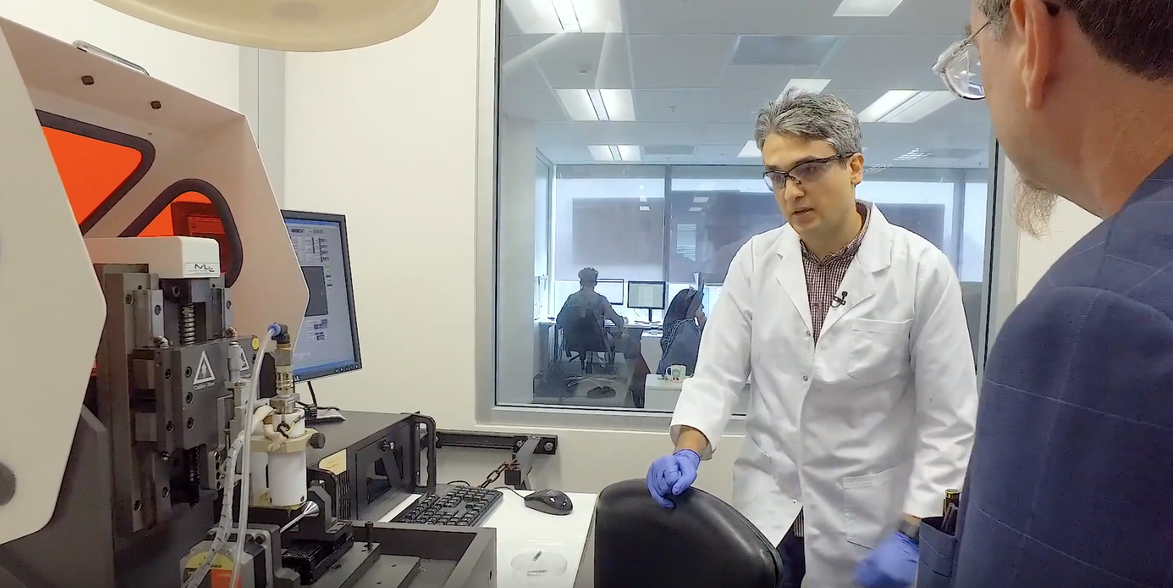
Gordon Wallace and Ali Jeirani looking into how to fabricate 3D printed stents

After delving into advancements in biomaterials and graphene, Wallace headed upstairs to the cell lab where Research Fellow at ACES, Eva Tomaskovic-Crook, revealed another important part of their work: the integration of living cells into printing protocols, which basically entails how scientists prepare the cells for printing.

They have several environments ready for the cells, from storing them in liquid nitrogen sample storage tanks–they have at least two Taylor Wharton LS750– to incubating them, which offer an environment where they nurture cells and provide the right growth conditions to expand. Incubators have a warm 37-degree environment ideal for maintaining cell growth. 

“Quality control of our cells is very important. We need to be sure that the cells maintain the ability to be pluripotent (pluripotent stem cells have the ability to undergo self-renewal and to give rise to all cells of the tissues of the body). We want to scale up the number of cells and to encapsulate them in the biomaterial.” suggested Tomaskovic-Crook.

Scaling up the number of cells is crucial because when they go into the bioprinting process they want to create a three-dimensional tissue with a high cell to biomaterial mass, not just have a few cells. According to the specialist, “it involves a process of going back and forth: scaling up the cells at the lab, then printing them, and bringing them back to the lab to interrogate the cells and see if they are still living, proliferating and turning into the cells we want them to.” 

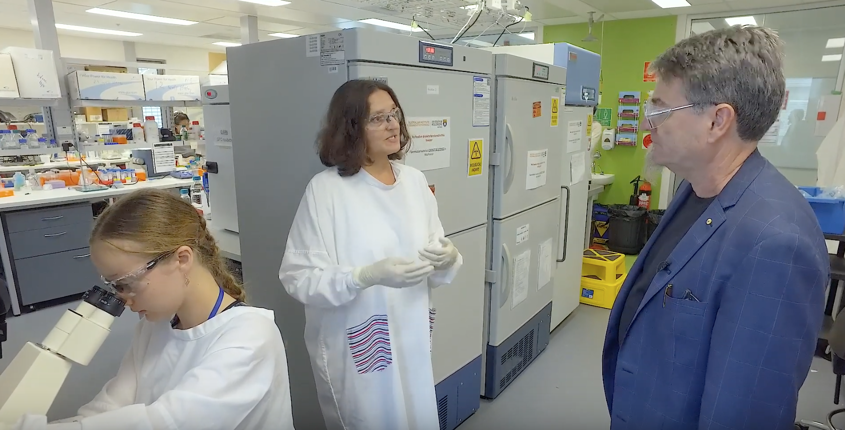
Gordon Wallace and Eva Tomaskovic-Crook talking about preparing cells for bioprinting 

Known for their expertise in advanced materials and device fabrication, ACES incorporates collaborators from across Australia and the world. ACES is generating options for the future, so being able to peek into some of the advanced materials and device fabrication for game-changing health and energy solutions is a privilege. Not only did Wallace explain some of the most breakthrough research in biomedicine, but he also showed viewers the machines that researchers work with on a daily basis. Wallace tends to emphasize that a big part of the Australian bioprinting community is about sharing research, insights, and knowledge to advance the field. The unique landscape of the country, with its cultural and linguistic diversity as well as residence to scientists from around the globe, makes it ideal for ideas and creativity to emerge.

You can tune in to see the first part of the virtual lab tour here.

The post An Inside Look into the ACES Lab (Part I) appeared first on 3DPrint.com | The Voice of 3D Printing / Additive Manufacturing.

Wipro 3D Launches Addwize to Accelerate Adoption of Metal 3D Printing

wipro addwize logoWipro 3D, the AM subsidiary of Wipro Infrastructure Engineering, recently launched an additive technology adoption and acceleration program called Addwize. One of the current hurdles to the adoption of metal AM is the learning curve of both the hardware and the new ecosystem. In our article on the post-processing of metal 3D prints, we discussed […]

Tis the season for Lightsabers! Builds by @chinbeard @sophywong and @KjDesigns365

This week there were awesome lightsaber builds released this week by Bill Doran, Sophy Wong and Kirby Downey. Check out their build videos on YouTube. Both feature custom designs that are based off the Adafruit PropMaker FeatherWing and Circuit Python.

Wanna build your own? Check out the learn guides for thorough tutorials on how to wire, code and assembly your own lightsaber. And may the force be with you!

Learn Guide: 3W LED Saber Build

Learn Guide: NeoPixel Saber Build

Learn Guide: HalloWing Ligtsaber

We recently released a new video on building our 3W LED propmaker lightsaber on YouTube. The learn guide was published back in June, 2019.

Exploring Exotic 3D Printing Materials Used in Prosthetics

Those familiar with 3D printing materials know that polylactic acid (PLA) is an extremely popular material. It’s strong, inexpensive, and easy to print with. PLA is is great for a wide range of applications, but there are other material options out there as well. Nylon is known for its toughness, but it is notoriously difficult to print successfully. Other plastics like polyvinyl alcohol (PVA) can be dissolved in water, making it the perfect choice for support material because it can be so easily removed.

In the prosthetics industry, 3D printing is starting to become a viable manufacturing process. Since a high level of quality is required, there are some situations where PLA and other common materials don’t quite cut it. Prosthetics and bionics companies are looking at using more exotic materials so that they can improve their products.

Above all else, prosthetic limbs must be lightweight and comfortable. If a prosthetic is powered either electrically or mechanically, it is considered a bionic prosthetic. It is beneficial for bionic products to be as simple as possible, so flexible parts may be desired to reduce the complexity. Bionic prosthetics can also feature sensory capabilities for user-feedback, so electronics may be required. All of these design criteria can be achieved by using special materials that do a little bit more than just your average PLA.

Carbon Fiber Reinforced Filament

Carbon fiber reinforced (CFR) filament is a 3D printing material that contains short strands of carbon. It is stiffer than most filaments, and it is a great material choice if weight and rigidity need to be optimized. One such application is in the socket for prosthetic legs. The socket is the part that connects the person’s leg to their prosthesis. People with leg amputations need their prosthesis to be both lightweight and rigid, and CFR filament fits both of these criteria.

This material consists of short carbon strands suspended in a plastic such as PLA or Nylon. These plastics are all classified as thermoplastics, which means they can be remelted. This material property can be quite beneficial. Typically, an amputee’s residual limb will change shape slightly over the course of months or years. This can lead to discomfort if their socket does not change shape. If modifications to the socket need to be performed, CFR filament can simply be heated up to soften it and then reshaped.

Carbon fiber composite material is different from CFR filament. It consists of woven carbon sheets glued together with epoxy. It does not soften with heat. This is because epoxy is classified as a thermoset polymer. That means it undergoes a chemical reaction as it cures which causes it to permanently harden.

Prosthetic sockets made from carbon fiber composite are in fact stronger than 3D printed carbon fiber material but they are expensive, hard to manufacture, and difficult to re-shape. This is why some prostheses are now made by 3D printing with CFR filament. With a 3D scan of the amputee’s residual limb, a socket can be 3D printed which very accurately captures every detail. This makes for a more comfortable fit. 3D printing a socket is much quicker and also less expensive than the traditional method of creating a prosthetic socket.

Flexible Filament

Unlike most 3D printing materials, thermoplastic polyurethane (TPU) is soft and flexible. This material is perfect for creating flexible joints, and it sees use in applications like prosthetic fingers and as soft liners for prosthetic sockets.

Prosthetic sockets which are made from rigid materials (such as CFR filament) can become uncomfortable if pressure is not evenly distributed. Introducing a soft inner liner can provide cushioning and support, improving comfort for the user. Because 3D printing can create complex shapes, a mesh-like structure can be printed which allows airflow throughout the socket. This ventilation is very necessary because moisture build-up can cause discomfort.

TPU is also being used in bionic hands as a material for flexible fingers. Instead of using a rigid mechanism, using flexible and compliant mechanisms to transfer forces can result in a more natural motion. Using flexible materials in compliant mechanisms reduces the number of parts, removes the need for lubrication, and greatly speeds up the assembly and manufacturing process.

Conductive Filament

3D printing is usually used to produce mechanical components, but certain filaments are electrically conductive and can be used in a variety of interesting electronics applications. Magalie Darnis (M.Eng), made this the topic of their master’s thesis. Magalie used a material known as ETPU to create 3D printed sensors.

ETPU combines carbon powder and TPU to develop a flexible and electrically conductive polymer that can be 3D printed. Although ETPU contains carbon, it is very different from CFR filament. This is because it uses graphene powder instead of short carbon fibers. Graphene easily conducts electricity, but it does not add much mechanical strength. Other types of conductive filaments exist, but they are rigid and sometimes brittle. In other applications, this may be desired, but for bionics, the flexibility that comes with ETPU allows for flexible, form-fitting sensors to be embedded in wearable products.

Currently, 3D printed sensors are only found in bionics prototypes, but ETPU has proven to be effective in applications such as touch sensors in bionic fingertips. To create a touch sensor, two ETPU surfaces are printed with a small air gap between them. These surfaces will move closer together when pressure is applied to the fingertip. When these surfaces make contact, it closes a circuit, and this signal can be used to let the user know when they’ve firmly grasped an object.

This binary (on/off) touch sensor is one of the most basic 3D printed sensors, and it can be modified to make more complex sensors such as deformation sensors, vibration sensors, and force sensors.

One of the main benefits of 3D printing sensors is that it simplifies and speeds up production. With 3D printed sensors, pre-built components do not need to be manually attached to an object. The sensors can instead be part of the printing process itself.

Want to find out how Shapeways can help fulfill your medical industry needs? Contact our team today to get a personalized consultation.

The post Exploring Exotic 3D Printing Materials Used in Prosthetics appeared first on Shapeways Magazine.

Chinese University of Hong Kong Studies 3D Printing for Heart Disease

In the recently published ‘Three-dimensional printing in structural heart disease and intervention,’ authors Yiting Fan, Randolph H.L. Wong, and Alex Pui-Wai Lee, all from The Chinese University of Hong Kong, explore the potential for 3D printing in the world of medicine, as well as cardiology—and more specifically, structural heart disease (SHD).

SHD causes issues like:

  • Aortic stenosis
  • Mitral regurgitation
  • Atrial septal defect
  • Left atrial appendage (LAA) clots

Conventional imaging is limited, while the emergence of 3D printed models allows medical professionals to progress from mentally reconstructing 2D images to gaining a more complex understanding of pathology.

As 3D printing continues to make its way into the realm of medicine, models are used for:

  • Guiding treatment
  • Procedural simulation
  • Facilitating hemodynamic research
  • Improving interventional training
  • Promoting patient-clinician communication

To create a medical model, images must be attained, data must be processed, and the object must be 3D printed.

“The most commonly used imaging sources for SHD are echocardiography, computed tomography (CT) and magnetic resonance imaging (MRI),” state the researchers. “Other modalities, such as positron emission tomography, single photon emission CT and cone beam CT, are less commonly used. All images should use the common Digital Imaging and Communication in Medicine (DICOM) format.

Pre-procedural simulation of MitraClip on 3D-printed model. (A) Digital model of a heart. Different colors stand for different cardiac components (grey: mitral valves; light pink: tricuspid valves; light gold: atrial septum, left atrium, left and right ventricle). (B) Multi-material 3D-printed heart model for pre-procedural simulation. The valves were printed with flexible material and the rests were printed with hard material. (C) The 5 holes drilled in the atrial septum represents the different position for different kinds of structural heart interventions. (D) The MiraClip device was released via delivery catheter through the atrial septum to the mitral valve. Blue circle: MitraClip; red circle: left atrial appendage occlusion (LAAO). S, superior; A, anterior; P, posterior; I, inferior; IVC, inferior vena cava; LAA, left atrial appendage; MV, mitral valve.

A range of materials are both popular and possible for fabricating medical models:

“Multi-material printing by material jetting is increasingly used to create cardiac structures. Different tissue components were printed with different textures. For instance, an aortic valve was printed with flexible printing material, and the calcifications attached to valves were printed with hard printing material, respectively,” report the authors.

Application of 3D printing for peri-device leak. (A,B,C) A case found with peri-device leak post TAVI and needed peri-device leak occlusion: (A) routine TEE post-TAVI showed peri-device leak (yellow circle); (B) simulation of peri-device leak occlusion on 3D-printed aortic root model derived from post-TAVI CT; (C) the 10-mm vascular plug was found to be best-fit for this case. (D,E,F,G) A case found with residual leak after ASD closure: (D) multi-material 3D printed model showed residual leak (blue circle) next to the ASD occluder (asterisk *); (E) the delivery catheter went through the leak position; (F) the device (two asterisks **) was released in situ. (G) The bicaval view of 3D-printed model showed stable release and stay of the chosen device. 3D, three-dimensional; TAVI, transcatheter aortic valve implantation; TEE, transesophageal echo; CT, computed tomography; ASD, atrial septal defect.

There continue to be ongoing challenges in the creation of medical models, however, ‘despite the enthusiasm in applying 3D printing cardiovascular medicine.’ While there is an obvious lack of technical standards, mainly due to the novelty of the technology, the authors point out also that there are still issues with affordability—along with ‘scant evidence on the added clinical benefit.’

Greater accuracy is needed, along with improved standardization of data acquisition, and post-processing techniques. While deeper research is required into the creation and use of models and surgical guides, so are comparisons for offering up better information and creating industry standards. The authors also recommended a more streamlined workflow.

“The mechanical properties of the 3D-printed materials, such as tensile strength, elasticity, flexibility, hardness, and durability have utmost importance for cardiovascular applications. The majority of cardiovascular applications reported so far have employed materials with properties that have not been meticulously compared with the cardiovascular tissue they are mimicking. Validation of 3D-printed material properties against actual human patient tissues is important to ensure that procedural simulation is realistic,” conclude the authors.

“Further effort in technical standardization, and clinical evaluation of added benefit and cost-effectiveness of 3D printing are needed to bring this promising technique to clinical reality.”

3D printed medical models are extremely beneficial to doctors and patients as they allow not only for diagnosing but have also continued to change medicine—allowing for procedures involving complex reconstructions, fabrication of surgical guides, and much more.

What do you think of this news? Let us know your thoughts! Join the discussion of this and other 3D printing topics at 3DPrintBoard.com.

[Source / Images: ‘Three-dimensional printing in structural heart disease and intervention’]

The post Chinese University of Hong Kong Studies 3D Printing for Heart Disease appeared first on 3DPrint.com | The Voice of 3D Printing / Additive Manufacturing.

Engineering fashion: GE Additive engineers on the haute-couture of 3D printing

In May this year, 3D printing made its red carpet debut. The vision of New York fashion designer Zac Posen, several unique fashion pieces caused a stir at the 2019 Met Gala. Worn by British supermodel Jourdan Dunn and Canadian actor Nina Dobrev, arguably the most iconic pieces in Posen’s collection were the so-called “rose gown,” […]

3D Printing Microstructures for New Drug Delivery Systems with SPHRINT

In the recently published, ‘SPHRINT – Printing Drug Delivery Microspheres from Polymeric Melts,’ authors Tal Shpigel, Almog Uziel, and Dan Y. Lewitus explore better ways to offer sustained release pharmaceuticals via 3D printed structures.

Currently, numerous materials are used to create microparticles for sustained drug release, with numerous choices in biodegradable materials, such as include poly(lactic acid) (PLA), polyglycolic acid (PGA), their copolymer poly(lactic-co-glycolic) acid (PLGA) and polycaprolactone.

“Numerous microparticle-based depots products are FDA approved, amongst them are: Zmax® (Azithromycin), Decapeptyl®/Trelstar® (Triptorelin), Vivitrol® (Naltrexone), Arestin® (Minocycline), Risperdal® Consta® (Risperidone), Sandostatin® LAR Depot (Octreotide), Nutropin Depot® (Somatropin), Lupron Depot® (Leuprolide), DepoCyt® (Cytarabine), DepoDur® (Morphine), Bydureon® (Exenatide) Somatuline LA (Lanreotide) [1] and recently approved ZILRETTA™ (triamcinolone acetonide),” state the authors.

Frames obtained from high-speed imaging capturing the evolution of the shape of a molten 30% IBU-PCL blend interacting with either a superoleophobic
surface (top) and preserving its spherical shape after 1.8 s or an aluminum surface (bottom), in which the droplet gradually flattens (See SI Video 4 and SI Video 5,
respectively). Scale bar: 500 µm. These frames emphasis the significance of using non-wetting surfaces responsible for the formation of spherical droplets.

With the advent of 3D printing and inkjet technology, researchers have experimented with numerous techniques. Many have encountered obstacles though, and challenges regarding methods relying on both inkjet printing and those that are solvent based. Because of that, the researchers created an affordable, yet solvent-free technique for fabricating polymer melts in this study—showing the capability of their SPHRINT technique.

(A) Optical images of (i) neat PCL (154 ± 3°), (ii) 30% IBU-PCL (171 ± 4°), (iii) neat PLGA (167 ± 6°), and (iv) 30% IBU-PLGA (169 ± 4°) microspheres, cooled at room temperature (RT). The values in brackets denote the “sphericity,” expressed as the contact angle values (mean ± S.E.M., n = 10). Scale bar: 200 µm. (B) SEM images of (i) 30% IBU-PCL and (ii) 30% IBU-PLGA microspheres, cooled at RT. Scale bars: 100 µm

Producing drug delivery microspheres from a polymer loaded with a sample drug like ibuprofen, the researchers experimented and conducted an analysis regarding the potential for fabrication of amorphous polymeric microspheres. The researchers evaluated microsphere size, morphology, and texture. Ultimately, they were able to produce ‘near-perfect microspheres.’

“We discovered intricate physical phenomena governing the mechanism of sphere-formation; beside process and performance efficiencies, which in turn render microsphere products more accessible,” stated the researchers. “SPHRINT printing eliminates the use of organic solvents and surfactants; it offers microspheres with reproducible size, shape, and morphology within and between the batches; and the produced microspheres can be easily collected owing to their spherical shape.”

Jetting rate and shear rate were calculated, along with an investigation of melt interaction with the superoleophobic substrate and sphere formation.

In terms of drug encapsulation efficiency, they found that values in connection with SPHRINT were on the highest scale. The authors were encouraged to find ‘stable, consistent, reproducible results.’

“… we believe that SPHRINT may turn microsphere production ubiquitous, allowing for desktop manufacturing of microspheres easily scalable to industrial quantities (with a production rate of 25 Hz, in 1 h, 4.7 g of PCL microspheres may be printed from a single printing head). Finally, SPHRINT may provide a new dimension in reservoir-injectable drug delivery technologies, enabling the employment of multifarious polymers for microsphere production and tuned release profiles,” concluded the researchers.

3D printing has been used in connection with a variety of different drug delivery systems, from experimenting with microreservoirs to hydrophilic matrices, and even spermbots. What do you think of this news? Let us know your thoughts; join the discussion of this and other 3D printing topics at 3DPrintBoard.com.

[Source / Images: ‘SPHRINT – Printing Drug Delivery Microspheres from Polymeric Melts’]

The post 3D Printing Microstructures for New Drug Delivery Systems with SPHRINT appeared first on 3DPrint.com | The Voice of 3D Printing / Additive Manufacturing.

Voxel8 Lands New CEO to Lead Multi-Material 3D Printing

After its co-founder, Harvard professor Jennifer Lewis, moved to a supervisory role, Voxel8 has found a new CEO. Friedrich von Gottberg will lead the Boston startup as President and CEO. 

Uppers 3D-printed by Voxel8’s Active Lab solution.

Voxel8 first made a splash at CES in 2015 with a desktop electronics 3D printer, dubbed the Developer’s Kit, capable of both printing plastic and depositing conductive inks. The company wowed attendees with the ability to 3D print a complete, functioning quad copter that could fly right off the print bed. 

The printer was meant to aid Voxel8 in discovering a killer app for the multi-material printing technology being developed in the lab of Jennifer Lewis. The lab has been involved in research that has included lightweight composites, stretchable sensors, solar panels and organic matter. Much of these achievements are dependent on a unique pneumatic printhead that lays at the heart of Voxel8’s printing process. 

Between then and now, the startup sold its desktop electronics 3D printer to the likes of Google ATAP, worked with such partners as the MITRE Corporation, and took on investors that included the CIA, via In-Q-Tel. In the process, the company switched gears, discontinuing the Developer’s Kit and launching the Active Lab, which deploys the material dispensing technology to 3D print plastic onto fabric, along with an inkjet head that provides a full range of color. The killer app, Voxel8 hopes, is athletic wear, specifically shoe uppers. To drive these efforts, the startup received funding from DSM and HP Tech Ventures, as well as existing investors ARCH Venture Partners and Braemar Energy Ventures.

Along the way, Jennifer Lewis shifted from her CEO role to an advisory position so that she could focus on her work at Harvard. Meanwhile, her co-founder, Travis Busbee, took over as CEO as the firm looked for Lewis’s replacement. 

Friedrich von Gottberg, President & CEO at Voxel8, Inc.

Prior to joining Voxel8, von Gottberg was with Cabot Corporation, where worked for 20 years. His last positions at Cabot were as senior vice president and president of Purification Solutions, before which he served as vice president and general manager of New Business Segments, as well as vice president and global director of R&D. Cabot is a large, publicly traded chemicals company known for its work with carbon and aerogel, among other materials. While its environmental record has not always been so hot (though it has made progress on this front), it manufactures activated carbon, used to remove pollutants from water, air and more.

The chemical company also makes elastomeric composites, colorants, and inks, fumed metal oxides and other materials, meaning that von Gottberg’s experience with these chemistries, along with his contacts in the industry, will serve Voxel8 in its mission to deliver mass production multi-material 3D printing. Meanwhile, Travis Busbee will return to his position as Chief Technology Officer. 

The post Voxel8 Lands New CEO to Lead Multi-Material 3D Printing appeared first on 3DPrint.com | The Voice of 3D Printing / Additive Manufacturing.