
Cardiovascular disease such as hypertension and others are frequently caused by hardened blood vessels, and finding a way to replace those vessels has been a challenge in the past. But researchers from the University of Colorado Boulder have developed a 3D printing technique that allows for localized control of an object’s firmness, which could open up new possibilities for the 3D printing tissue. One future possible application of this technology is in the creation of artificial arteries and organ tissue. The research is documented in a paper entitled “Orthoganal programming of heterogeneous micro-mechano-environments and geometries in three-dimensional bio-stereolithography.”
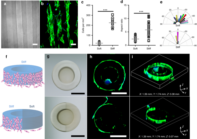
The research features a layer-by-layer printing method with fine-grain, programmable control over rigidity, which allows the researchers to mimic the complex geometry of highly structured yet pliable blood vessels.
“The idea was to add independent mechanical properties to 3D structures that can mimic the body’s natural tissue,” said Xiaobo Yin, an associate professor in CU Boulder’s Department of Mechanical Engineering and the senior author of the study. “This technology allows us to create microstructures that can be customized for disease models.”
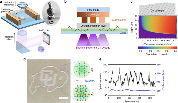
To overcome the traditional challenges involved in engineering blood vessels, the researchers found a way to take advantage of oxygen’s role in setting the final form of a 3D printed structure.
“Oxygen is usually a bad thing in that it causes incomplete curing,” said Yonghui Ding, a postdoctoral researcher in Mechanical Engineering and the lead author of the study. “Here, we utilize a layer that allows a fixed rate of oxygen permeation.”
By keeping tight control over oxygen migration and its subsequent light exposure, the researchers can control which areas of an object are solidified to be harder or softer, while keeping the overall geometry the same.
“This is a profound development and an encouraging first step toward our goal of creating structures that function like a healthy cell should function,” Ding said.
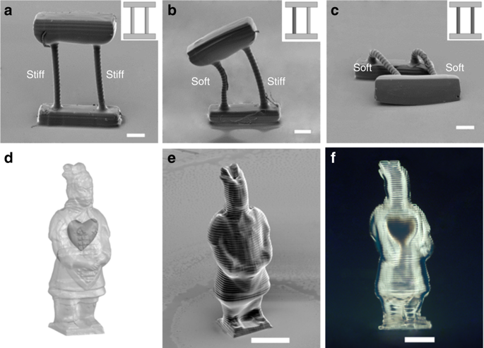
The researchers demonstrated their technique by 3D printing three different versions of a simple structure: a top beam supported by two rods. Each structure was identical in shape, size and materials, but varied in rod rigidity: soft/soft, hard/soft and hard/hard. The hard rods supported the top beam while the soft rods allowed it to collapse. The researchers then repeated the exercise with a small Chinese warrior figure, making the outside hard but the inside soft.
The 3D printer used by the researchers is capable of printing biomaterials as small as 10 microns, or one-tenth the width of a human hair. The researchers believe that they can further improve their technique with future work.
“The challenge is to create an even finer scale for the chemical reactions,” said Yin. “But we see tremendous opportunity ahead for this technology and the potential for artificial tissue fabrication.”
One in every four deaths in the United States is caused by heart disease, totaling over 600,000 deaths per year. It’s the leading cause of death for both men and women, but the discovery of a way to 3D print healthy blood vessels could make a tremendous difference. Having said that there are many other tissue printing technologies that are making strides. Tunable geometries and gradient materials are already possible with other technologies and in other materials in bioprinting. We’re saddened that so many media have misreported this story and overstated the claims as expressed by the researchers in their work. This paper gives us a great new path forward to tissue engineering but the media should be careful to read the papers that they write about. For SLA and tissue engineering this is a good step forward but it is wrong to speculate so widely and make unassociated claims especially when the researchers in question express themselves so succinctly and clearly in the paper.
Authors of the study include Hang Yin, Yonghui Ding, Yao Zhai, Wei Tan and Xiaobo Yin.
Discuss this and other 3D printing topics at 3DPrintBoard.com or share your thoughts below.
57 Replies to “Tunable Bioprinted Tissue Using SLA May Lead to 3D Printed Tissue Engineering Using This Method”
Comments are closed.