Continuous Chaotic 3D Printing: Using Chaos Theory to Control Microstructures

Researchers continue to experiment with materials and extrusion techniques, introducing the use of chaotic flows for improved microarchitectures in the recent study, ‘‘Using chaotic advection for facile high-throughput fabrication of ordered multilayer micro- and nanostructures: continuous chaotic printing.’

The authors present a unique form of fabrication with a method that allows for more precise control of the structure and surface area being printed through the use of what they call ‘continuous chaotic printing.’ The process relies on a combination of chaos theory and fluid dynamics, in which the flow of material develops into complex fractals. To achieve this, the team created chaotic material flows by reorienting and splitting fluid as it is mixed, as shown in the figure below.

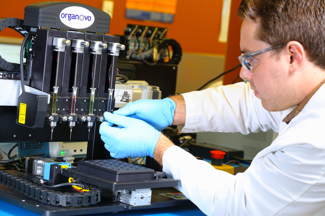
With a focus on multiple materials, multiple layers, and fabrication of varying cells in proximity to each other on larger surfaces, their goal was to test the potential for chaotic printing in a variety of applications, but with a special focus on bioprinting and biomedical research. Building on previous research with chaotic flow, the authors experimented with a chaotic printer (paired with a Kenics static mixer), fabricating fibers made from alginate.

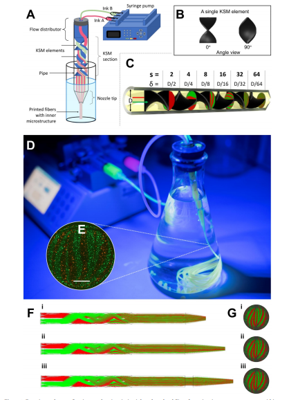
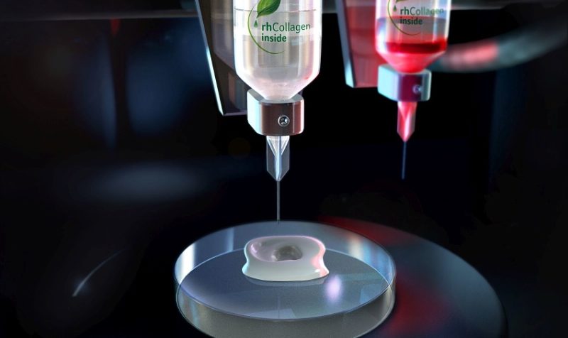
Experimental setup. Continuous chaotic printing is based on the ability of a static mixer to create structure within a fluid. The Kenics static mixer (KSM) induces a chaotic flow by a repeated process of reorientation and splitting of fluid as it passes through the mixing elements. (A) Schematic representation of a KSM with two inlets on the lid. The inks are fed at a constant rate through the inlets using syringe pumps. The inks flow across the static mixer to produce a lamellar structure at the outlet. The inks are crosslinked at the exit of the KSM to stabilize the structure. Our KSM design includes a cap with 2 inlet ports, a straight non-mixing section that keeps the ink injections independent, a mixing section containing one or more mixing elements, and a nozzle tip. The lid can be adapted to inject several inks simultaneously. (B) Two rotated views at 0◦ and 90◦, of a single KSM element. (C) 3D design of a KSM with 6 elements and schematic representation of the flow splitting action, the increase in the number of striations, and the reduction in length scales, in a KSM-printhead. The resolution, namely the number of lamellae and the distance between them (δ), can be tuned using different numbers of KSM elements. (D) Actual continuous chaotic printing in operation. The inset (E) shows the inner lamellar structure formed at the cross-section of the printed fiber (the use of 4 KSM elements originates 16 striations). Scale bar: 250 µm. (F) Longitudinal or (G) cross-sectional microstructure of fiber obtained using different tip nozzle geometries. Images show CFD results of particle tracking experiments where two different inks containing red or green particles are coextruded through a printhead containing 4 KSM elements. The lamellar structure is preserved when the outlet diameter is reduced, from 4 mm (inner diameter of the pipe section) to 2 mm (inner diameter of the tip), through tips differing in their reduction slope.

An unlimited number of inks can be used in chaotic printing, but for this study, the researchers used basic techniques, with sodium alginate as a base. Experiments were then performed with composite inks featuring suspensions like polymer microparticles, graphite microparticles, mammalian cells, or bacteria. Ultimately, the team reported they were able to print ‘fine and well-aligned microstructures’ at high extrusion speeds of 1–5 m of fiber/min.

“This printing strategy is also robust across a wide range of operation settings. We conducted a series of printing experiments at different inlet flow rates to assess the stability of the printing process. As long as the flow regime is laminar and the fluid behaves in a Newtonian manner, the quality of the printing process is not affected by the flow rate used in a wide range of flow conditions,” explained the authors.

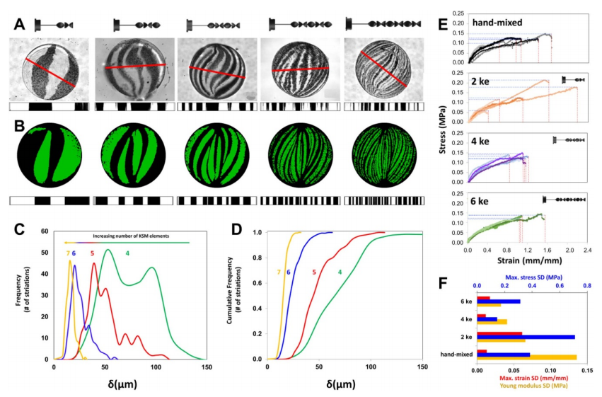
Evaluation of the striation profiles and mechanical properties of chaotically printed alginate/graphite fibers. (A) Lamellar microstructure of fibers produced with printheads containing 2, 3, 4, 5, or 6 KSM elements. The thickness of each lamella, along the red line, was determined by image analysis using Image J (shown below each cross-sectional cut). Scale bar (red): 2 mm. (B) The microstructure at each cross-section was reproduced by CFD simulations, and the thickness and position of each lamella was calculated. (C) Striation Thickness Distribution (STD) and (D) cumulative STD for constructs printed using 4, 5, 6, and 7 KSM elements. (E) Comparison of stress-strain curves of fibers fabricated by extrusion of pristine alginate and graphite without chaotic mixing (marked as hand-mixed) or with chaotic printing using 2, 4, or 6 KSM elements (marked as 2, 4, or 6 ke). (F) Comparison of the standard deviation of tensile properties (i.e. maximum stress, maximum strain, and Young’s modulus for the same set of fibers; 5 fibers per treatment).

The researchers also noted the following while printing:

  • Stable fibers were printed using a cone-shaped nozzle tip with an outlet diameter of 1 mm, with flow rates ranging from 0.003 to 5.0 ml min−1.
  • Printheads with varying geometries did not affect structures being printed.
  • Computational fluid dynamics (CFD) showed that the inclination of the nozzle tip also did not affect the materials.

Continuing to emphasize the ‘robust’ qualities of chaotic printing—especially when used with small nozzles—the researchers were able to control resolution, noting also that, due to the deterministic qualities of chaotic flows, fabrication of structures was ‘fully predictable.’  Resolution was in part illustrated by the number of gill-like ridges, or ‘lamellae,’ created through the chaotic mixing process.

“As the number of elements used to print increased, the number of lamellae observed in any given cross-sectional plane of the fiber also increased, while the thickness of each lamella decreased,” explained the researchers. “Therefore, users of continuous chaotic printing will have more degrees of freedom to determine the multi-scale resolution of a construct, as this is no longer mainly restricted by the diameter of the nozzle (or the smallest length-scale of the nozzle at cross-section).”

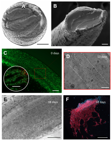
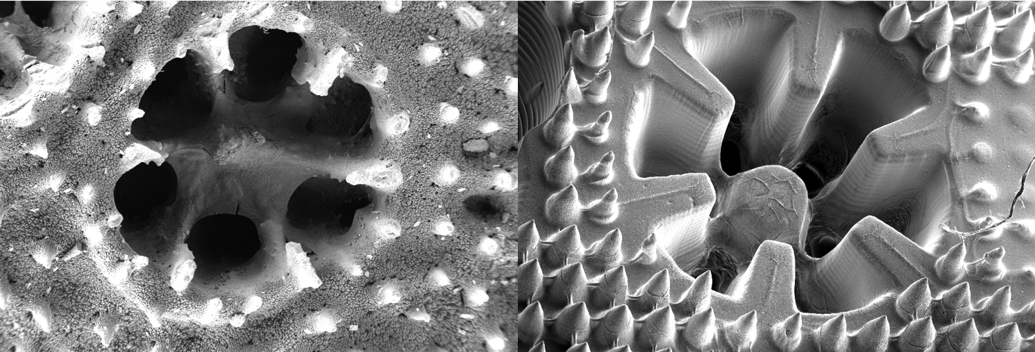
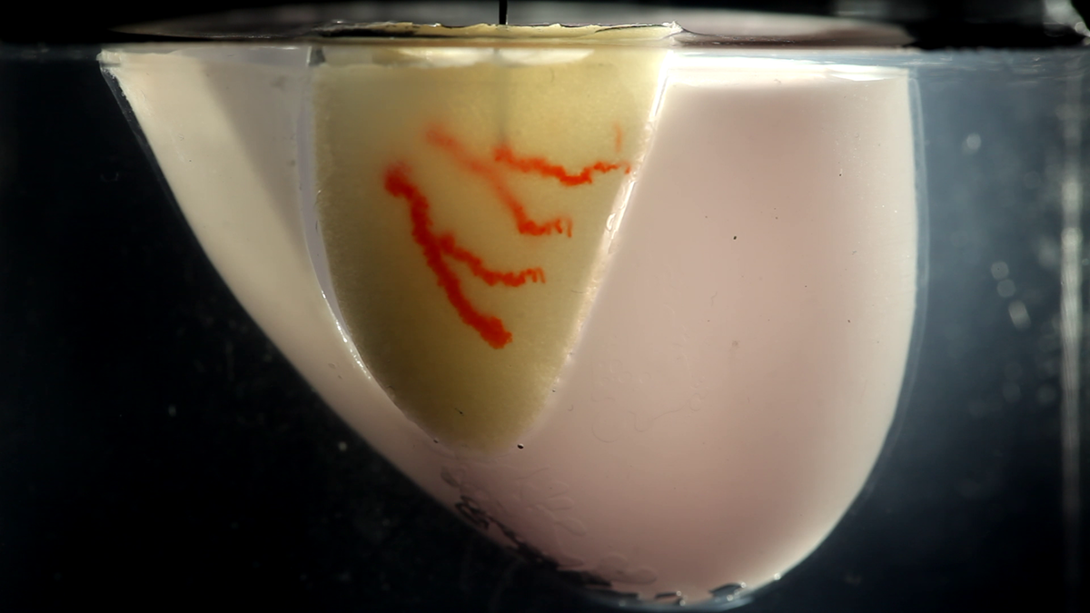
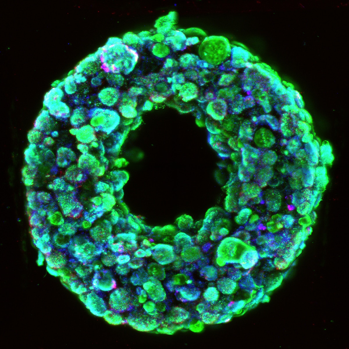
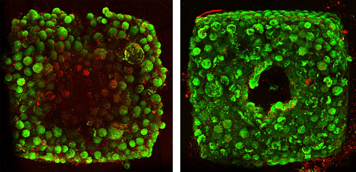
Bioprinting of living micro-tissues: (A) Optical and (B) SEM micrographs of the cross-sectional view of a construct in which C2C12 cells are chaotically bioprinted in an alginate/GelMA hydrogel using a 3-KSM printhead; Scale bars: 500 µm and 50 µm, respectively. (C) Longitudinal view of a chaotically bioprinted construct; a high cell viability is observed at the initial time, as revealed by a live/dead staining and fluorescence microscopy. Scale bar: 500 µm. Inset shows a cross-sectional cut. Scale bar: 500 µm. (D) Cells spread along the chaotically printed striations, preserving their original positions after 13 d of culture. Scale bar: 200 µm. (E) Optical microscopy view of a segment of fiber containing C2C12 cells 18 d after printing. Scale bar: 500 µm. (F) Close-up of a region stained to reveal F-actin/nuclei, showing the cell spreading and the formation of interacting cell clusters. Cell nuclei can be identified as blue dots. Actin filaments appear in red. Scale bar: 200 µm.

Further, as the researchers began experimenting with bioprinting, they were able to create alginate fibers rich with cells, and lightly enriched with protein to encourage ongoing sustainability. This is one of the greatest challenges in tissue engineering. Even though the correct materials, techniques, and concepts may be in place, if researchers cannot keep the cells alive long enough, they must go back to the drawing board.

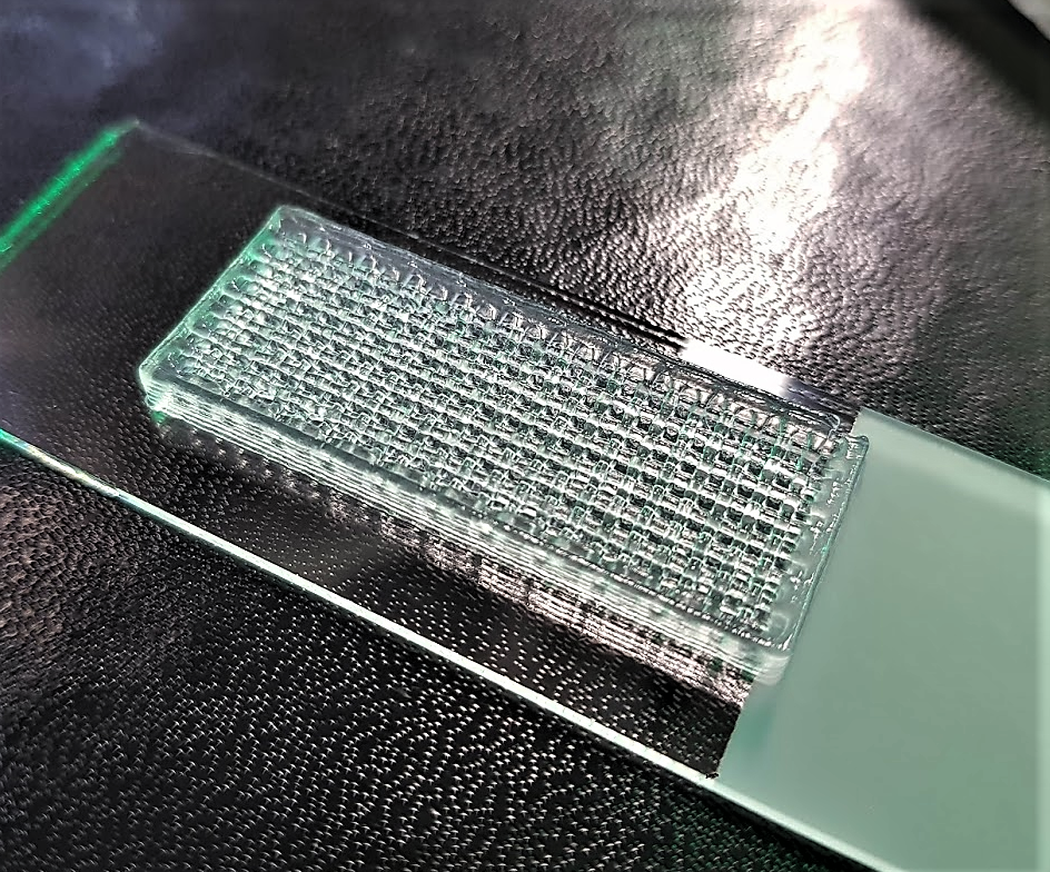
Altogether, the team suggests that it has developed a completely novel way to control the resolution of an extrusion-based printing system. As shown in the image below, not only can this continuous chaotic printing process control print resolution through the diameter of the nozzle, but at the microscale as well through the chaotic mixing techniques deployed.

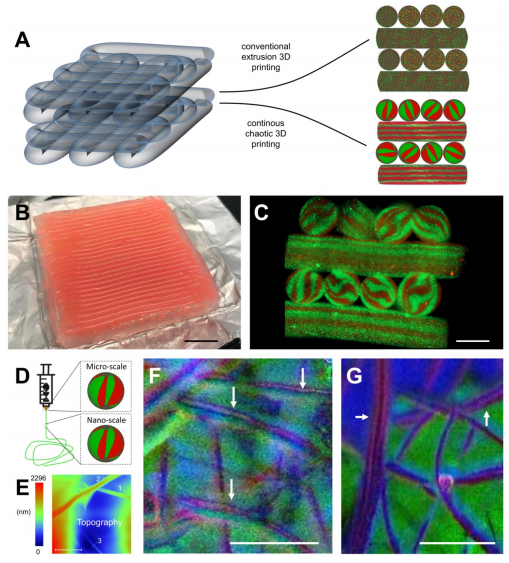
Development of multi-scale architectures based on 3D continuous chaotic printing: (A)–(C) 3D printing of hydrogel constructs using a KSM-printhead integrated to a commercial cartesian 3D printer. (A) Schematic comparison of the lack (prepared using conventional extrusion techniques) and presence of internal lamellar microstructures (developed using continuous chaotic printing). (B) Printing of a long fiber arranged into a macro-scale hydrogel construct (3 cm × 3 cm × 4 mm). Scale bar: 5 mm. (C) Transverse cut of the macro-construct showing the internal microstructures. Scale bar: 1 mm. (D)–(G) Chaotic printing of fibers coupled with electrospinning. (D) Schematic representation of the coupling between continuous chaotic printing and an electrospinning platform; an ink composed of a pristine alginate ink (4% sodium alginate in water) and an ink composed of a polyethylene oxide blend (7% polyethylene oxide in water), were coextruded through a chaotic printhead and electrospun into a nanomesh. (E) AFM image showing the diameter of three individual nanofibers ((1) 0.82 µm, (2) 1.05 µm, and (3) 0.437 µm) within the electrospun mesh. Scale bar: 5 µm. (F), (G) photo-induced force microscopy (PiFM) reveals the lamellar nature of the nanostructure within a nanofiber (white arrows) originated using (F) a 2-element KSM printhead, and (G) a 3-element KSM printhead. Scale bar: 1 µm.

The researchers noted that not only could this have a profound impact on multimaterial printing technologies, but on bioprinting as well:

“Our results demonstrate the unrivaled ability of chaotic printing to deploy cells within high SAV fibers. As available bioprinting and bioassembly technologies approach the resolution and SAV of chaotic printing, they also tend to require long fabrication times and mechatronically coordinated control systems,” concluded the researchers. “In addition to multicellular, high SAV constructs, chaotic printing offers other breakthroughs in regards to currently available multimaterial printing technologies that, typically, require optimized inks that must be deployed under a specific and narrow range of conditions.”

[Source / Images: ‘Using chaotic advection for facile high-throughput fabrication of ordered multilayer micro- and nanostructures: continuous chaotic printing’]


The post Continuous Chaotic 3D Printing: Using Chaos Theory to Control Microstructures appeared first on 3DPrint.com | The Voice of 3D Printing / Additive Manufacturing.

3D Printing Webinar & Virtual Event Roundup, May 31, 2020

With so many events going virtual due to the ongoing COVID-19 pandemic, there’s also been an increase in the number of webinars that companies in the additive manufacturing industry are holding. To make things easier for our readers, since there’s so much online content to choose from these days, 3DPrint.com is compiling all of these available webinars, and the virtual events, into a weekly roundup for you, starting today.

Freeman Technology Webinar

Characterization Tools for Evaluating Polymer Powders for Laser Sintering Webinar

This Tuesday, June 2nd, UK-based Freeman Technology, a Micromeritics company that creates systems for measuring the flow properties of powder materials, will host a webinar at 9 am ET titled “Characterization Tools for Evaluating Polymer Powders for Laser Sintering.” Enrico Gallino, Senior Engineer – Material Specialist at Ricoh UK Products Ltd, will speak about evaluating an AM powder characterization methodology, and will also discuss the results of screening the relevant properties, such as flowability, shape, and thermal properties, of a variety of materials.

“As additive manufacturing (AM) technology transitions from the fabrication of prototypes to serial production of end-use parts, the understanding of the powder properties needed to reliably produce parts of acceptable quality becomes critical,” the webinar site states.

“Achieving the optimal quality for parts does not only depend on setting the right process parameters. Material feedstock also plays an important role when aiming for high performance products. In the case of selective laser sintering, polymer powders are used as a raw material. Therefore, controlling the quality and correctly characterizing the particles used in the process is a key step to successfully apply polymer AM techniques and also to expand the range of material that can be process with this technology.”

Click here to register.

Dassault Systèmes Webinar

Dassault Systèmes be will holding a live webinar on Thursday, June 4th at 10 am ET, titled “Intuitive 3D Designs with CATIA® and SOLIDWORKS® on Mobile Devices.” Participants will have the chance to learn how beneficial flexible design workflows can be when delivering products to market, faster, across many different industries. There will be a live demonstration, using tablets and PCs, on how combining CATIA and SOLIDWORKS on the 3DEXPERIENCE platform will allow your business to add engineering details with simple parametric modeling, create organic surfaces with subdivision (Sub-D) modeling, generate complex patterns and shapes quickly, optimize and evolve designs using an algorithmic approach, and more – all from your own device. The demonstration will be followed by a live Q&A session.

“Discover our portfolio of ready-to-go online Design and Engineering applications in action, which enable you to design from your laptop, your smartphone or tablet! Enjoy increased agility without compromising best-in-class design and engineering capabilities,” the webinar site states.

“With its growing app portfolio and secure cloud technology, the 3DEXPERIENCE platform enables you to manage all facets of your product development process while reducing infrastructure costs, IT overhead, software maintenance and complexity. All 3DEXPERIENCE solutions work together seamlessly making data management, sharing and collaboration easy.”

Click here to register.

3DHEALS 2020 Global Summit

The 3DHEALS conference is going virtual this year, as the 3DHEALS 2020 Global Summit runs from 11 am-9:30 pm ET June 5th and 6th. Offering powerful networking and effective programming on a global stage, this popular bioprinting conference – sponsored by Whova and Zoom – brings together influencers and audiences from over nine countries, offering opportunities and insights that can be beneficial to stakeholders. With over 70 speakers, more than four workshops, startup events, simulated in-conference experience, an interview series hosted by Dr. Jenny Chen, and more, this is one you won’t want to miss.

“3DHEALS2020 is designed to cater to a wide range of professionals, ranging from healthcare early adopter, manufacturers, engineers, legal professionals and policymakers, C-Level executives, entrepreneurs, investors, and more. We aim to create an effective program that maximizes the attendee’s experiences and decreases the barriers in communication among stakeholders,” the event site states.

Click here to register.

Will you attend these events and webinars, or have news to share about future ones? Let us know! Discuss this and other 3D printing topics at 3DPrintBoard.com or share your thoughts in the comments below.

The post 3D Printing Webinar & Virtual Event Roundup, May 31, 2020 appeared first on 3DPrint.com | The Voice of 3D Printing / Additive Manufacturing.

Marine Biologist Modifies Bioprinting for the Creation of Bionic Coral

Corals are dying globally. In the face of climate change and global warming, we can expect some severe consequences, which in turn directly affects marine life. In what is panning out to be a mass extinction event, coral reefs have been dangerously threatened by toxic substances and excess carbon dioxide for years, causing the certain death of may of these diverse marine invertebrates. Once the coral is dead, the reefs will also die and erode, destroying important marine life, that would otherwise feed and spawn on it.

Considering that scientists have predicted that nearly all coral reefs will disappear in 20 years, it is crucial that we protect corals and learn from them. For the expanding field of biotechnology, untapped resources like corals hold great potential, as bioactive compounds for cancer research or simply as an inspiration for the production of bioenergy and bioproducts.

In an interview with 3DPrint.com, interdisciplinary marine biologist Daniel Wangpraseurt, from the University of California San Diego (UCSD)’s Department of NanoEngineering, explained how bioprinting technology was a pivotal point in his work to develop bionic 3D printed corals as a new tool for coral-inspired biomaterials that can be used in algal biotechnology, coral reef conservation and in coral-algal symbiosis research. 

“For many years I have been studying how corals optimize light management and discovered that there are lots of interesting evolutionary tricks, such as different growth forms and material properties, so I became interested in copying these strategies and developing artificial materials that could host living microalgae, just like corals do in nature,” revealed Wangpraseurt.

Daniel Wangpraseurt

As one of the most productive ecosystems globally, coral reefs use photosynthesis to convert carbon dioxide into energy that they in turn use for food. Even though light provides the energy that fuels reef productivity, key nutrients such as nitrogen and phosphorus are also required, but are found in very low quantities in warm tropical oceans where coral reefs are generally found, making scientists wonder how these marine animals have managed to create a competitive habitat with such limited resources.

A laser beam is intensely scattered by elastic coral tissue and aragonite skeleton. (Credit: Daniel Wangpraseurt)

Wangpraseurt described that, while different corals have developed a plethora of geometries to achieve such capabilities, they are all characterized by an animal tissue-hosting microalgae, built upon a calcium carbonate skeleton that serves as mechanical support and as a scattering medium to optimize light delivery toward otherwise shaded algal-containing tissues.

“Taking what we learned about corals and biomaterials, we began working on a project to develop a synthetic, symbiotic system using a 3D bioprinting approach. We know corals have both animal cells and algal cells, and, so far, we have mimicked the animal part of the corals, that is, the physical and chemical microhabitat that partially controls the activity of the algal cells.”

At UCSD, Wangpraseurt expects to continue recreating coral-inspired photosynthetic biomaterial structures using a new bioprinting technique and a customized 3D bioprinter capable of mimicking functional and structural traits of the coral-algal symbiosis. Along with fellow researchers from UCSD, the University of Cambridge, the University of Copenhagen and the University of Technology Sydney, and thanks to a grant from the European Union’s Horizon 2020 research and innovation program, and the National Institutes of Health (NIH), the team reported the results of their work on bioinspired materials that was published in the journal Nature Communications earlier this year.

“We want to go further and not just develop similar physical microhabitat but also modulate cellular interactions, by mimicking biochemical pathways of symbiosis. We hope that this allows us to not only optimize photosynthesis and cell growth, but also to gain a deeper understanding of how the symbiosis works in nature. By doing so, we can improve our understanding of stress phenomena such as coral bleaching, which is largely responsible for global coral death.”

Living colonies of Symbiodinium are visible within the 3D bioprinted tissues (Credit: Daniel Wangpraseurt)

So, how did bioprinters become the go-to technology for this project? Wangpraseurt explains that, while working as a researcher at the University of Cambridge’s Department of Chemistry Bio-Inspired Photonics lab, he noticed that scientists were using cellulose as a biomaterial with interesting optical responses. He was wondering how he could use cellulose to develop a material with very defined architectural complexity. 

“In the beginning, the main aim was to develop a coral-inspired biomaterial, that has a similar optical response as natural coral, and then to grow algae on it or within it. Thereby, we started off with simple techniques, using conventional 3D printers; however, it wasn’t very easy to recreate the spatial resolution we needed for corals.”

Inspired by 3D bioprinting research in the medical sciences, Wangpraseurt reached out to scientists at the UCSD NanoEngineering lab that were developing artificial liver models, and who later became collaborators in the project.

A laser beam is intensely scattered by elastic coral tissue and aragonite skeleton (Credit: Daniel Wangpraseurt)

The team went on to develop a 3D printing platform that mimics morphological features of living coral tissue and the underlying skeleton with micron resolution, including their optical and mechanical properties. It uses a two-step continuous light projection-based approach for multilayer 3D bioprinting and the artificial coral tissue constructs are fabricated with a novel bioink solution, in which the symbiotic microalgae are mixed with a photopolymerizable gelatin-methacrylate (GelMA) hydrogel and cellulose-derived nanocrystals (CNC). Similarly, the artificial skeleton is 3D printed with a polyethylene glycol diacrylate-based polymer (PEGDA).

Close up of coral polyps and living photosynthetic biomaterials. Living colonies of Symbiodinium are visible within the 3D bioprinted tissues (Credit: Daniel Wangpraseurt)

Based in San Diego, Wangpraseurt has spent months trying to recreate the intricate structure of the corals with a distinguished symbiotic system that is known to grow as it creates one of the largest ecosystems on the planet. 

“We used a 3D bioprinter that had been developed for medical purposes, which we modulated and further developed a specific bioink for corals. A lot of the work was related to the optimization of the material properties to ensure cell viability. Having the right bioink for our algal strains was crucial as if we were to use mixtures commonly used for human cell cultures, the cells will not grow very well and can die rapidly.”

The implications of the newly developed 3D printed bionic corals capable of growing microalgae are many. Wangpraseurt said he plans to continue working on bionic corals and potentially scale up the process for his startup, called mantaz, as well as for commercial properties; or to develop coral-inspired materials at a larger scale to have a more immediate impact on efforts related to coral reef restoration, and also for biotechnology.

SEM images of the skeleton structure of the coral Stylophora pistillata and the coral-inspired, 3D-printed material (Credit: Daniel Wangpraseurt)

Wangpraseurt is looking to scale the bioprinting system to have a more immediate impact on algae biotechnology, bioenergy, and bioproducts. He claims that he and his colleagues can “customize the environment of the algae and fine-tune the production of a certain bioproduct to potentially tap into the algae bioproduct market and scale the system for bioenergy production.” 

“Another interest of mine is to further develop a 3D bioprinted synthetic coral-algal symbiosis system, which can provide important insight into the mechanisms that lead to coral death, but can also result in the development of future technology for coral reef restoration.”

The researcher talks about coral reefs with a reverent passion that today goes beyond his lab work. When he is not moving the research along at USCD, Wangpraseurt is working with his social enterprise in Panama, as he and his team try to restore coral reef ecosystems to help coastal communities in the tropics, including local fishermen, by harvesting algae biomass that can be sold for different purposes, such as natural fertilizer, which contributes to an organic and sustainable chain of production. Furthermore, the coral-inspired aspects of Wangpraseurt’s research and startup company are really coalescing to enable him and his team to understand how corals work and, in turn, how we can learn from them for the benefit of our planet.

Nutrient sampling at a polluted reef in Panama (Image: Daniel Wangpraseurt)

The post Marine Biologist Modifies Bioprinting for the Creation of Bionic Coral appeared first on 3DPrint.com | The Voice of 3D Printing / Additive Manufacturing.

Aalto University Develops a Novel Bioink for Cardiac Tissue Applications

Finland is one of Europe’s most forested nations. Over 70 percent of the country’s boreal forest is covered with spruce, pine, downy birch, and silver birch. But beyond the splendor of the Finnish woodlands, all these trees have one thing in common, and that is nanocellulose. A light solid substance obtained from plant matter which comprises cellulose nanofibrils (CNF) and is considered a pseudo-plastic that possesses the property of specific kinds of gels that are generally thick in normal conditions. Overall, it is a very environmentally friendly and non-toxic substance that is compatible with the human body and has the potential to be used for a range of medical applications.

In 2018, the Department of Bioproducts and Biosystems at Aalto University, located just outside Helsinki, began searching for new ideas to revitalize one of the country’s traditional economic engines, forests (which are handled sustainably thanks to renewable forest resources). At the time, they noticed that one of the possible applications could be working with nanocellulose. Forward two years and the researchers have come up with a new bioink formulation praising nanocellulose at its basis.

Thanks to the structural similarity to extracellular matrices and excellent biocompatibility of supporting crucial cellular activities, nanocellulose-based bioprinting has clearly emerged for its potential in tissue engineering and regenerative medicine. The qualities of the generally thick and fluid light substance make it an excellent match to develop bioinks that are both suitable and scalable in their production, but also have consistent properties. However, there have been major challenges in processing nanocellulose.

As described by Aalto University researchers in a recently published paper in the science journal ACS Publication, the unresolved challenges of bioink formulations based on nanocelluloses are what stops the substance from becoming one of the preferred components for 3D bioprinting structures. This is why Finnish researchers focused on developing a single-component bioink that could be used to create scaffolds with potential applications in cardiac biomedical devices, while fundamentally dealing with some of the limitations of using nanocellulose-based bioinks.

A co-author of the paper and a doctoral candidate at Aalto’s Department of Bioproducts and Biosystems, Rubina Ajdary, told 3DPrint.com that “other than natural abundance and as a renewable resource, nanocellulose has demonstrated to have an outstanding performance in tissue engineering.” She also suggested that “recent efforts usually consider the use of nanocellulose in combination with other biopolymers, for example, in multicomponent ink formulations or to encapsulate nanoparticles. But we were interested in investigating the potential of monocomponent nanocellulose 3D printed scaffolds that did not require crosslinking to develop the strength or solidity.”

In fact, the Biobased Colloids and Materials (BiCMat) research group at Aalto University, led by Orlando Rojas, proposed heterogeneous acetylation of wood fibers to ease their deconstruction into acetylated nanocellulose (AceCNF). As a unique biomaterial opportunity in 3D scaffold applications, the team considered using nanocelluloses due to the natural, easy to sterilize, and high stability porosity of the substance, and chose to introduce AceCNF for the generation of 3D printed scaffolds for implantation in the human body. The team then went on to evaluate the interactions of the scaffolds with cardiac myoblast cells.
“Most modifications make the hydrogels susceptible to dimensional instability after 3D printing, for instance, upon drying or wetting. This is exacerbated if the inks are highly diluted, which is typical of nanocellulose suspensions, forming gels at low concentrations,” went on Ajdary. “This instability is one of the main reasons why nanocellulose is mainly combined with other compounds. Instead, in this research, we propose heterogeneous acetylation of wood fibers to ease their deconstruction into acetylated nanocellulose for direct ink writing. A higher surface charge of acetylated nanocellulose, compared to native nanocellulose, reduces aggregation and favors the retention of the structure after extrusion even in significantly less concentration.”
This is exactly why it was important for the researches to develop a single component bioink. Nanocellulose has shown promises when combined with other biopolymers and particles. However, Ajdary insists that benefits including similarity to the extracellular matrix, high porosity, high swelling capacity, ease of surface modification, and shear thinning behavior of cellulose, encouraged them to study the potential of monocomponent surface-modified nanocelluloses.

Acetylated nanocellulose (Credit: Aalto University School of Chemical Engineering)

The team at Aalto University used the sustainable and widely available nanocelluloses to make several formulations of bioinks and evaluate them, including unmodified nanocellulose CNF, Acetylated CNF (AceCNF), and TEMPO-oxidized CNF.
To 3D bioprint the hydrogels, researchers used Cellink bioprinters, something Ajdary attributed to the user-friendliness of the device and because it provided a lot of flexibility to test different types of hydrogels and emulsions produced in the research group.
In this new process, the single-component nanocellulose inks were first 3D printed into scaffolds using Cellink’s BIO X bioprinter, which is equipped with a pneumatic print head was used to extrude single filaments and form the 3D structures. Then freeze-dried to avoid extensive shrinkage, and sterilized under UV light. After sterilization the scaffold was ready and cells seeded on the samples.
“3D structures of acetylated nanocellulose are highly stable after extrusion in far less concentrations. The lower concentration in wet condition facilitates the scaffold with higher porosity after dehydration which can improve the cell penetration in the structure and assist in nutrient transport to the cells as well as in the transport of metabolic waste,” specified Ajdary.
The researchers claim that the method was successful as the 3D printed scaffolds were compatible with the cardiomyoblast cells, enabling their proliferation and attachment, and revealing that the constructs are not toxic. Although still in research stages, these bioinks and technique can be used for the inexpensive, consistent fabrication and storage of constructs that can be applied as base materials for cardiac regeneration.
What is novel in this study is the particular focus on single-component nanocellulose-based bioinks that open up a possibility for the reliable and scale-up fabrication of scaffolds appropriate for studies on cellular processes and for tissue engineering. Since this is an ongoing research, we can expect to read more published material from Aalto University researchers as they continue testing their unique technique even further.

Scaffolds corresponding to 3D printed AceCNF (Credit: Aalto University)

The post Aalto University Develops a Novel Bioink for Cardiac Tissue Applications appeared first on 3DPrint.com | The Voice of 3D Printing / Additive Manufacturing.

Former CEO of Organovo Urges Stockholders Against Merger with Tarveda

Gaining a competitive advantage is at the core of most businesses. That is why the financial world has a section dedicated exclusively to study the behind-the-scenes of mergers and acquisitions (M&A). Between 1893 and 1904, the first wave of M&As became commonly known as the “great merger movement” in the US business scene, particularly the manufacturing sector. We’ve come a long way since then, and the two words became quite consolidated among financial experts and economic gurus as they decisively found M&As as a way to increase market shares, eliminate the competition or increase business diversification. However, contrary to what most people think, and according to a Harvard Business Review report, the failure rate for M&As is between 70% and 90%. And a KPMG study particularly determined that 83% of merger deals did not boost shareholder returns. Meaning they are not the best choice for most companies. That could be the case with bioprinting pioneer Organovo.

Last January, the company, announced a merger with Tarveda Therapeutics, a privately-held clinical-stage biopharmaceutical firm. However, at the time, the founder and former Chief Executive Officer (CEO) of Organovo, Keith Murphy, spoke against joining forces with Tarveda as part of a strategic alternative in the development of its human liver tissue for transplant. And just this Monday, Murphy decided to issue a new letter to stockholders discouraging them to vote in favor of the proposed merger with Tarveda, alleging that the firm’s financial position is “dismal” as well as the company’s “lack of synergies” and “highly questionable future prospects,” further reinforcing why a merger would be a terrible outcome for Organovo stockholders.

Under the terms of the original deal, announced in the company’s annual report published on December of last year, Tarveda would merge with a wholly-owned subsidiary of Organovo in an all-stock transaction and upon completion of the merger, the merged company would operate under the name Tarveda Therapeutics and trade on the Nasdaq Stock Market under the ticker symbol TVDA. Without any mention of Organovo’s bioprinting technology, we were left to wonder at the time whether the company’s pioneer technology and research activities would cease to exist.

Just like in January, this new letter provides details of what Murphy considers is wrong with Tarveda, even describing it as a company that “possesses unattractive technology that needs a bailout, via a reverse merger, just to survive.”

Murphy’s insistence on stockholders to vote “against” the merger is quite valid since the actual merger is still subject to customary closing conditions, which means that the merger still requires the approval of stockholders. Last December, Organovo directives suspected that the merger would be completed sometime in late 2020, however, stockholders may be persuaded either way in this waiting game.

Although personally attached to the biotechnology firm, Murphy resolves to take this drastic measure in order to speak directly to stakeholders, describing, not only, Tarveda’s “weak financial position with unexciting technology and unattractive prospects”, but also recognizing that Organovo is today “in its own precarious position due to recent mismanagement.” Concluding that he firmly believes that “the transaction being irrationally championed by the Board runs completely counter to stockholders’ best interests.”

Researcher working at the Organovo lab (Image: Organovo)

Bioprinting startups are emerging, and researchers’ interest in technology worldwide is helping many firms compete to develop better machines or upgrade previously successful ones. Organovo was one of the big driving forces behind bioprinting technology in the 2000s. However, recently, other companies have proven to be more successful, while Organovo’s stock price has been depressed for the past five years, going from USD 5.04 on June 6, 2015, to USD 0.27 on March 25, 2020.

On this last point, Murphy argues in his letter that “Organovo’s stock has been a relatively safer harbor, trending with the NASDAQ overall. Considering Organovo has natural downside protection due to being valued near its book value, I believe that waiting—and not consummating a reckless merger—may be the best course until the current storm has passed [that is, the novel coronavirus that began in December 2019].” While suggesting that, in contrast, “Tarveda is a company in need of additional capital beyond Organovo’s current cash in order to fund clinical trials to a successful point.”

In April 2017 Murphy stepped down as CEO of Organovo to create a new company, Viscient Biosciences, to develop drugs using human 3D bioprinted tissues. Nonetheless, he remained close to Organovo as chairman of the board and company advisor, even proposing a merger at one point between Viscient and Organovo to unlock 3D bioprinting potential.

In fact, Murphy maintains in his letter that “my assessment is unaffected by the Board’s decision to not pursue a merger with Viscient Biosciences (“Viscient”). While I firmly believe that the Board failed to create value for stockholders when it rebuffed what could have been a very synergistic merger with Viscient, I am fully aligned with you and recognize that there are many other viable combination partners out there. However, Tarveda is by no means an attractive deal partner.”

A few other reasons Murphy found discouraging against the merger involve Tarveda’s loss of its longtime Chief Scientific Officer (CSO), who was also President of Research & Development, in 2019, which he considered “hardly the sign of a strong technology.” As well as Tarveda’s downward trajectory following a prior record of poor investments, and that the company was running out of money in late 2019, with no venture capital funds or public market investors apparently eager to invest.

Massachusetts-based company Tarveda has been focused on developing Pentarin, a precision oncology medicine that selectively accumulates anti-cancer payloads within solid tumors. The company claims that following the closing of the merger, they intend to continue to focus on advancing two clinical-stage oncology programs, PEN-866 and PEN-221, and on the further development of novel conjugates from its proprietary miniature drug conjugate platform.Moreover, at the closing of the merger, they estimated that the combined company will have approximately 35 million dollars of cash on hand that is expected to provide sufficient funding into the second half of 2021 to achieve key upcoming clinical data milestones on both clinical programs.

Nonetheless, towards the end of the letter, Murphy offers three other options that he considers superior to the Tarveda transaction:

  1. The Board can abandon the merger to re-focus on organic growth via bioprinting.
  2. The Board should reconstitute its membership so that if the Board does not see the value of Organovo’s bioprinting opportunity following an abandoned or defeated merger, they should step down.
  3. The Board can run an improved Strategic alternatives process and rather than combining with a company that is in retreat, running low on capital and can only scrape together several months of cash in connection to the merger, the Board could admit its mistake and aim to find a combination partner that has upward trending progress of any type or the ability to leverage the company’s world-class bioprinting technology.

As Murphy concludes his arguments, he urges stockholders to review the terms of the merger closely, considering that they are “built on hopes and speculation about products and developments that I believe the circumstances prove have not been able to interest investment funds and have already lost support from VC [venture capital] funds.” He also believes that the “Board has not earned the right to have stockholders trust its judgment or its recommendation with respect to this merger. The Board’s record of value destruction speaks for itself.”

Keith Murphy

Organovo’s 3D bioprinting technology platform was once leading studies of 3D printed liver tissue, which was determined to even be capable of surviving and functioning inside an animal test subject. However, beyond 2017, reporting on biotechnology research from Organovo was hard to come by, and the company began to slip under the radar. Immediately after Murphy stepped down as CEO, Organovo holdings stock fell 5.2%, and a year later it dropped more than 28% as investors began growing impatient with the company’s slow pace of growth for its technology platform and products, which continued to deplete cash reserves. The company faces an uncertain future, and this new letter sheds new light on a merger that might or might not happen. Either way, stockholders will have to make their decision. Murphy already has. If you wish to read the letter by Keith Murphy follow this link.

The post Former CEO of Organovo Urges Stockholders Against Merger with Tarveda appeared first on 3DPrint.com | The Voice of 3D Printing / Additive Manufacturing.

Polbionica Could Become the Next Success Story in Organ Bioprinting

Last year, a scientific team in Warsaw, Poland, bioprinted the world’s first prototype of a bionic pancreas with a vascular system. Led by clinical transplantation expert and inventor, Michał Wszoła, the specialists seek to introduce 3D bioprinting of the bionic pancreas to clinical practices worldwide in just over three years. The work, conducted at Polbionica, a spin-off company from the Foundation of Research and Science Development, will bring to market the research to 3D bioprint scaffolds using live pancreatic islands or insulin-producing cells to create a bionic pancreas, like the bioinks, bioreactor and the g-code files necessary to print bionic pancreas.

With more than 40 million people suffering from type I diabetes worldwide, this project holds a lot of promise. In Europe alone, seven million people are afflicted with the disease, with 700,000 of them undergoing serious complications.

The statistics alone offer a troubling overall pan of the disease. Even more so because, as Wszoła suggested in an interview with 3DPrint.com, hypoglycemia unawareness is a life-threatening complication that causes sudden death and is one of the major problems for type I diabetes; and the only method leading to a complete cure is a pancreas or pancreatic islet transplantation. But less than 200 pancreatic transplantations are carried out annually in Europe, which means that hundreds of people die while waiting for a transplant.

Polbionica is working to develop the key building blocks that support the development of the first bionic pancreas suitable for transplantation: bioink A for bioprinting bionic pancreas, bioink B for bioprinting vasculature, a novel bioreactor for growing organs, and a g-code file with specific bioprinting commands.

The company developed its own bioinks for this project and for bioprinting other organs of the body, while another bioink was used in 3D bioprinting of vessels with endothelial cells. Moreover, to carry out their research, they used Cellink‘s BioX bioprinter.

Bioreactor (Image: Polbionica)

According to Wszoła, the organ based on bioprinted 3D cell-laden bioinks, functional vessels, and pancreatic islets would restore the body’s ability to regulate blood sugar levels and revolutionize the treatment of diabetes.

For now, the scientific team has the ability to bioprint a living organ of 3x5x3.5 centimeters, which consists of more than 600,000 islets equivalent that are retrieved from the donor and considered to be the suitable amount to cure a person with diabetes.

“Our next step is to replace the pancreatic islets with stem cell-derived alpha and beta cells. With this approach, the patient would not have to wait for donor cells since the pluripotent stem cells being used are derived from their own tissues,” indicated Wszoła, who is also a transplant and general surgeon. “So far, studies on animals proved that the use of established products was safe.”

Scientists at work at the lab (Image: Polbionica)

“In order to reverse diabetes in humans, we need to have about one billion stem cells because efficacy to transform them into insulin-producing cells varies between 15% and 40%. I don’t believe that we will be able to solve the problem of brittle diabetes with transplantation of stem cell-derived islets (alpha and beta cells mixed into 3D organoids) alone,” he stated. “We should remember the lesson learned from pancreatic islet transplantation, whether we use original islets derived from a donor pancreas or produced from a patients’ stem cells, it will not solve the problem. In my opinion, we have to give those new islets a special nest, which involves an extracellular matrix through our bioinks and vessels with oxygen supply.”
Researchers at Polbionica have recently performed studies on mice proving that the bioprinted pancreatic petals using bioinks were well tolerated by the animals without any extended foreign body reaction to them. In April they will move onto studies with pigs and are planning studies with bigger animals together with Artur Kaminski, head of the Department of Transplantology and Central Tissue Bank at Warsaw Medical University.
“We expect clinical trials will be performed in Warsaw with the cooperation of our partners MediSpace Medical Centre and Warsaw Medical University. However, to begin this stage, we still have to overcome a few hurdles, like product stability, animal trials, approval from authorities as well as funding. If all that happens, just a few patients will be involved in the first stage of the clinical trial, mainly those who cannot receive any other treatment, and we have to remember that for the majority of people with diabetes, intensive insulin intake with CGM control is sufficient,” described Wszoła.
In 2012, diabetes expenses around the world accounted for 11% of the total health care expenditure. The Polish state needs close to one billion euros every year for diabetes. According to Wszoła, their potential competition, working on developing artificial pancreas is only offering a bridge treatment. Polbionica wants to go beyond that: their bionic pancreas could be a living organ that is a breakthrough in the treatment of type 1 diabetes.
He, along with his team hopes that their final product and know-how will solve problems related to the shortage of organs, postoperative complications and immunosuppression after transplantation, and above all, will be a chance to completely cure type 1 diabetes.
Moreover, the positive development of the organ production technology would significantly affect the general health of society, largely eliminating the problem of diseases associated with end-stage organ failure, reducing treatment costs, the need for social care, and professional absenteeism, while improving the quality of life of patients, and speeding up the process of introducing new drugs into the market.
“Bioprinting can have a great impact on the development of medicine, however, like every technology, it also has some limitations. We must remember that we are handling living cells, and the stress and other conditions which cells undergo during the bioprinting process has an influence on its function. Besides, we still have to work on better materials to build organs, materials that will keep cells together and allow them to function properly, materials with special strength, viscosity, and elascity,” claimed Wszoła.
The technology established by Polbionica even could let researchers bioprint vascularized organ models with cancer tumors to conduct research on the efficacy of newly implemented drugs. It may even revolutionize drug implementation routes and help diminish the need to perform animal studies.
“The field of drug testing can highly benefit from bioprinitng, with our technology we are now able to bioprint different pathologic models, such as pancreatic and liver cancers, melanomas, large bowel and breast cancer. We can also mimic microenvironments within tumors, print vessels and observe them in the lab when we add drugs and perform different analysis. In short, we can give a lot of answers and have an insight on drug development like never before.”

Polbionica is implementing the project as part of the Prevention Practises and Treatment of Civilization Diseases (STRATEGMED) program, funded by the Polish National Center for Research and Development. With experts in the fields of biotechnology, chemistry, mechatronics, bioprinting, and medicine, the team is moving forward quite rapidly in an area that to date has no cure, new technology can help patients reduce the burden of managing the condition, especially with regards to measuring their blood sugar levels and administering insulin, however, breakthroughs are not common. And although still in animal trials, the team is looking forward to the day when they will bioprint a bionic pancreas with living cells and tissues using their own bioinks.

The post Polbionica Could Become the Next Success Story in Organ Bioprinting appeared first on 3DPrint.com | The Voice of 3D Printing / Additive Manufacturing.

FRESH News: SLAM Used to Fabricate Complex Hydrogel Structures With Gradients

There has been plenty of research on creating 3D printed hydrogels and using them to fabricate functional tissues. Biopolymer hydrogels, with properties that can be tailored and controlled, can be crosslinked to replicate tissue structures, and extrusion-based 3D printing is often used. But, the use of biopolymer hydrogels as 3D printing bioinks is tough, due to issues like low viscosity and trouble controlling microstructure variations. Some researchers have turned to embedded 3D printing methods, but this comes with its own laundry list of problems, such as having difficulty extracting the final product.

UK researchers Jessica J. Senior, Megan E. Cooke, Liam M. Grover, and Alan M. Smith from the University of Huddersfield and University of Birmingham created a method called suspended layer additive manufacturing, or SLAM, that can extrude low viscosity biopolymers into a self‐healing fluid‐gel matrix. The team recently published a paper on their work, titled “Fabrication of Complex Hydrogel Structures Using Suspended Layer Additive Manufacturing (SLAM).” It is worthwhile to note here that FRESH (Freeform Reversible Embedding of Suspended Hydrogels) is a super remarkably similar if not identical technology. So far we’re not getting involved with calling out which term should win here but we are leaning towards using FRESH because it will make it simpler for everyone going forward.

A schematic showing the production of a 3D bioprinted scaffold by use of SLAM.

The abstract states, “There have been a number of recently reported approaches for the manufacture of complex 3D printed cell‐containing hydrogels. Given the fragility of the parts during manufacturing, the most successful approaches use a supportive particulate gel bed and have enabled the production of complex gel structures previously unattainable using other 3D printing methods. The supporting gel bed provides protection to the fragile printed part during the printing process, preventing the structure from collapsing under its own weight prior to crosslinking. Despite the apparent similarity of the particulate beds, the way the particles are manufactured strongly influences how they interact with one another and the part during fabrication, with implications to the quality of the final product. Recently, the process of suspended layer additive manufacture (SLAM) is demonstrated to create a structure that recapitulated the osteochondral region by printing into an agarose particulate gel. The manufacturing process for this gel (the application of shear during gelation) produced a self‐healing gel with rapid recovery of its elastic properties following disruption.”

SLAM works like this: shear cooling a hot agarose solution throughout the sol–gel transition creates the fluid-gel print bed, as fluid gels behave like liquids once stress is applied. Then, the solution is put into a container in order to support the scaffold. Hydrogel and cells are mixed to produce a bioink, which is added to a bioprinter cartridge and extruded into the self-healing, fluid-gel matrix. The bioink is then suspended in its liquid state, and solidification is induced through crosslinking and cell media, which also provides the cell scaffold with metabolites. The construct is released from the supporting gel through low shear washing with deionized water.

The method prevents the initiation of gelation during 3D printing, which allows for great layer integration and “the production of constructs from two or more different materials that have dissimilar physicochemical and mechanical properties,” which creates a part with anisotropic behavior.

“To demonstrate clinical application, we recently created a structure that recapitulated the osteochondral region (the microstructure of which changes across a hard/soft tissue interface) as directed by microcomputed tomography (micro‐CT) imaging to provide accurate dimensions and was tailored to support specific cell phenotypes by controlling the microenvironment,” the researchers explained. “These complex scaffolds feature mechanical gradients that were similar to those found within the ECM and play a crucial role in preventing mechanical failure between interconnecting tissues as well as maintaining cell phenotype.”

The researchers needed to consider the mechanical properties of the fluid-gel print bed during SLAM 3D printing, as “they can impact on construct resolution and complexity.”

“Another embedded printing technique, freeform reversible embedding of suspended hydrogels (FRESH), developed by Hinton et al., uses a gelatin slurry support bath, however the rheological behavior of such material as a suspending agent for 3D bioprinting has not been investigated in depth,” the researchers wrote.

Additionally, they prepared fluid gels at different concentrations of agarose in order to find the best formulation for 3D printing uniform particle sizes, and investigated using needles with differing inner diameters, and a low viscosity dye solution, to find the optimal print resolution.

Optimizing print parameters within agarose fluid gel. A–G) Resolution of bioink printed within fluid bed support using multiple needle diameters and H–J) diffusion of dye through the gel at given time points.

“As the needle inner diameter was increased, the potential resolution decreased. Further, with larger needles, the printable filament thickness was more variable. This is likely due to both more material being extruded and also greater deformation of the fluid‐gel print bed. In very low viscosity solutions of low Mw (molecular weight), diffusion is also a limiting factor for resolution,” the researchers explained.

A) Intricate lattice prior to (left) and following extraction (right) from the bed. B) T7 intervertebral disc as CAD file (left) and lateral (middle) and apical (right) views. C) Intricate bulk structure in the form of a gellan spider. D) Carotid artery as CAD file (left) and during printing (right). D) Tubular structure (left) demonstrating material durability (middle) and perfusibility.

Alginate, collagen, gellan gum, and i‐carrageenan bioinks were used to demonstrate how many complex structures could be made. An intricate lattice structure showed off the scale and complexity that SLAM can achieve, while a T7 intervertebral disc was manufactured to show how the system can print large bulk structures and a spider was an example of 3D printing smaller, more intricate parts.

Several hollow and bifurcating structures, like a carotid artery model and a thick-walled tubular structure, were printed to show how the system can create geometries which are impossible without a 2D collector.

“These structures highlight the capability of this technique for freeform fabrication as large overhanging structures can be printed without the need for additional support structures,” the researchers explained.

The SLAM method can also deposit multiple layers laterally, horizontally, and within “a previously deposited extrude,” which allows constructs to be fabricated with the same biochemical and mechanical gradients that can be found in native tissues. The technique also uses microextrusion to continuously dispense bioinks, which caused better dispensing precision than the use of inkjet printing, and allows for more freedom with cell densities inside the bioink.

“Previous studies have indicated that the use of inkjet printers enables a reduction in print‐induced shear stresses applied to suspended cell populations compared with microextrusion methods, however, there are key features of the supporting bed utilized for SLAM that enable shear minimization utilizing microextrusion,” the researchers wrote.

Due to SLAM’s supporting fluid‐gel bed, low viscosity bioinks can be used – material viscosity can cause shear induction in bioinks, so it’s optimal for fabricating hydrogels.

“Using our system, it has therefore been demonstrated that the issues associated with cell shearing during microextrusion can be easily reduced, achieving admirably low shear stresses on cells that rival those seen during other forms of biofabrication including drop‐on‐demand techniques such as inkjet printing,” the team wrote.

A,B) Bilayer scaffolds using combinations of collagen‐alginate and collagen‐gellan. C) Large collagen‐core gellan‐shell scaffold and D) small collagen‐core alginate‐shell scaffold. E) Schematic of diagram showing control of cell behavior with attachment motif bearing complexes in the upper collagen gel and no attachment motifs for cell suspension within an alginate gel. F) Micro‐CT showing gradient porosity within a lyophilized collagen‐alginate scaffold. G) Confocal micrographs of Hoechst/actin cell staining of HDFs attached in the collagen layer and suspended in the alginate regions of a dual layer scaffold. H) Stress versus showing variations in gel strength and elasticity across a collagen‐alginate scaffold.

The SLAM method can also incorporate multiple biopolymer hydrogels into a single structure, which is important to “satisfy the mechanical, chemical, and biological variations that occur throughout native tissue.” The researchers demonstrated this capability by 3D printing an osteochondral construct, with ex vivo chondrocytes deposited into a gellan gum layer and osteoblasts into one of gellan‐hydroxyapatite. But they went even further, and used SLAM to 3D print integrated structures with different chemistries and gelatin mechanisms.

“Ionotropically gelled (alginate, gellan, and ι‐carrageenan) and thermally gelled (collagen) biopolymers were successfully integrated to form interfacing, dual‐phase scaffolds,” the researchers wrote.

The materials blended enough that mechanical failure did not occur – an environment that closely mimics the native tissue environment.

“Furthermore, this technique of printing integrated layered structures is not only compliant to printing different materials layer upon layer, but also deposition of a second material into the center of another. For example, in addition to producing layered constructs, it was possible to create 3D printed core–shell structures comprising a cylindrical core of collagen encapsulated within a gellan or alginate cylinder with various dimensions,” they continued.

“Another advantage of being able to deposit scaffold material precisely is that cell behavior can be spatially manipulated. Polymers such as collagens that are saturated with integrin binding domains allow cell attachment to the scaffold, whereas alginate and gellan do not naturally possess cell attachment motifs and instead, encapsulate cells with minimum attachment to the surrounding material.”

To learn more about the team’s use of SLAM to 3D print multilayer gradient scaffolds, I suggest you read the paper – they can explain it far better.

“In summary, we have demonstrated that the SLAM technique can be used to overcome the problems associated with using low viscosity bioinks in extrusion‐based bioprinting,” the researchers concluded. “The method enabled the successful fabrication of bulk, intricate, dual phase, and phase‐encapsulated hydrogels from a variety of biopolymer materials that are currently widely investigated in regenerative medicine. Furthermore, it was shown that controlled spatial gradients in mechanical and chemical properties can be produced throughout a single part with interface integrity between different materials. This allows for physicochemical properties of the structure to be designed accordingly with the ability to control porosity, mechanical gradients, cell distribution, and morphology.”

Discuss this and other 3D printing topics at 3DPrintBoard.com or share your thoughts below.

The post FRESH News: SLAM Used to Fabricate Complex Hydrogel Structures With Gradients appeared first on 3DPrint.com | The Voice of 3D Printing / Additive Manufacturing.

LulzBot Releases It’s First Bioprinter

Bioprinting is revolutionizing the way 3D printed tissues can be used to mimic in vivo conditions. The fields of regenerative medicine, pharmaceutical development, and cosmetic testing are benefiting from this technological disruption, enabling researchers and companies to better predict efficacy and toxicology of potential drugs early on in the drug discovery process. But it’s no wonder this technology is so enticing, since bringing a new drug to market, with current methods, could cost $350 million dollars and can take more than a decade from start to finish. On the North American front, Colorado-based manufacturer Aleph Objects, the developer behind the LulzBot 3D Printers, announced today a new open-source bioprinter: the LulzBot Bio.

After almost ten years of manufacturing 3D printers, LulzBot finally decided to move into the bioprinting market. The new machine, which is now available for pre-order on the site and will begin shipping in November, enables 3D printing with materials such as unmodified collagen, bioinks, and other soft materials, and is the company’s first-ever Fluid Deposition Fabrication (FDF) 3D printer. FDF is a newfangled name for the FRESH process which we wrote about here and here.  According to LulzBot, unlike its pneumatic counterparts, the Bio’s syringe pump system allows for precise stopping and retraction, preventing unintentional extrusion and stringing while printing intricate models, like vasculature.

The new LulzBot Bio

The printer has a Free Software design that removes proprietary restrictions, providing, what the company considers, a versatile platform for innovation that grows with everchanging discoveries and advancements. LulzBot reports a commitment to freedom of design in general, developing machines that come with freely licensed designs, and specifications, allowing for modifications and improvements to both software and hardware. In this respect, they have partnered with organizations, such as the Open Source Hardware Association, Free Software, and Libre Innovation. The Bio’s free software and open hardware design give researchers the ability to innovate together, letting the machine be easily adjusted for new materials and processes.

“For researchers, you don’t know what materials or processes you’ll be using in six months, let alone one year from now, so you need hardware that can be adjusted quickly and easily, without proprietary restrictions,” said Grant Flaharty, CEO and President of Aleph Objects.

The LulzBot Bio touchscreen for easy control

The LulzBot Bio comes with nearly everything needed to start bioprinting right away, including extensively tested, preconfigured material profiles in Cura LulzBot Edition, the recommended software for the LulzBot printers; Petri dishes; Life Support gel (by FluidForm); alginate, and tools. It also enables printing with unmodified collagen, something that has proven extremely difficult and is considered one of the most promising materials for bioprinting applications, since it is the human body’s major structural protein and is prominent in biological structures.

Actually, printing with unmodified collagen is currently done using the FRESH method, short for Freeform Reversible Embedding of Suspended Hydrogels, which was developed and refined by the Regenerative Biomaterials and Therapeutics Group at Carnegie Mellon University, in Pittsburgh. The LulzBot Bio is actually FRESH-certified, which means it uses thermoreversible support gels to hold soft materials during printing. Then, the temporary support gel is then dissolved, leaving the print intact.

“Other bioprinting techniques often require materials to be chemically altered or mixed with other materials to make them 3D printable,” explained Steven Abadie, CTO of Aleph Objects. “Because of the excellent biocompatibility of collagen, being able to 3D print with it in its original form brings us that much closer to recreating models that mimic human physiology.”

As stated by the company, the LulzBot Bio has already been instrumental in 3D printing some of the first-ever fully functional human heart tissue. This was achieved by a team of researchers at Carnegie Mellon, led by Adam Feinberg, that used the new device to 3D print heart tissue containing collagen and producing parts of the heart at various scales, from capillaries to the full organ.

“What we’ve shown is that we can print pieces of the heart out of cells and collagen into parts that truly function, like a heart valve or a small beating ventricle. By using MRI data of a human heart, we were able to accurately reproduce patient-specific anatomical structure and 3D bioprint collagen and human heart cells,” inidcated Adam Feinberg, principal investigator of the Regenerative Biomaterials and Therapeutics Group at Carnegie Mellon and co-founder of FluidForm.

FluidForm, powered by Carnegie’s research, has been working on the science behind the FRESH technology for quite some time. Now, Aleph Objects has taken the concept straight to the hardware, manufacturing this new machine, which they expect will be the first step to open up bioprinting to the broader market for exponential innovation.

Last June, LulzBot had already announced its collaboration with FluidForm, to combine their expertise and offer new bioprinting solutions. The LulzBot Bio has also been used by Newell Washburn, professor of biomedical engineering and chemistry at Carnegie, and a team of his colleagues to demonstrate how a new machine-learning algorithm could optimize high quality, soft material 3D prints.

According to company execs, the LulzBot Bio will satisfy the needs of many industries, for example, biotechnology, pharmaceuticals, cosmetics, medical devices, and life sciences. It could be ideal for producing bioprinted tissue for pre-clinical testing or used to recreate physiology to study diseases. It certainly seems like a great start to a new printer and perhaps the beginning of the company’s immersion in the bioprinting world.

[Images: LulzBot]

The post LulzBot Releases It’s First Bioprinter appeared first on 3DPrint.com | The Voice of 3D Printing / Additive Manufacturing.

SWIFT: Uzel and Skylar-Scott are Paving the Way for the Future of Bioprinting

A few weeks ago Mark Skylar-Scott and Sébastien Uzel, researchers working in Jennifer Lewis’ Lab at Harvard´s Wyss Institute for Biologically Inspired Engineering and John A. Paulson School of Engineering and Applied Sciences (SEAS), came up with a breakthrough new technique that could one day provide organ tissues for therapeutic use. The method, called SWIFT (sacrificial writing into functional tissue), allows 3D printing to focus on creating the vessels necessary to support a living tissue construct.


All organs need blood vessels to supply their cells with nutrients, but most lab-grown organoids lack a supportive vasculature. This is were the SWIFT method comes into play, 3D printing vascular channels into living tissues. Two weeks ago, 3DPrint.com went into some of the main details of the research, but now we have gone straight to the source and spoken with two of the co-first authors of the paper which came out on September 6 in Science Advances, to understand the process behind the method, as well as the collaborative work shaping the future of Harvard’s bioengineering aspirations.

“Inspired by the 3D bioprinting techniques emerging from the Lewis lab and the community in general, Mark [Skylar-Scott] and I decided that is was time to tackle, head-on, the challenge of cell function and density, and tissue volume, which were keeping us from reaching organ manufacturing at therapeutic scale,” revealed Uzel. “Using patient-derived organoids or 3D cell spheroids as our building blocks appeared like a natural choice. They are cellularly dense and exhibit great functional and architectural similarities with the organs they are meant to mimic.”

A branching network of channels of red, gelatin-based “ink” is 3D printed into a living cardiac tissue construct composed of millions of cells (yellow) using a thin nozzle to mimic organ vasculature.

Uzel went on to explain that “the idea of this SWIFT printing process really took shape when we speculated that once jammed into a dense slurry, those organoids would behave as predicted by the science of colloid suspensions and therefore could serve as a supporting living matrix for the free form templating of perfusable vessels. The rest was many months of testing and optimization!”

Both researchers and their colleagues found a way to pack living cells tightly enough together to replicate the density of the human body. Actually they assembled hundreds of thousands of organ building blocks (OBBs) composed of patient-specific-induced pluripotent stem cell-derived organoids, which offer a pathway to achieving tissues with the requisite cellular density, microarchitecture, and function required. At the same time, they introduced vascular tunnels via embedded 3D bioprinting in between the OBBs to mimic blood vessels that are needed to deliver fluids, like nutrients and oxygen, that are vital to survival.

As an example, the group of researchers created a perfusable cardiac tissue that fuses and beats synchronously over a seven-day period. The SWIFT biomanufacturing method enables the rapid assembly of perfusable patient and organ-specific tissues at therapeutic scales. What is so novel about the new lab-grown heart tissue is that it beats, just like a normal human heart, and has an embedded network of the blood vessels that would be needed to survive if it was ever transplanted into a patient. It still needs to be tested before it can be used in humans, and their channels aren’t yet truly blood vessels, but if the innovation works for heart tissue, the experts expect SWIFT
could also be used for other organs.

Living embryoid bodies surround a hollow vascular channel printed using the SWIFT method.

“We believe that this new technique addresses the technical roadblocks of cell density and manufacturing scalability. From a biology standpoint, making each building block more functional and performant, meaning being able to contract stronger in the context of cardiac tissues, for instance, is among the challenges that need to be overcome and will require gaining even more insights in pluripotent cell differentiation and how it can be recapitulated in vitro. We will also need to better emulate the multicellular and hierarchical complexity of the vessels as found in the human body,” proposed Uzel.

The researchers consider that on the manufacturing side of the process, the cost of reagents for scaling up cell culture and differentiation will have to be drastically reduced for de novo organ manufacturing to be a viable option looking into the long term.

When it comes to considering SWIFT as one of the main advances in the last few years towards bioprinting organs, Skylar-Scott claims “it would be presumptuous to say that SWIFT came out of a vacuum”.

“There have been many great works in this decade that have applied 3D printing to generate perfusable tissues, and our work builds on these efforts. What really does get us excited about SWIFT is how we have brought the matrix for embedded printing ‘to life’, and, by using organoids, we hope that SWIFT may serve as a bridge between the bottom-up self-assembly of developmental biology, and the top-down directed assembly of 3D printing,” Skylar-Scott asserted. “We can say, with reasonable certainty, that any successful engineering of a complex organ from scratch will require a combination of these two approaches.”

“The recent progress in the field of bioprinting has brought us a lot closer to the eventuality of 3D printed organs. The field is moving faster than we expected. Just five years ago, we were afraid to use “the big O word” [organs], but we are now, as a field, beginning to tentatively see a path forward,” he continued.

SWIFT is one of the projects at Harvard that could ultimately be used therapeutically to repair and replace human organs with lab-grown versions containing patients’ own cells. There is actually so much research at Wyss and SEAS, from scaling up tissue engineering to engineering miniature kidneys, it’s even one of the first places where researchers entirely 3D-printed an organ-on-a-chip with integrated sensing. Moreover, the creation of highly-organized multicellular biological tissues and organoids is structurally diverse and complex, so tissue manufacturing techniques require extreme precision, making us wonder what type of bioprinter the researchers are using. According to Skylar-Scott, they “exclusively use custom made printers and extruders” in the lab, that “for the purposes of wacky experimentation, they offer the most versatility by far.” He also suggests that these printers are large and expensive, “but, for many processes, including SWIFT, we’re confident that it can be replicated with commercially available or open-source alternatives.”

As part of the SWIFT project evolution, collaborations are underway with Wyss Institute faculty members Christopher Chen, Professor of Biomedical Engineering and director of the Tissue Microfabrication Laboratory at Boston University and Sangeeta Bhatia, Professor at MIT’s Institute for Medical Engineering & Science (IMES) and Electrical Engineering & Computer Science (EECS), to implant these organ-specific tissues created by SWIFT into animal models and explore their host integration, as part of the 3D Organ Engineering Initiative, co-led by 3D printing pioneer and Wyss core faculty member, Jennifer Lewis, and Chen.

“We are currently working on rodent models for our initial in vivo phase. Along with perfecting our technique and improving the performance of printed tissues, we are investigating how small vascularized SWIFT-printed cardiac constructs integrate within the animal and connect to the existing blood stream. Once confident that the SWIFT tissues behave appropriately in small animals, the hope is to move to larger chunks of tissue to be tested on larger animals, in preparation for tests in humans in the long run,” revealed Uzel.

The collaborative work to make SWIFT a reality is a great example of integrating various disciplines and professionals into bioprinting projects.

“A process like SWIFT combines various expertise, from developmental biology to materials science or mechanical engineering. The strength of the lab is that it is built around great talents in all those disciplines. The Lewis lab is roughly divided into bioprinting and non-bioprinting work, but the two groups share technologies, techniques, and printing inks very frequently,” said Scott.

Tissues created without SWIFT-printed channels display cell death (red) in their cores after 12 hours of culture (left), while tissues with channels (right) have healthy cells.

He went on to explain that “it is unlikely that 3D printing can print all length-scales of an organ – from centimeter-scale ventricles to micrometer scale capillaries. So, we specifically designed the SWIFT process so that it can work with ‘organoids’ being built by the stem cell and developmental biology communities. By bridging the 3D printing and organoid fields, we believe there is a great potential for collaboration, and have already heard from researchers interested in using SWIFT to test scaling up their organoid systems. This interest has come from all sorts of specialists in different organs, including kidney, liver, heart, and brain.”

With so much going on, a typical day at the lab for Uzel and Skylar-Scott is not so typical. Although most of the daily tasks involve a combination of cell culture, printing ink formulation and characterization, CAD design and fabrication of printing and perfusion systems, tissue maintenance, imaging, and analysis. At busy times, Skylar-Scott says they could have upwards of four hours of work per day just to keep their cells fed, which has led to many long nights and weekends in the lab.

Similar to most academic labs, graduate students and postdocs all have two or three projects running in parallel. “For SWIFT, we had to culture so many cells for a single print, that we were only running about one print per week. Since staring at cells doesn’t make them grow faster, it is often helpful to have a second project to focus on,” joked Skylar-Scott. For example, they are currently working on new 3D printer hardware technology and focused on testing the SWIFT printed tissues in vivo so they can begin to test for additional function. All in a days work.

[Image Credit: Wyss Institute at Harvard University, John A. Paulson School of Engineering, Mark Skylar-Scott and Sébastien Uzel]

The post SWIFT: Uzel and Skylar-Scott are Paving the Way for the Future of Bioprinting appeared first on 3DPrint.com | The Voice of 3D Printing / Additive Manufacturing.

CollPlant Biotechnologies Raise $5.5 Million

Regenerative medicine company CollPlant announced new fundraising of $5.5 million in convertible loans intended to support the advancement of their research projects in the fields of medical aesthetics and 3D bioprinting of tissues and organs. On September 9, the company revealed a new private placement with Ami Sagi, CollPlant’s largest shareholder, and other US accredited investors with many years of extensive experience in 3D printing.

The Israel-based clinical-stage company CollPlant is focused on developing and commercializing tissue repair products for orthobiologics, and advanced wound care markets. Their products are based on their rhCollagen (recombinant human collagen), which is produced with CollPlant’s proprietary plant-based genetic engineering technology for use in tissue repair products. Last year the company even entered into an agreement with United Therapeutics to use their BioInk‘s in the manufacture of 3D bioprinted lungs for future transplant in humans.

CollPlant BioInk

The initial closing of the new capital raise took place on September 3, when Mr. Sagi purchased $2 million of the convertible loans through a non-brokered private placement. The remaining $3.5 million in convertible loans were purchased by the US accredited investors.

Since CollPlant is headquartered outside of the US, the convertible loans totaling $5.5 million, automatically convert into the company’s American Depositary Shares (ADS), a US dollar-denominated equity share of a foreign-based company available for purchase on an American stock exchange. In the case of CollPlant, at a conversion price of $4 per ADS following approval of the transaction by CollPlant’s shareholders. Both Sagi and the US investors will also receive three-year warrants to purchase up to an aggregate of 1,625,000 ADSs. Sagi has already agreed to fund an additional $1 million following the execution of a license and/or a co-development agreement between CollPlant and a strategic business partner.

“We are now focused on facilitating our development programs of dermal fillers and regenerative breast implants. Our collaboration with United Therapeutics, which is using our BioInk technology for 3D printing lungs, is progressing, and we continue to expand our business collaborations with large international healthcare companies that seek to implement our revolutionary regenerative medicine technology. We are very pleased to have entered into this transaction with Mr. Sagi and the other investors,” stated Yehiel Tal, CEO of CollPlant.

Bioink for 3D printing

CollPlant is one of two companies developing biotechnology in Israel. The up and coming firm launched its new headquarters and R&D center in Rehovot (Israel) last May, for the development of its product pipeline, including the BioInks for 3D bioprinting of tissues and organs, and dermal fillers for medical aesthetics that can be injected into wrinkles.
“In the medical aesthetics market, we are moving forward with the development of a new dermal filler product line, addressing the need for more innovative aesthetic products to treat wrinkles. CollPlant is advancing collaborations with leading companies in this segment. Our new product line will be based on the combination of hyaluronic acid, a naturally-occurring, moisture-binding compound, with our plant-based, tissue regenerating rhCollagen,” detailed Tal while announcing the financial results for the company’s first quarter ending last March.
CollPlant works with collagen, a protein found in tissues such as tendons, skin, blood vessels and bones, and producing it from tobacco plants genetically engineered with five human genes. Its first successful products approved for sale in Europe are used for tendonitis and wound care, and according to company Chairman Jonathan Rigby, they are seeking to commercialize their products in the United States. The company is working hard at reaching their long term goals in regenerative medicine, including transplantable lungs for patients with serious medical conditions, bone repair and chronic wound closure.
Most recently, the firm announced the creation of their 3D bioprinted implants for the regeneration of breast tissue and the successful production of the first prototypes. According to company officials, the implants will be comprised of CollPlant’s proprietary type I recombinant human collagen and additional materials. Loaded with fat cells taken from the patient, these implants are intended to promote breast tissue regeneration. Eventually, the scaffold is designed to degrade and be replaced by newly grown natural breast tissue, that is free of any foreign material.
“The implants we are developing leverage our 3D bioprinting technology and the unique properties of our recombinant human collagen, that has an excellent safety profile. We believe that our technology can eliminate the high risk for adverse events associated with permanent breast implants and provide a revolutionary alternative. This technology is already raising interest from leading companies in this segment,” claims Tal.
CollPlant has made significant progress over the past two years thanks to the combination of their breakthrough technology, new R&D center, and developing new product lines for aesthetics and wound markets, enabling the company to move forward with more products and partnerships.
[Images: CollPlant]

The post CollPlant Biotechnologies Raise $5.5 Million appeared first on 3DPrint.com | The Voice of 3D Printing / Additive Manufacturing.