Dinosaurs: First 3D Model of Embryonic Sauropod Reveals New Facial Features

In the eroded badlands of Argentina’s northern Patagonia, sedimented layers of Upper Cretaceous deposits at Auca Mahuevo offer a one of a kind view of the largest nesting site of fossilized sauropod dinosaur eggs. The intact hatching ground of the long-necked, large herbivores that roamed this exceptionally preserved land 80 million years ago was discovered in 1997 and, since then, has revealed many secrets about the reproductive habits of sauropods and their anatomical development. This dinosaur egg “sanctuary” in the lateral swamps of large streams and rivers where dinosaurs ceremoniously deposited their eggs would, later on, be gently covered by water, causing the muddy sheet-floods to bury the eggs and nests. The preserved fossils contain some of the most interesting remains ever found, from tiny embryonic bones to patches of delicate fossilized skin, and even a skull and teeth of one of the creatures.

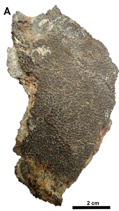
Sediment filling the egg with the embryonic skull in situ. (Image courtesy of Martin Kundrat and the journal Current Biology)

Twenty-three years after the groundbreaking discovery, researchers report the first 3D images of the preserved embryo of a sauropod. A new scientific study published in the journal Current Biology on August 27, 2020, described the first near-intact embryonic sauropod skull analyzed from first-hand observations of 3D virtual high-resolution models. The new findings, led by Martin Kundrat of the Paleo BioImaging Lab at Pavol Jozef Šafárik University, in the Slovak Republic, add to the understanding of the development of sauropod dinosaurs, a group characterized by long necks and tails and small heads, and suggests that they may have had specialized facial features as hatchlings that changed as they grew into adults.

“The specimen studied in our paper represents the first 3D preserved embryonic skull of a sauropod sauropodomorph,” said Kundrat, who is also an Associate Professor in Evolutionary and Developmental Biology at the Pavol Jozef Šafárik University. “The most striking feature is head appearance, which implies that hatchlings of giant dinosaurs may differ in where and how they lived in their earliest stages of life. But because it differs in facial anatomy and size from the sauropod embryos of Auca Mahuevo, we cannot rule out that it may represent a new titanosaurian dinosaur.”

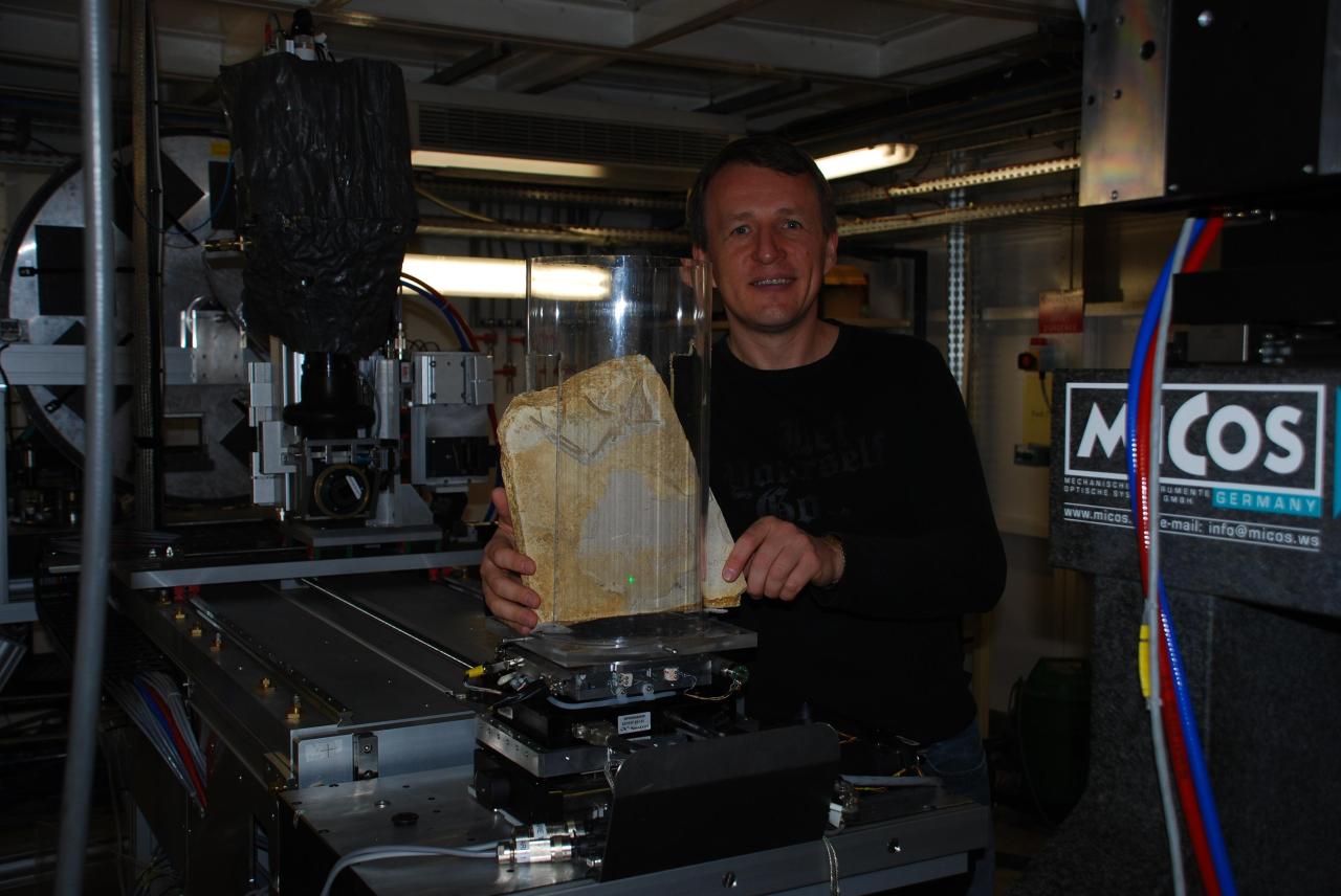
Martin Kundrat at the European Synchrotron Radiation Facility (ESRF) in Grenoble. (Image courtesy of Martin Kundrat)

The scientists recognized a well-exposed skull inside a fragmented egg, preserved in three dimensions with most bones virtually intact and articulated. Although the skull is visibly exposed on its left side, the 3D morphology and internal structure of all the preserved bones became accessible to the researchers through virtual replicas produced using scanning and imaging tools at the European Synchrotron Radiation Facility (ESRF) in Grenoble.

In the study, Kundrat’s team used imaging technology called synchrotron microtomography to study the inner structure of bones, teeth, and soft tissues of the embryonic dinosaur. The scans allowed Kundrat and co-author Daniel Snitting, from Sweden’s Uppsala University, to find hidden details, including tiny teeth preserved deeply in tiny jaw sockets. They also discovered many previously unknown anatomical details in the cranial bones, including embryonic braincase components that kept their original shape and what appear to be the remains of temporal muscles.

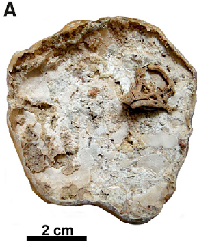
The outer fragment of the original egg. (Image courtesy of Martin Kundrat and the journal Current Biology)

According to the researchers, sauropodomorph embryology remains one of the least explored areas of the life history of dinosaurs. But these new 3D models allowed investigators to reconstruct the most plausible appearance of the skull in titanosaurian sauropods before hatching, with useful details for taxonomic or developmental comparisons among related dinosaurs.

The preserved embryonic skull inside the fragmented fossil egg was scanned using the ESRF’s beamline ID 19, a multi-purpose long (145 m) imaging beamline. The scans were collected with propagation phase-contrast synchrotron microtomography using a pink beam with two different energies. Once the scanned data of the specimen was complete, the researchers turned to Mimics, a medical 3D image-based engineering software from Materialise, one of the leading providers of additive manufacturing software in Belgium, for the segmentation and 3D rendering of the skull.

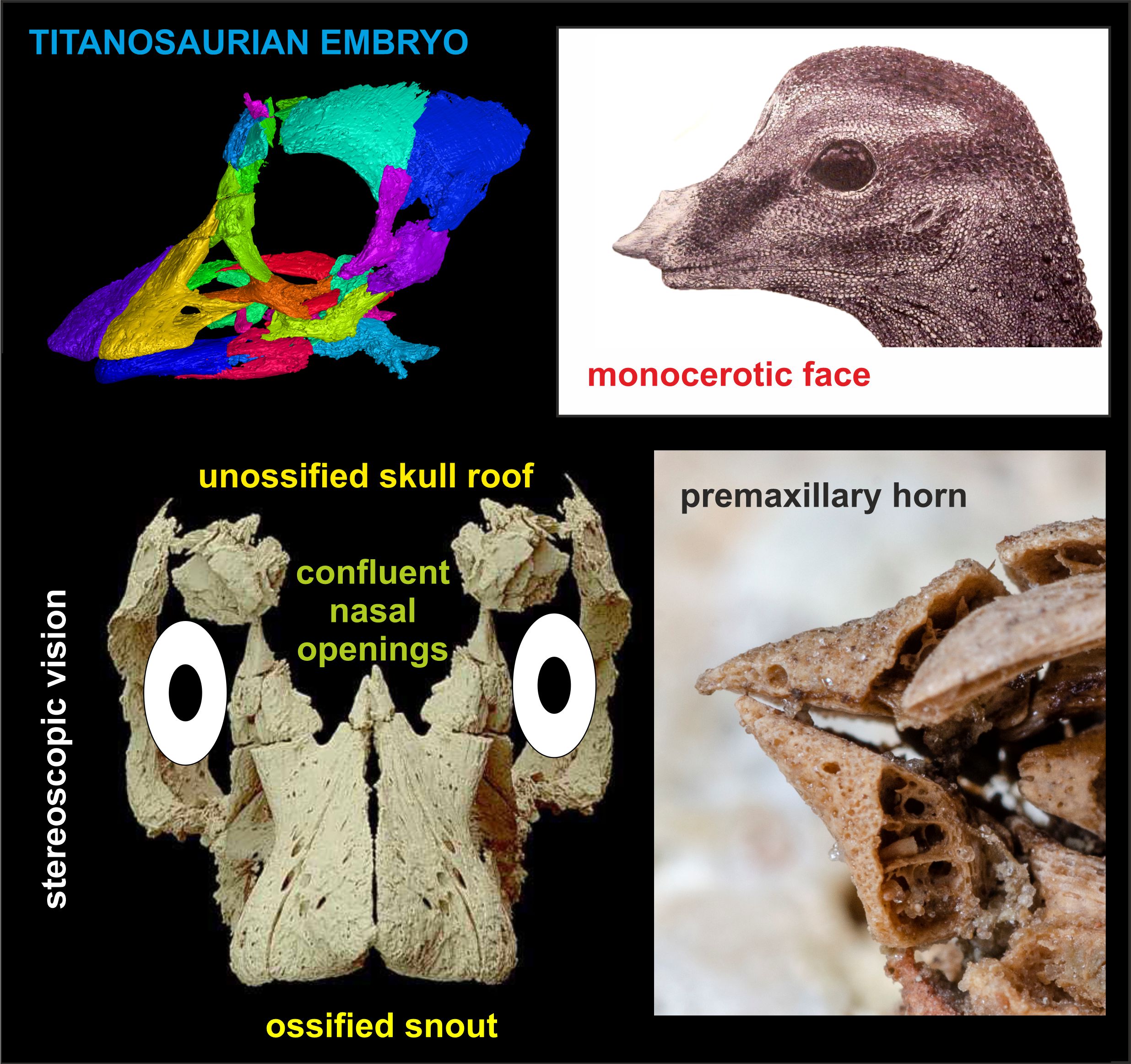
On the left: digital reconstruction of the cranial bones and reconstruction of the skull in anterior view showing incomplete skull roof. On the right: reconstruction of the head appearance by Vladimir Rimbala and premaxillary horn of embryonic skull. (Image courtesy of Martin Kundrat and the journal Current Biology)

Finally, the scientists reconstructed the internal structure and vasculature of the premaxilla (a pair of small cranial bones at the very tip of the upper jaw) thanks to German software provider Volume Graphics’ VGStudio Max 2.2, one of the most advanced software platforms for industrial CT data analysis and visualization.

Once the researchers had the 3D models, they were able to analyze the details in the sauropod’s prenatal cranial ossification. Kundrat and the study’s co-authors suggest “an alternative head appearance for babies of these Patagonian giants,” with a specialized head and face that transformed as the young dinosaurs grew and matured into adults. In fact, the visually enlightening findings suggest that the baby sauropods may have hatched out of the egg with the help of a thickened epidermal prominence rather than using a boney “egg-tooth.” The team also uncovered evidence that the titanosaurian hatchlings emerged with a temporary single-horned face, retracted openings on the nose, and early binocular vision.

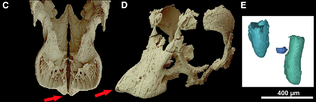
Left: the craniofacial region in ventral view showing the premaxillary and maxillary alveoli and the rostral premaxillary projection forming a basis of the horn-like process. Middle: the skull in antero-ventral view. Right: 3D rendered first mesial premaxillary teeth. (Image courtesy of Martin Kundrat and the journal Current Biology)

“Our study revealed several new aspects about the embryonic life of the largest herbivorous dinosaurs that lived on our planet. A horned faced and binocular vision are features quite different from what we expected in titanosaurian dinosaurs,” added Kundrat. “Dinosaur eggs are for me like time capsules that bring a message from the ancient time. This was the case of our specimen that tells a story about Patagonian giants before they hatched.”

The work is expected to enlighten the understanding of dinosaurs and how they lived. This newly unveiled reconstruction enabled experts to recreate anatomical aspects based on intact cranial features never seen before, revealing biological and geochemical characters that distinguish the new specimen from previously described titanosaurian embryos from Auca Mahuevo.

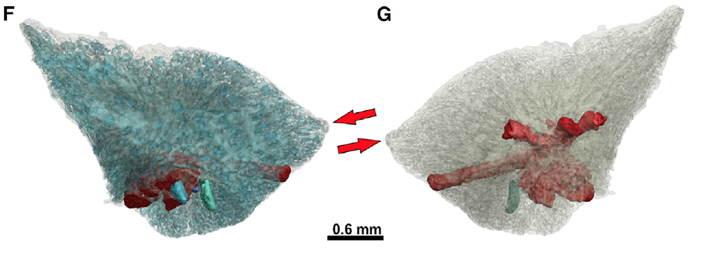
3D rendering of the opaque and semi-transparent premaxilla in medial and lateral views. (Image courtesy of Martin Kundrat and the journal Current Biology)

Although the egg fragment was originally illegally exported from Argentina and brought to researchers’ attention only later when study co-author Terry Manning, a Paleo Technician in Arizona, realized the unique preservation and scientific importance of the specimen, it is now housed in the Museo Municipal “Carmen Funes” in Plaza Huincul, just miles from the Auca Mahuevo fossil site in Argentina under the curation of paleontologist Rodolfo Coria, who is also a co-author of the study.

For decades, the extraordinary discovery of the Late Cretaceous sauropod dinosaur nesting ground has fascinated researchers worldwide. The dozens of intact eggs opened a window to understanding the life of the giant sauropods, and particularly their reproductive habits and early life. Now, 3D imaging and scanning technology can help uncover new traits and anatomy of these dinosaurs, with details never before seen.

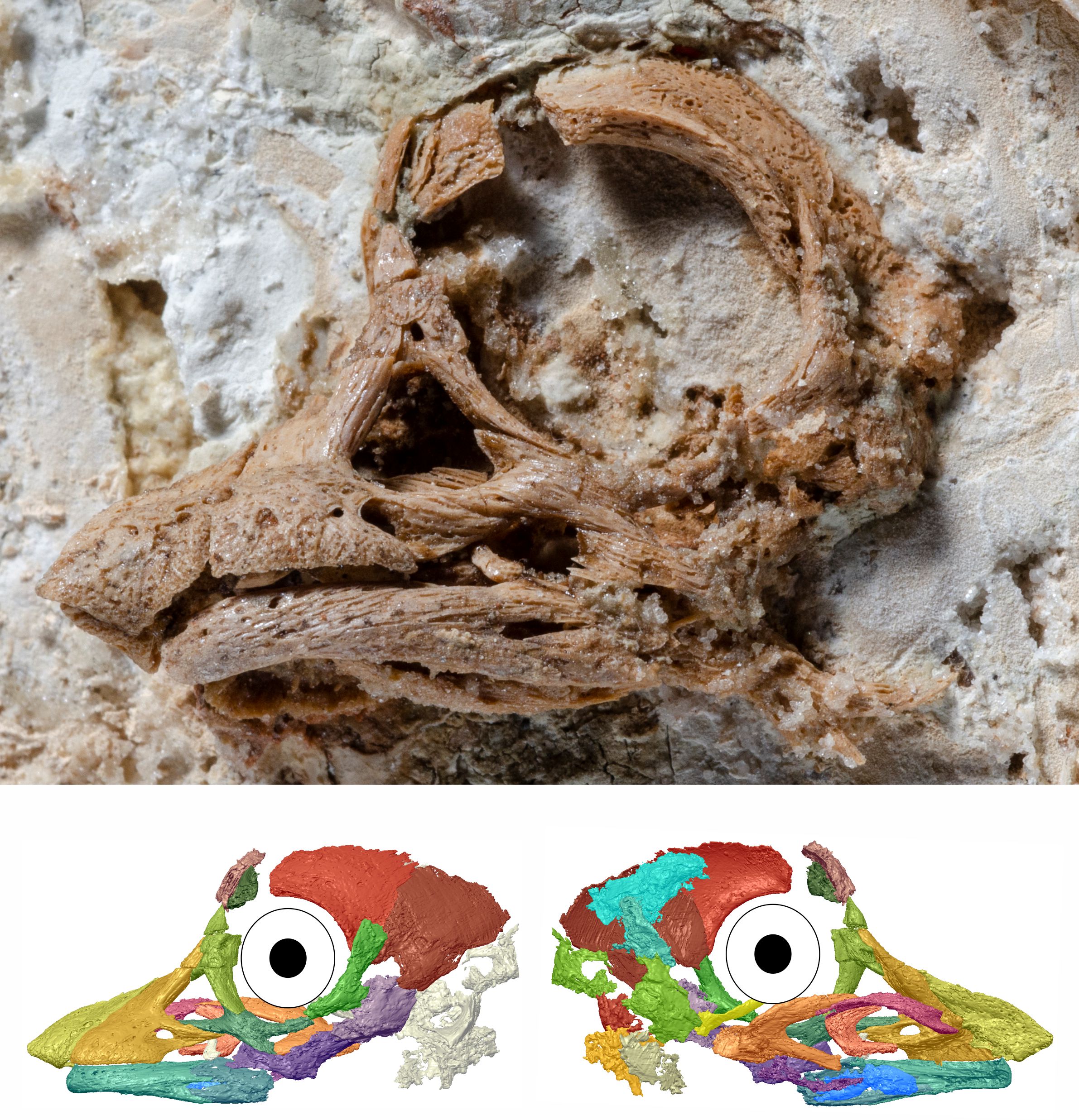
A magnified perspective of the embryonic Titanosaurian skull along with a skull reconstruction. (Image courtesy of Martin Kundrat and the journal Current Biology)

The post Dinosaurs: First 3D Model of Embryonic Sauropod Reveals New Facial Features appeared first on 3DPrint.com | The Voice of 3D Printing / Additive Manufacturing.

New Imaging Technique Helps to Understand Bioprinted Glioblastoma Tumors

With cancer as the second leading cause of death globally, hundreds of researchers continue their efforts to fight the disease. Years of oncology investigations have suggested that each person’s cancer has a unique combination of genetic changes – such as mutations in DNA – yet some types are considered to be more combative, uncontrollable, and fatal than others. In the case of glioblastomas, a very aggressive brain tumor, the median survival time is between 15 to 16 months in people who get surgery, chemotherapy, and radiation treatment. However, a median means that only around half of all patients with this tumor survive to this length of time. Experts suggest that since glioblastoma is the deadliest form of brain tumor, less than 10 percent of people who are diagnosed with it will survive more than five years.

Glioblastomas grow very fast inside the brain. The National Cancer Institute indicates that its cells copy themselves quickly, and a lot of blood vessels feed these tumors. We have reported quite a few researchers in the last months that are developing bioprinting techniques to work on different ways to tackle the disease. Now a team of scientists has created a new imaging technique that enables the study of 3D printed brain tumors.

In a recently published paper in Science Advances, Xavier Intes, a professor of biomedical engineering at Rensselaer Polytechnic Institute, New York, joined a multidisciplinary team from Northeastern University, in Boston, and the Icahn School of Medicine at Mount Sinai, New York, to demonstrate a methodology that combines the bioprinting and imaging of glioblastoma cells cost-effectively that more closely models what happens inside the human body.

“There is a need to understand the biology and the complexity of the glioblastoma,” said Intes, who is also the co-director of the Center for Modeling, Simulation and Imaging for Medicine (CeMSIM) at Rensselaer. “What’s known is that glioblastomas are very complex in terms of their makeup, and this can differ from patient to patient.”

To create their 3D tumor cell model, a team, led by Guohao Dai, an associate professor of bioengineering at Northeastern University and corresponding author on the study, made bioinks out of patient-derived tumor cells and printed them along with blood vessels. That vasculature allowed the printed tissue to live and mature, enabling researchers to study it over a matter of months.

As detailed in the paper, an integrated platform enabled generating an in vitro 3D bioprinted glioblastoma multiforme (GBM) tumor model with perfused vascular channels that allow long-term culture and drug delivery, as well as a 3D imaging modality – a second-generation mesoscopic fluorescence molecular tomography (2GMFMT) imaging system – that enables researchers to noninvasively assess longitudinal fluorescent signals over the whole in vitro model. And according to Northeastern University, this work could help medical professionals better understand how the tumor grows and to speed up the potential discovery of new drugs to fight it.

The study indicated that each imaging session exposes laser light on samples, and cells undergo stressful conditions during these long imaging processes, which reduces the cell viability. Thereby they selected an imaging modality not only for the shortest possible image acquisition time but also without potential photodamage. The 2GMFMT offers the least stress on cell culture allowing frequent imaging sessions without compromising tissue integrity.

“This is a very difficult brain tumor to treat,” said Dai. “And it’s also difficult to do research on the brain tumor, because you cannot really see what’s happening.”

Associate professor Guohao Dai (Image: Matthew Modoono/Northeastern University)

Dai also described that animal studies (typically done in mice or rats) to understand a tumor’s development, are expensive, time-consuming, and don’t allow for day-to-day observations of the same tumor in living tissue. Dai’s lab, specializing in 3D printing live tissue, grew a three-dimensional model to act as brain tissue for tumor cells to infiltrate so that they would be able to study glioblastomas more directly.

“We use human brain blood vessel cells, and connect them with all the neurons, pericytes, astrocytes, the major cell types in the human brain,” Dai said. “A water-based substance known as a hydrogel serves as a matrix to hold these cells in place. Then we use 3D printing to stack them in three-dimensional fashion.”

In the middle of the structure, which is only a few millimeters thick, the researchers place glioblastoma tumor stem cells. The cells are derived from brain tumor patients thanks to Hongyan Zou, a neurosurgeon and professor of neuroscience at Mount Sinai’s medical school and head of the Zou Lab at the Icahn School of Medicine.

“We can observe how the brain tumor cells aggressively invade, just like what we see in patients,” Dai went on. “They invade everywhere. We treated the tumor with the same kind of drug you give to a patient when they undergo chemotherapy. We monitored this chemotherapy over two months, and what we found was that the chemotherapy was not able to kill the tumor.”

To get an accurate picture of what’s happening inside the 3D model without disrupting it, Intes used a laser to scan the sample and quickly create a 3D snapshot of the cellular structure, an imaging technique developed in his lab. This combination of techniques allowed them to evaluate the effectiveness of a commonly used chemotherapy drug, temozolomide (TMZ). Initially, the tumor shrank in response to the drugs, but then it grew back swiftly and aggressively. This indicates that the drug did not work in the long term, which seems to line up with the experience of patients with glioblastoma.

The TMZ chemotherapy treatment traveled through the channels provided by the bioprinted blood vessels. The team claims that in the body, drug delivery to glioblastoma cells is especially complicated because of the blood-brain barrier, a wall of cells that blocks most substances from reaching the brain. It appears that the team’s method provides a more accurate evaluation of a drug’s effectiveness than directly injecting the therapy into the cells.

Moreover, Dai suggested that they need to develop and screen other chemotherapy drugs, and this model may be able to speed up that process, since this method could be used to weed out unsuccessful drugs early, ensuring that only the most promising ones move to animal, and eventually human, trials.

Dai considered that “you have a tremendous amount of time and cost associated with animal research,” but “with our 3D glioblastoma model and imaging platform, you can see how the cells respond to radiation or chemotherapy very quickly.”

They conclude that beyond the necessity to guide the development of new drugs, efficient model systems that enable fast and predictive evaluations of candidate drugs are a critical need. To provide biological relevant experimental settings in which drug efficacy can be assessed, a suitable tumor growth environment and long-term culture capabilities are required.

The publication offers a detailed recount of the new technique that according to Intes, could allow researchers to evaluate the effectiveness of multiple drugs at the same time. According to Rensselaer’s School of Engineering, Intes pointed out that it is not yet realistic though for studying the effectiveness of certain therapeutics on a person’s tumor because of the short time that clinicians often have to provide treatment.

“We developed a new technology that allows us to see, first, if the cells are growing, and then, if they respond to the drug,” conveyed Intes.

If glioblastomas are the most common malignant brain tumor and one of the hardest to treat, then this team is certainly moving in the right direction. Glioblastoma tumor growth is considered to almost always outpace chemotherapy and radiation treatments, so the remaining available treatments are primarily experimental. With so much uncertainty with regards to a successful treatment, new techniques, like this one, offer an encouraging message. Researchers are hurrying to mimic the conditions of tumors thanks to 3D bioprinting and the ability to generate bioinks out of patient-derived tumor cells. Moving from lab research to actual clinical trials might take a long time, but at least the technology is providing the strong foundations needed to understand the true nature of these malignant tumors.

The post New Imaging Technique Helps to Understand Bioprinted Glioblastoma Tumors appeared first on 3DPrint.com | The Voice of 3D Printing / Additive Manufacturing.

Interview with Len Wagner of Deer Valley Ventures

Len Wanger

Len Wagner

Len Wagner is Chief Technology Manager at Impossible Objects, Inc. He is also currently a Managing Partner at Deer Valley Ventures, investing in early stage advanced and additive manufacturing companies. He gives good insight into both the technical world as well as the financial realm within additive manufacturing. He also is involved as a chairman of the FabLab Association for the Museum of Science and Industry.

What has lead you to this point?

I started my career in computer graphics. I was focused on the software that produced the images. I did a lot of graphics simulations. This lead to CAD and finite element analysis. In 1992 I was a researcher and was able to work on one of the first 3D printers. I was able to help researchers to visualize their data. Later on I was able to work on the financial side as I run a fund. I sit between these two things. I am also involved in a lot of STEM education as well and it has been important for me to give back. It took a while, but I figured out I had a skill of explaining the technical side to the business side. It took a while to realize that was important.

What kind of developments have recently disrupted this marketplace?

We have seen a big movement in the industry. We have moved from prototyping to manufacturing. We are at the very beginning stages of this. Customers and vendors are doing things to make this transition. It is a very different set of requirements from making prototypes to actual production levels. We at Impossible Objects are somewhat betting on this. The word disruption is funny. It is a slow methodical process to move in this field. Manufacturing moves very conservatively and methodically. More parts are moving toward digital manufacturing and additive manufacturing. If you talk to a large aircraft manufacturer like Boeing, a modern aircraft has hundreds of parts. A small percentage of these parts are continuously being made with additive manufacturing. Good steady progress is important. The full life cycle of material properties is important to understand.

Can you explain your work and day to day operations for Impossible Objects?

I run the engineering group as the Chief Technology Manager. The main function we have is designing and building new machines. We want to improve the process with new materials and machinery. We work on process development and I also help to make an automated machine that may assist with these types of process developments. I also work with customers for them to work with machines.

Impossible Objects

You have an interesting mix of skills in terms of venture capital as well as engineering. Can you give some insight into how you operate within both worlds?

It really comes down to building teams and having communication skills. It is important to build the communication skills. It’s important to translate the cultures. Engineers have a certain way of speaking and it is important to be able to explain things in terms of the business side and that realm of communication.

I feel the future of the additive sector lies within the precision of 3 dimensional imaging techniques. What are your thoughts on this?

I think it is important to measure the quality control of a product. 3D optical scanning at a cheap rate is not really on the market just yet and I think there is a great market need for it. Why is there not an open source package that is oriented towards this?

Fab Lab Association

People compare the additive manufacturing industry to the early days of computers. Do you agree or disagree and why?

I largely agree but it is not a perfect analogy. I agree that the transformation for being able to do manufacturing cheaper and faster at a small scale is similar to how programming costs went down extremely over time. Authoring is hard to do in 3D. Thinking in 3D is difficult. I also do not think there is a Moore’s law of Additive Manufacturing. I do think the ability to change the manufacturing sector is large.

Museum of Science and Industry Fab Lab

Can you explain some of the work you do as chairman of the FabLab Association for the Museum of Science and Industry?

With the FabLab Network, I am an advisor to the board of the MIT FabLab Network. The Museum of Science and Industry has a FabLab and it is great to expose people to one aspect of the maker movement. Schools and organizations are allowed some exposure and experience to this environment. There is an educational aspect of the Museum as well. This also invigorates people. It sparks the interest in people as well. I help to raise funds and I advise the lab. The equipment has become relatively cheap so schools can have access to these items. It is important for us to teach educators how to use these types of machines. It is important to give people access as well as give people mentorship.

What are some key points that companies should be focusing on in terms of the additive manufacturing strategies?

We must focus on material properties. It also is important to know the speed of prints. It is also important to have the economics down pat as well. Lastly, I think these machines have to work within your larger manufacturing environments. We are adding a camera to slice every image of all levels that have been printed. It is also to take advantage of digital manufacturing and mass customization.