Chinese University of Hong Kong Studies 3D Printing for Heart Disease

In the recently published ‘Three-dimensional printing in structural heart disease and intervention,’ authors Yiting Fan, Randolph H.L. Wong, and Alex Pui-Wai Lee, all from The Chinese University of Hong Kong, explore the potential for 3D printing in the world of medicine, as well as cardiology—and more specifically, structural heart disease (SHD).

SHD causes issues like:

  • Aortic stenosis
  • Mitral regurgitation
  • Atrial septal defect
  • Left atrial appendage (LAA) clots
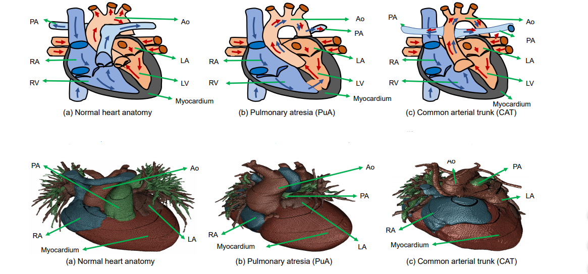
Conventional imaging is limited, while the emergence of 3D printed models allows medical professionals to progress from mentally reconstructing 2D images to gaining a more complex understanding of pathology.

As 3D printing continues to make its way into the realm of medicine, models are used for:

  • Guiding treatment
  • Procedural simulation
  • Facilitating hemodynamic research
  • Improving interventional training
  • Promoting patient-clinician communication

To create a medical model, images must be attained, data must be processed, and the object must be 3D printed.

“The most commonly used imaging sources for SHD are echocardiography, computed tomography (CT) and magnetic resonance imaging (MRI),” state the researchers. “Other modalities, such as positron emission tomography, single photon emission CT and cone beam CT, are less commonly used. All images should use the common Digital Imaging and Communication in Medicine (DICOM) format.

Pre-procedural simulation of MitraClip on 3D-printed model. (A) Digital model of a heart. Different colors stand for different cardiac components (grey: mitral valves; light pink: tricuspid valves; light gold: atrial septum, left atrium, left and right ventricle). (B) Multi-material 3D-printed heart model for pre-procedural simulation. The valves were printed with flexible material and the rests were printed with hard material. (C) The 5 holes drilled in the atrial septum represents the different position for different kinds of structural heart interventions. (D) The MiraClip device was released via delivery catheter through the atrial septum to the mitral valve. Blue circle: MitraClip; red circle: left atrial appendage occlusion (LAAO). S, superior; A, anterior; P, posterior; I, inferior; IVC, inferior vena cava; LAA, left atrial appendage; MV, mitral valve.

A range of materials are both popular and possible for fabricating medical models:

“Multi-material printing by material jetting is increasingly used to create cardiac structures. Different tissue components were printed with different textures. For instance, an aortic valve was printed with flexible printing material, and the calcifications attached to valves were printed with hard printing material, respectively,” report the authors.

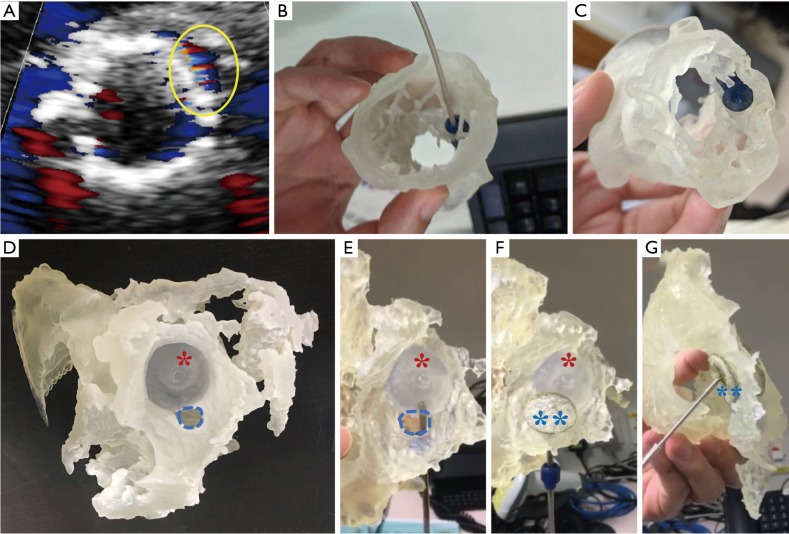
Application of 3D printing for peri-device leak. (A,B,C) A case found with peri-device leak post TAVI and needed peri-device leak occlusion: (A) routine TEE post-TAVI showed peri-device leak (yellow circle); (B) simulation of peri-device leak occlusion on 3D-printed aortic root model derived from post-TAVI CT; (C) the 10-mm vascular plug was found to be best-fit for this case. (D,E,F,G) A case found with residual leak after ASD closure: (D) multi-material 3D printed model showed residual leak (blue circle) next to the ASD occluder (asterisk *); (E) the delivery catheter went through the leak position; (F) the device (two asterisks **) was released in situ. (G) The bicaval view of 3D-printed model showed stable release and stay of the chosen device. 3D, three-dimensional; TAVI, transcatheter aortic valve implantation; TEE, transesophageal echo; CT, computed tomography; ASD, atrial septal defect.

There continue to be ongoing challenges in the creation of medical models, however, ‘despite the enthusiasm in applying 3D printing cardiovascular medicine.’ While there is an obvious lack of technical standards, mainly due to the novelty of the technology, the authors point out also that there are still issues with affordability—along with ‘scant evidence on the added clinical benefit.’

Greater accuracy is needed, along with improved standardization of data acquisition, and post-processing techniques. While deeper research is required into the creation and use of models and surgical guides, so are comparisons for offering up better information and creating industry standards. The authors also recommended a more streamlined workflow.

“The mechanical properties of the 3D-printed materials, such as tensile strength, elasticity, flexibility, hardness, and durability have utmost importance for cardiovascular applications. The majority of cardiovascular applications reported so far have employed materials with properties that have not been meticulously compared with the cardiovascular tissue they are mimicking. Validation of 3D-printed material properties against actual human patient tissues is important to ensure that procedural simulation is realistic,” conclude the authors.

“Further effort in technical standardization, and clinical evaluation of added benefit and cost-effectiveness of 3D printing are needed to bring this promising technique to clinical reality.”

3D printed medical models are extremely beneficial to doctors and patients as they allow not only for diagnosing but have also continued to change medicine—allowing for procedures involving complex reconstructions, fabrication of surgical guides, and much more.

What do you think of this news? Let us know your thoughts! Join the discussion of this and other 3D printing topics at 3DPrintBoard.com.

[Source / Images: ‘Three-dimensional printing in structural heart disease and intervention’]

The post Chinese University of Hong Kong Studies 3D Printing for Heart Disease appeared first on 3DPrint.com | The Voice of 3D Printing / Additive Manufacturing.

Scientists Use 3D Printed Models to Further Congenital Heart Disease Studies

In the recently published ‘Accurate Congenital Heart Disease Model Generation for 3D Printing,’ researchers explore 3D printing for diagnosis, treatment, and planning in congenital heart disease (CHD) patients. CHD usually presents itself at birth and can be difficult to analyze, even with 3D medical images—and despite many different studies by scientists around the world. The researchers note that 3D printing has been ‘widely adopted’ in clinical settings lately, offering for improvements in the following:

  • Clinical decision making
  • Interventional planning
  • Communication between physicians and patients
  • Improving medical education

(Top) Examples of large structure variations in CHD. In normal heart anatomy (a), PA is connected to RV. However, in pulmonary atresia (b), PA is rather small and connected to descending Ao. In common arterial trunk (c), Ao is connected to both RV and LV, and PA is connected to Ao. (Bottom) Pulmonary atresia and common arterial trunk examples in our dataset, with large variations from normal heart anatomy.

In the dataset for this research, combining deep learning and graph matching for whole heart and great vessel segmentation in CHD, patients ranged in age from one month to 21 years old—while most were from one month to two years old. Out of 16 cases, the study covers 14 types of CHD to include the most common 8, which are atrial septal defect (ASD), atrio-ventricular septal defect (AVSD), patent ductus arteriosus (PDA), pulmonary stenosis (PS), ventricular septal defect (VSD), co-arctation (CA), Tetrology of Fallot (ToF), and transposition of great arteries (TGA).

Overview of the proposed framework combining deep learning and graph matching for whole heart and great vessel segmentation in CHD.

The researchers explain that there has already been a prolific amount of research using the multi-modality whole heart segmentation method, with ‘state-of-the-art performance’ found in combining 3D U-net for segmentation and a simple convolutional neural network for label position prediction. Another technique involves using the basic simple convolutional neural network for label position prediction, while another handles blood pool and myocardium in blood pool segmentation.

 “Considering the significant variations in heart structures and great vessel connections in CHD, almost all the existing methods cannot effectively perform whole heart and great vessels segmentation in CHD,” state the researchers.

Motivated instead by the promise of graph matching, they used deep learning and graph matching, collecting 68 CT images overall, with an 11.9 percent higher Dice score. The framework consisted of:

  • Region of interest cropping
  • Chambers and myocardium segmentation
  • Blood pool segmentation
  • Chambers and myocardium refinement
  • Graph matching

Segmentation results were printed on a Sailner J501Pro for evaluation—a process that took the researchers about three to four hours. The researchers assessed the 3D printed model as correct, and with clear shape and connections, with minor refinements necessary such as thin coronary vessels.

“We also printed out part of the segmentation results with minor manual refinement and showed that it can be applied to clinic use,” concluded the researchers.

3D printed models have proven to be more than just helpful in many different areas of the medical realm, especially as they are able to offer so much in terms of education not only for students but also for patients and their families, as well as helping in the preoperative stages—and during the actual operation too with surgical planning models.

(Top) Visualized comparison between the state-of-the-art method Seg-CNN [12] and our method. The differences from the ground truth are highlighted by the red circles. (Bottom) Examples of 3D printing models using our method with some minor manual refinement.

[Source / Images: ‘Accurate Congenital Heart Disease Model Generation for 3D Printing’]

The post Scientists Use 3D Printed Models to Further Congenital Heart Disease Studies appeared first on 3DPrint.com | The Voice of 3D Printing / Additive Manufacturing.

Interview with Dr. Vahid Serpooshan, Who Created a ‘Heart Attack Patch’ With the Help of 3D Printing

According to the World Health Organization, cardiovascular disease is the number one cause of death globally. An estimated 17.9 million people died from cardiovascular disease in 2016, representing 31% of all global deaths. Treatments for all manner of cardiovascular impairments are therefore a high priority. A very prevalent ailment in that category is a heart attack which happens to over 735,000 Americans a year

A team of researchers led by Dr. Vahid Serpooshan, an assistant professor of Biomedical Engineering and Pediatrics at Georgia Institute of Technology and Emory University School of Medicine, have created a patch with a regenerative protein to treat the infarcted myocardium (cardiac muscle, a part of the heart that gets damaged during a heart attack). According to Georgia Institute of Technology the patch will cover the infarcted tissue with a collagen patch infused with a  protein called FSTL1, which is tailored to fit an area a little larger than the dead tissue. Dr. Serpooshan used bioprinters to develop the patch using a collagen gel. The patch is about to start its pre-clinical trial phase at Emory University’s School of Medicine in Atlanta, and a clinical trial is already ongoing in Europe for a version of this patch device. So it could be a couple more years before they are available for patient treatment. Dr. Serpooshan’s Lab (which has two locations at both Emory University and Georgia Tech) uses a multidisciplinary approach to design and develop micro/nano-scale tissue engineering technologies with the ultimate goal of generating functional tissues and organs. 3DPrint.com reached out to talk to Dr. Serpooshan about his team’s groundbreaking work. 

Professor Vahid Serpooshan sets up the medical 3D printer that will print a patch engineered to strengthen heart muscle damaged in a heart attack. (Photo by Rob Felt, Georgia Institute of Technology)

How has 3D printing helped to engineer the patch?

3D printing has been a huge help in manufacturing the patch in so many ways. It enabled us to incorporate vasculature inside the patch, something that would not have been available with traditional tissue engineering techniques, since they offer really limited potential to create complex, functional vasculature in thick, 3D tissue constructs. Additionally, it allowed us to have a controlled deposition of different cells, biomaterials, and molecules to form heterogenous, complex 3D structures that closely mimic native tissue structure. Also it provided us with patient -and disease- specificity: we utilize MR or CT imaging data obtained from patients to create 3D printed tissue constructs that precisely match the patient’s diseased or damaged tissue. Finally, bioprinting technologies have enabled tissue engineers to markedly enhance resolution and precision in various tissue manufacturing endeavors, making precision medicine even more promising.

What 3D printer do you use to create the patch?

We are mainly using two types of 3D bioprinters for this project. A BioAssemblyBot bioprinting system, made by Advanced Solutions Inc. (an American company in Louisville, Kentucky). This is the ONLY six-axis robotic arm printer in the market, with a resolution down to approximately 20 to 50 μm (microns). The second bioprinter is a BIO-X from Swedish biotech company Cellink, a pioneer in bioprinting.

An illustration of the heart with the cardiac patch already in place (Serpooshan Lab)

How long will it take to manufacture the patch?

Biomanufacturing of a cardiac patch at a clinically-relevant scale would take about 15 to 30 minutes, depending on the number of cell types involved, the vasculature design, and other factors. This enables the clinics, hopefully in the near future, to have heart attack patients come to the clinic, conduct CT or MR imaging, prepare 3D digital model of the damaged tissue, and sending it to the bioprinter to manufacture a patch device customized for that patient.

Vahid Serpooshan designing the myocardial infarct patch (Photo by Rob Felt, Georgia Institute of Technology)

Is there already a procedure in place to apply the patch?

Actually, there already is a procedure to apply the patch. We use a left thoracotomy approach to get access to the surface of the heart and suture the patch onto the epicardial layer of the heart, covering the myocardial infarction (also called a heart attack) area. There are currently some ongoing works trying to minimize the aggressiveness of this surgical procedure, potentially using catheter-based techniques.

What are the benefits of using collagen in the patch?

Collagen is the most abundant protein of the human body. This protein highly supports cell viability, proliferation, and function. It is a biodegradable protein which is a major advantage for in vivo applications, facilitating timely integration of the implant with the host tissue and avoiding long-term immunogenic complications. For our 3D bioprinting works, we use gelatin methacrylate (gelMA). Gelatin is derived from collagen (a hydrolyzed collagen) and offers similar advantages. The main advantages of gelMA for bioprinting of cardiac patches include: acceptable compatibility with cardiac cells (cardiomyocytes), tunable biomimetic mechanical properties (stiffness), controllable degradation rate, and of course, great printability (rheology properties).   

The collagen gel used to develop the 3D printed patch. (Photo by Rob Felt, Georgia Institute of Technology)

What percentage of heart disease patients will benefit from this device?

This therapy is designed for a specific group of heart attack patients, approximately one third of patients who either arrive too late to the clinics or are resistant to current catheter-based reperfusion methods and pharmacologic therapies.

If approved, Dr. Serpooshan’s patch could help over 200,000 patients per year.

3D Printing News Briefs: July 17, 2018

In Today’s 3D Printing News Briefs, we’re covering a lot of business and a little medical news. AMFG is partnering with a top UK bearings manufacturer to help automate its digital manufacturing workflows, while Segula Technologies has begun an industrial 3D printing partnership with digital manufacturing company Multistation. Techniplas has completed a deployment of Sharebot 3D printers to its 14 manufacturing facilities around the world, and the winners of the SkillsUSA Additive Manufacturing Competition have been announced. Finally, a pediatric cardiologist used the Sinterit Lisa to create a 3D printed model of a newborn boy’s heart to plan his risky surgery.

Bowman International Announces Partnership with AMFG

Bowman’s bearings

Automation software specialist AMFG, which recently launched a new AI software platform, has partnered with Bowman International, one of the top bearings manufacturers in the UK, as it works to grow its 3D printing capabilities through its Bowman Additive Production (AP) division. Bowman AP has several MJF and SLS 3D printers available for its use, and uses 3D printing to design and produce its end-part bearings, which has helped increase their load bearing capacity by up to 70%.

In the meantime, Bowman International’s goal is to use AMFG’s AI-powered production automation software to oversee production of said bearings, by automating production job scheduling, optimizing digital CAD files for production with printability analyses, and creating a custom digital part catalog.

“We’re very pleased to be partnering with AMFG and using their automation software to scale our already expanding AM facility,” said Jacob Turner, the Head of Additive Production at Bowman International. “Additive manufacturing is transforming the way bearings are manufactured, and we aim to continue to be at the forefront of innovating the production of bearings using AM. AMFG’s automation software will enable us to achieve this by significantly increasing the efficiency of our production processes.”

Multistation Partners with Segula Technologies

Another newly announced 3D printing partnership is the one between international engineering group Segula Technologies and Paris-based 3D printing company Multistation. The two are working together to further develop the potential of 3D printing in the industrial sector, which will allow both companies to increase their offerings and provide customers with excellent services along the AM value chain. Segula will bring its design, product-process qualification, and technology integration in industrial environments to the table, while Multistation will share and apply its expertise in AM design and simulation by determining any potential parts that could be 3D printed instead of fabricated with a more traditional method of manufacturing.

“Additive manufacturing is an integral part of a value chain within which Multistation provides a comprehensive offering; Segula Technologies was an obvious partner of choice to enable our Additive Consulting division to address manufacturers’ concerns more effectively,” said Yannick Loisance, the CEO of Multistation. “We will thus be able to supply them not just with software packages, machines and materials, but also with a more comprehensive range of high-quality engineering services that are suited to a host of different business sectors.”

Techniplas Adds Sharebot 3D Printers to Its Manufacturing Facilities

This fall, Italian professional-grade 3D printer manufacturer Sharebot joined the open innovation program at Techniplas, a top automotive design and manufacturing provider. Now, as part of its own continuing digital transformation, Techniplas has deployed Sharebot 3D printers to all of its 14 manufacturing facilities across five continents. This move will allow the company to 3D print the majority of the manufacturing products it uses every day on-site, which will equal major cost and time savings as Techniplas previously used only third-party providers for this task.

With Sharebot 3D printers installed in all of our manufacturing facilities worldwide, we are taking decisive steps toward fabricating the majority of our manufacturing line assembly tools, jigs, fixtures, gauges and even robotic arm attachments in-house. Based on our experience with Sharebot printers thus far, we expect to significantly reduce our development time and annual assembly line tooling costs in each manufacturing facility over time,” said Techniplas COO Manfred Kwade.

Winners of the SkillsUSA Additive Manufacturing Competition Announced

For the fourth year running, advanced manufacturing technology industry organization SME and Stratasys have co-sponsored the SkillsUSA Additive Manufacturing Contest, held during the annual SkillsUSA National Leadership and Skills Conference in Louisville. The winners of this year’s student contest, which asks contestants to solve real world problems with 3D printing, were just announced. This year, entrants had to design an adaptive device for a veteran, who had endured a traumatic thumb amputation, so he could keep playing his PlayStation 3. Prizes include RAPID + TCT conference passes, SOLIDWORKS’ 3D-CAD design software, SME Education Foundation scholarships (for high school participants), a one-year Tooling U-SME subscription, and a MakerBot Mini 3D printer.

“The SkillsUSA contest is designed to help students and educators realize the power of additive manufacturing to drive innovation. This year’s competition was particularly meaningful as it directly resulted in enhancing a veteran’s life with a custom solution not possible without additive manufacturing,” said Gina Scala, the Director of Marketing, Global Education at Stratasys.

The high school winners include:

  • Gold medal: Getty George and Sam Green, Martin Luther King High School, Riverside, California
  • Silver medal: Noah Logan and Johnathan Urbani, Stafford Tech Center, Rutland, Vermont
  • Bronze medal: Andrew Daddone and Layke Martin, Frederick County Career & Tech Center, Frederick, Maryland

The college winners include:

  • Gold medal: Adolfo Vargas and Alexander Kemnitz, Central Community College-Hastings, Hastings, Nebraska
  • Silver medal: Deema Al Namee and Aric Donerkiel, Vermont Technical College, Randolph Center, Vermont
  • Bronze medal: William Swaner and Ashton DeZwarte, Tenneseee College of Applied Tech-Nashville, Nashville, Tennessee

Watch a video about the 2018 competition here, and check out the winning designs here; you can also view SME’s Flickr album for more competition photos.

Surgeon 3D Prints Pediatric Heart Model with Sinterit Lisa

Desktop SLS 3D printer manufacturing Sinterit has seen its flagship Lisa 3D printer, which went through a recent upgrade, used to save lives in multiple ways, from fighting wildfires and protecting the faces of children to providing assistance in a tough pediatric cardiac surgery.

“Delivering desktop SLS 3D printer for more than three years caused that our clients send us tonnes of useful and exciting cases. Writing about all of them is hard, if not impossible, but when 3D printing helps saving lives, especially those most fragile, we feel proud, and also a duty to share it with you,” Michał Krzak, Sinterit’s Marketing Communication Manager, told 3DPrint.com.

A newborn’s heart can weigh barely 20 grams, and fits in the palm of an adult’s hand, so you can imagine that surgeries on such a delicate organ are exceedingly difficult. Jarosław Meyer-Szary, MD, from the Department of Pediatric Cardiology and Congenital Heart Defects at the University Clinical Center in Poland recently turned to Sinterit’s Lisa 3D printer to save the life of Kordian, an infant less than one month old suffering from a potentially fatal heart disease called interrupted aortic arch.

Meyer-Szary created 3D printed, life-size model of Kordian’s tiny heart, and SLS technology was able to recreate each intricate artery and vein. The model not only helped him plan the surgery ahead of time, but also helped Kordian’s mother gain a more thorough understanding of her son’s condition. Kordian is now a thriving and happy 18 month-old, thanks to Sinterit’s SLS technology.

Discuss these stories and other 3D printing topics at 3DPrintBoard.com or share your thoughts in the comments below.