Ophthalmology: Researchers Explore Progress in Bioprinting

A large group of researchers came together to author Bioprinting in Ophthalmology: Current Advances and Future Pathways, published recently regarding their findings on bioprinting within the field of ophthalmology. While they understand the promise 3D printing with cells has in so many applications, there is serious potential for ‘highly delicate organs’ like the eyes and the heart. Here, they review some of the strides made in bioprinting over the past 20 years, and specifically in ophthalmology.

As the researchers point out, over 30 percent of people around the world have visual impairment issues of some kind. Because the eye presents such an easy access point, however, doctors have excellent access for performing medical procedures and supplying implants. This means that the eyes are also very conducive to treatments with bioprinting.

3D printing so far has been responsible for a wide range of developments in optics, whether for lenses in smart phones, or a variety of different printing systems to include those for fabricating models of the eye for surgeons. Although bioprinting allows for tissue engineering and the potential for transplants, the benefits of 3D medical models alone are enormous as they give medical professionals access to visual aids for more accurate diagnoses, treatment, and education for both patients and their families.

Medical models of the eye also serve as invaluable training devices for procedures for medical students, and for surgeons who may be performing unique surgeries never attempted. They may even use the models in the operating room. Most of these models today are created with the 3D Systems Z650 printer.

(a) Schematic view of the cross-section of our physical model eye; (b) two printed parts provided main structure of the physical model eye; (c) use of the physical eye model for assessing the fundus range of the viewing system; (d–f) pictures of the angle bars photographed under 128D lens, 60D lens, and 60D lens with model eye tilt; (g–i) other three eye models printed and fabricated with different anterior chamber and total axial length.

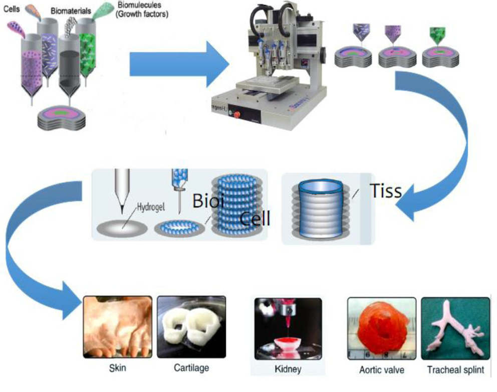
The authors point out that because there are still so few ‘workable materials’ for ophthalmology in additive manufacturing, there is still substantial room for further innovation:

“The printing of artificial lenses, glaucoma valves and other medical implants developed in customized processes will be a reality in the future,” stated the authors.

“It is believed that printing of artificial lenses, glaucoma valves and other medical implants with customization and on-demand supply will be possible in the coming years. Further, numerous next generations ophthalmological products are likely to be benefited with this technology.”

Smart-phone technology, the impetus for many different applications today, also allows for an interesting ‘alternative use of 3D printing,’ as a variety of different devices can be attached to mobile phones for examination of areas like the ocular anterior segment, giving medical professionals easy access to detect conditions like cataracts, uveitis, ulcers, and other defects.

“These devices are more than ten times cheaper than standard ocular imaging devices,” state the authors.

(A) 3D printed retinal imaging adapter on a smartphone; (B) an image of a glaucomatous disc captured with the smartphone retinal imaging adapter; (C) an image of the same glaucomatous disc captured with a standard fundus camera; (D) 3D printed smartphone slit lamp microscope, (E) an image of a patient with a white cataract captured on a smartphone with the 3D printed slit lamp microscope.

Bioprinting systems for ophthalmology are still difficult to come by, due to the lack of suitable materials, mechanical limits, speed in production, and affordability; however, the researchers are convinced that because so many innovations are being continually presented, ‘the development of a fully functional artificial eye’ is imminent.

“Overall, it can be concluded from the research endeavors in 3D printing in ophthalmology that this technology has the potential to improve the treatments of vision impaired patient by helping the doctors in performing risky surgery,” concluded the researchers. “The only need for this is to explore the innovative trends in customization of the medical devices which are highly desirable in-terms of market demand. Ultimately, the printed ophthalmological devices can heal the poor vision and other ocular diseases.”

Bioprinting continues to make steady impacts in the medical field, and while so many fascinating innovations have been made in ophthalmology, from orbital implants to prosthetic eyes, researchers continue to branch out into nearly every area of human health with medical models, and a variety of other implants and devices to change patient’s lives around the globe. Find out more about bioprinting efforts in ophthalmology here.

[Source / Images: Bioprinting in Ophthalmology: Current Advances and Future Pathways]

Swiss Hospital Will Use axial3D’s Software Platform to Improve Patient Care with 3D Printed Medical Models

[Image: axial3D]

The award-winning, Belfast-based medical 3D printing and healthcare technology firm axial3D is focused on helping the global healthcare industry adopt 3D printing by using its patient-specific medical models to improve surgical outcomes, assist patients and doctors in better understanding ailments and treatments, and facilitate pre-operative planning.

Now, on the heels of a new partnership with Tallahassee Memorial HealthCare, the company has announced that it is collaborating with top Swiss medical center University Hospital Basel (USB) in order to improve process management and patient care and outcomes at the hospital’s interdisciplinary 3D Print Lab.

“3D printed models have been shown to help surgeons complete complex life-saving surgeries that would be otherwise impossible,” axial3D’s Ryan Kyle told 3DPrint.com. “University Hospital Basel’s new collaboration with axial3D will help to deliver high-quality 3D printed models much quicker than before.”

The hospital, which has about 7,000 people on staff, is northwest Switzerland’s biggest healthcare facility. Its 3D Print Lab uses patient image data to fabricate realistic anatomical models, and other objects, using a variety of different materials and 3D printing methods. Now it will be using axial3D’s new cloud-based platform, axial3Dassure, to support its 3D printing program.

[Image: University Hospital Basel]

By using axial3Dassure, USB will optimize its 3D Print Lab in order to provide a greater level of performance and patient care. The software, which has an end to end workflow, provides features like processing and quality management, so that hospitals and medical centers can meet their expanding business needs through its powerful analytics. The new axial3Dassure platform will also help support collaboration within the hospital’s 3D Print Lab with such features as email notifications and task-driven workflows.

Daniel Crawford, axial3D

“We are very excited to be working with the team at University Hospital Basel. They are a leading force in medical 3D printing, not just in Europe, but globally, and this alliance will ensure the expertise they have developed can support our company’s growth by informing the ongoing development of axial3D’s software solutions,” said axial3D’s CEO and Founder Daniel Crawford. “With a growing requirement for 3D printing within healthcare, a centralized management platform is necessary for any 3D print lab, which plans to scale and grow in the coming years. University Hospital Basel has taken strides in its commitment to improving outcomes for patients through technology advances in the form of this collaboration.

“Our software will help the hospital gain insight into the statistics and figures usually hidden within data, ultimately allowing them to measure clinical impact and value 3D printing is having for patients. The workflow management capability will allow the hospital to speed up the creation, processing, and delivery of 3D printed models, while ensuring auditability, reliability and standardization.”

By using axial3Dassure software, USB will be able to increase efficiency and improve compliance and productivity. The hospital’s 3D Print Lab, which includes over 20 desktop and industrial 3D printers, will now be better equipped to manage communication, quality control, tracking, and workflow management.

In addition, USB will benefit from the company’s orthopaedic auto-segmentation software module, which is embedded within the axial3Dassure platform. This module will help lower the amount of time that is typically required during pre-production of 3D printing orthopaedic models.

Finally, by partnering with axial3D, USB will be able to speed up the creation, processing, and delivery of its 3D printed surgical guides.

“Our initial focus for the use of 3D printed surgical guides was within the Department of Cranio-Maxillofacial Surgery where 3D printing has now become routine,” explained Philipp Brantner, Senior Physician of Radiology and the Co-Director of the 3D Print Lab at University Hospital Basel. “Having access to onsite printing has revolutionized how we treat those patients, some who arrive with life-threatening injuries that require immediate action. The functionality that we now get provided will allow us to speed up production and treat patients more effectively and efficiently.”

What do you think about this news? Discuss this story and other 3D printing topics at 3DPrintBoard.com or share your thoughts in the Facebook comments below.

Researchers Compare Human Cadavers and 3D Printed Anatomical Models to Determine Print Accuracy

3D pelvis model in Meshlab.

Surgeons often turn to innovative technology, like 3D printed anatomical models, to get a closer, more detailed look inside a patient’s body ahead of complex procedures. In particular, these models can help trauma surgeons determine the best approach to fixing complex fractures. But just how accurate are these 3D printed models when it comes to matching the look of human bone?

Accuracy is very important when it comes to the fitting of surgical guides and plates, as it’s difficult to characterize and analyze these fractures ahead of time, even with the help of CT scans. But a collaborative group of researchers from the Netherlands just completed a validation study to test the accuracy of 3D printed anatomical models for surgical planning purposes.

Their results were published in a paper, titled “Validation study of 3D-printed anatomical models using 2 PLA printers for preoperative planning in trauma surgery, a human cadaver study,” in the European Journal of Trauma and Emergency Surgery; co-authors are Lars Brouwers from Elisabeth-Tweesteden Hospital, Arno Teutelink with Bernhoven Hospital, and Fiek A. J. B. van Tilborg, Mariska A. C. de Jongh, Koen W. W. Lansink, and Mike Bemelman from Elisabeth-Tweesteden Hospital.

“Surgeons generally need years of practice to transform a two-dimensional (2D) image into a three-dimensional (3D) image in their mind in order to get a proper understanding of the fracture patterns. CT software however easily enables volume rendering of 2DCT into a 3D reconstruction,” the study’s introduction reads.

“3D printing has become increasingly utilized in the preoperative planning of clinical orthopaedics, trauma orthopaedics and other disciplines over the past decade [2]. 3D-printed models are readily accessible due to the wide availability of 3D printing techniques and 3D printers. 3D printing contributes to a better understanding of the surgical approach, reduction and fixation of fractures, especially in complex fractures such as acetabular fractures.

“However, it is unclear how a 3D-printed model relates to a human bone. To our knowledge, there is no literature that validates the accuracy of 3D-printed models in a preoperative planning strategy when applied to real human bones.”


The team dissected nine human cadavers to acquire three specimens each of a pelvis, hand, and foot, and inserted Titanium Kirschner (K-) wires in them to mark important anatomical landmarks. In order to convert CT scans in the DICOM file format to STL, the team used a Siemens Somatom Definition AS 64-slice CT to scan the specimens at a slice thickness of 0.6 mm, before moving on to the next stage of image post-processing.

3D model of a pelvis after CT scanning with all measurements between the five marker points performed on the Philips Intellispace Portal.

Phillips Intellispace Portal software was used to render the the DICOM data into 3D reconstructions, and then the data was digitally cleaned and saved as STL files, the landmarks of which were measured by two independent reviewers using open source Meshlab. The files were then imported and the G-code generated, and then the models were 3D printed, in a ratio of 1:1, on both an Ultimaker 3 and a Makerbot Replicator Z18 using PLA material.

Then, the independent observers measured the distances between the K-wires on the 3D printed models and the human cadaver specimens, in addition to Meshlab, 2DCT, and the 3D reconstructions. These distances were measured a second time one month later, with the exception of the specimens, as these had to be disposed of quickly. In addition to analyzing the observers’ data, the team also completed some calculations to provide an overview of the print process settings.

According to the study, “The least decrease in average distance in millimetres was seen in “the 3D printed pelvis 1”, − 0.3 and − 0.8% on respectively the Ultimaker and Makerbot when compared with cadaver Pelvis (1) The 3D model of “Hand 2” showed the most decrease, − 2.5 and − 3.2% on the Ultimaker and Makerbot when compared with cadaver hand (2) Most significant differences in measurements were found in the conversion from 3D file into a 3D print and between the cadaver and 3D-printed model from the Makerbot.”

Cadaver hand with titanium K-wire marker points next to its 3D printed model. The K-wires are visible on the 3D printed model.

The team concluded that 3D printing can be used to create accurate medical models that are “suitable” for pre-op planning; they also determined that the Ultimaker 3 was just a little more accurate than the Replicator Z18. The researchers recommend that any medical professionals who use 3D printed models for surgical planning first test out the accuracy of their own 3D printing processes.

Discuss this and other medical 3D printing topics at 3DPrintBoard.com, or share your thoughts below.

[Images: Brouwers et. al.]