Omid Afarinan is Bioprinting in Iran

When we were assembling our bioprinting world map, we omitted some companies. We are adding to that map and will continue to do so until we have everyone. One of the firms that we did not initially find is Omid Afarinan, also known as 3D Bio. This is an Iranian firm that makes bioprinters and bioinks. We have thus far seen comparatively little 3D printing and bioprinting activity in Iran, so we were more than happy to do an interview with the firm to find out more (as well as apologies for forgetting them in the world map!).

What does your company do?

Our company works on the whole bioprinting chain from tissue design to application stage. The focal point of our efforts in this chain is design, fabrication and development of commercial bioprinters. All research and development attempts are defined based on the targeted tissue or in other words the tissue need and market trend. Based on this fact, the target tissue determines the customization of the bioprinters, the choice of living cells and bioinks creating a multi-disciplinary ecosystem of scientific fields.

Where do you hope to be in five years?

Our mid-term goal in the next five years is turning into a leading company in bioprinting research and development with several specialized laboratories and the sole bioprinting service hub. Achieving the first transplantable living tissue would be the ultimate goal in this period.

Why should someone choose to work with you?

Our team has the capability of creating a wide range of customizations in bioprinter design both on the hardware systems and software. This capability leads to customer-made versions and tissue-specific printers. Our bioprinters are of high quality and competitive to its foreign counterparts from the accuracy and cost point of view. Our exemplary teamwork among various experts and the existence of a multi-profession environment led to current achievements and also resulted in training talented trainees. Our team welcomes specialized groups and eager students especially in the field of molecular and cellular biology to strengthen its abilities.

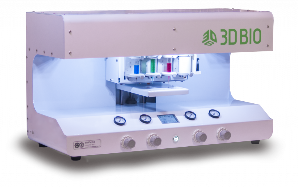
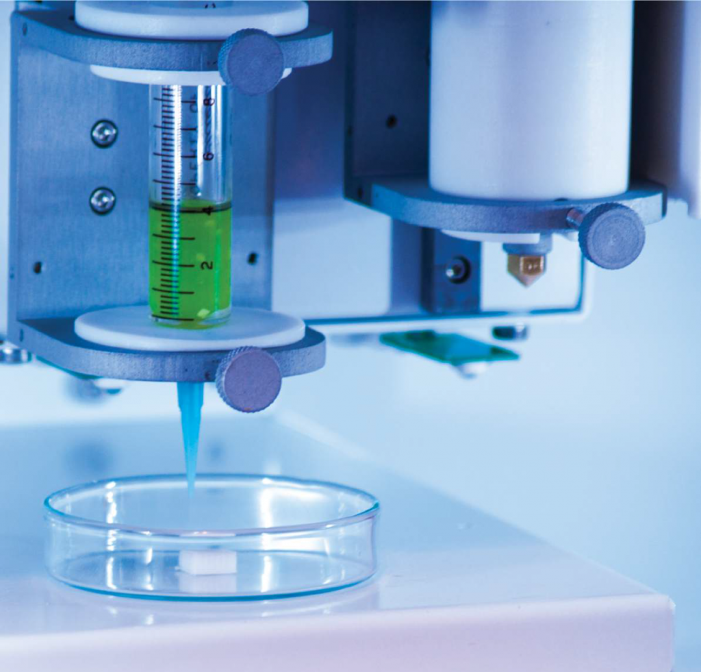
What are the differences between the Biofab and the Pioneer series?

The main difference between these series is their printing mechanism. The Pioneer bioprinter is extrusion-based using screws while Biofab utilizes pneumatic actuators for printing. This, in turn, makes Biofab more capable especially in the case of accuracy. The Pioneer version possesses the ability of unparalleled control over print heads and can print a variety of hydrogels. Moreover, the Biofab version supports a wide range of biomaterials and viscous cell suspensions for printing.

Both series provided in two versions, with 2 or 4 printing heads.

What kind of bioinks have you developed?

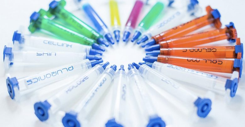
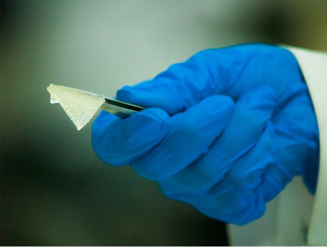
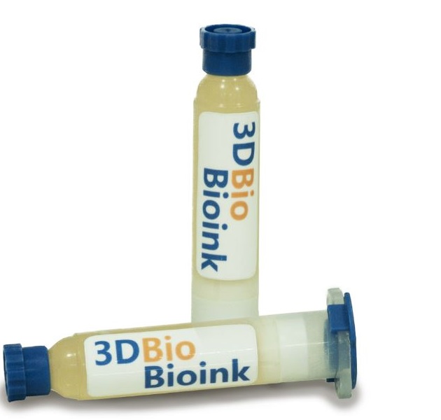
Omid Afarinan, as the first national company in Iran, has gathered experts from different fields of science with the aim of producing novel bioinks. Omid Afarinan bioinks are cost-effective, have high printability, mimic extracellular matrix (ECM) and provide a suitable environment for cell proliferation, growth, and differentiation. Some of the new unique bioinks of the 3D-Bio Team are PCL, PCL/Starch, Alginate and Alginate/Gelatin bioinks. Soft-Ink is the newest bioink of the company; a biodegradable bioink based on pure Alginate and Gelatin which can support growth and proliferation of any cell type of soft tissues. One of the main features of this bioink is high printability and uniformity with the ability to adjust the stiffness of its printed matrix.

In addition to a variety of bioinks from thermoplastic materials to hydrogels, the company also produces custom-made bioinks for specific applications.

What are customers doing with your printers?

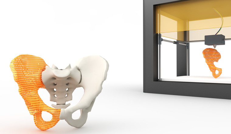
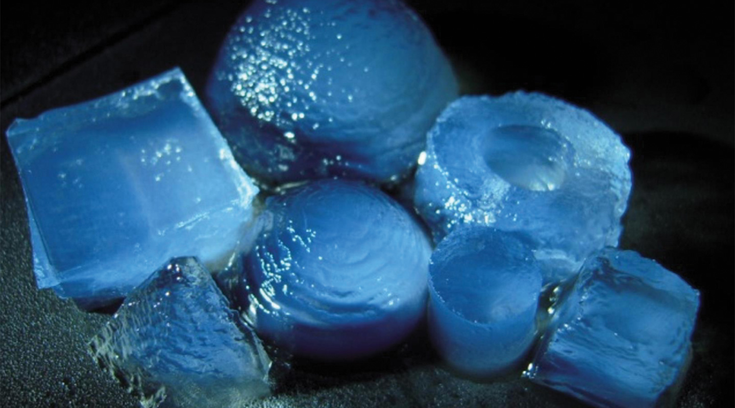
The customers mainly use the printer for conducting state-of-the-art researches especially on creating regenerative tissues for transplantation and studying the behavior of living tissues. In particular, our current customers work on hard tissue as bone scaffold especially maxillofacial and soft tissue including cartilage, skin, cornea and heart. The study on drug delivery, cosmetic research and cancer treatment are other aspects of what our customers do with bioprinters.

What short term successes do you see occurring in bioprinting?

Successes in bone and cartilage tissues are promising in recent years. This is due to the fact that such tissues have low cell densities. This is more pronounced in the bone tissue where acellular scaffolds can be used. Skin printing comes next in the list on the soft tissue side, for being a flat and multi-layered tissue. Also, Bioinks are being developed in parallel but at a slower pace. These short-term successes would pave the path for tissues with more complex geometries.

Where is bioprinting challenging?

The challenges exist in both pre- and post-printing stages of bioprinting. At pre-printing stage, the challenge is on the printability of biomaterials and Bioinks. In other words, making the materials printable and the suitability of the printing is of prime importance. Overcoming this challenge leads to printed functional tissues that could mimic real ones. On the post-printing stage, the challenges mainly arise from the complexity in living tissues especially vascularization and post-printing processes such as cell culture and migration that makes the printed tissues ready for their applications. These challenges and issues need close collaborations between different experts to resolve in short and long-term periods.


What advice would you have for a researcher wanting to get into bioprinting?

The first advice is that the researcher should wear iron shoes. In other words, facing many difficulties is unavoidable and gaining achievements may take time. So, patience and being hopeful is the key point. Furthermore, bioprinting requires a multi-disciplinary group including engineers, biologist and doctors and no one can individually succeed in this path. Finally, utilizing a standard and reliable bioprinter could be beneficial and time-saving for any researcher. 

The post Omid Afarinan is Bioprinting in Iran appeared first on 3DPrint.com | The Voice of 3D Printing / Additive Manufacturing.

China: Complex GelMA-based Scaffolds Improved with the Addition of Nanoclay

Chinese scientists are delving further into successful bioprinting in ‘3D printing of complex GelMA-based scaffolds with nanoclay,’ exploring why photo-crosslinkable gelatin methacrylate (GelMA) has become so enticing for researchers attempting to engineer tissue. In a realm rife with obstacles, however, GelMA is no exception—constricted by viscosity issues and long cross-linking time.

The authors decided to bolster the ink further with nanoclay, in the hopes of being able to print stable, complex scaffolds. During this study, they evaluated windows for printability, issues with porosity and mechanical strength, and biocompatibility.

Obviously, without cell viability, there is no bioink and there are no spectacular innovations to write about. A wide range of hydrogels have been used successfully, with alginate commonly involved due to rapid crosslinking speed. Here, however, the researchers explain that alginate is not always conducive to attachment of cells or good function. Gelatin methacrylate (GelMA), however, is known to crosslink easily during light exposure. The researchers point out also that it maintains the biocompatibility of gelatin.

In attempting to overcome multiple issues with the use of GelMA, such as low viscosity and extensive time required for cross-linking, they examined the use of pre-crosslinking, post-crosslinking, in-situ crosslinking, and two-step crosslinking. Ultimately, the consensus was that all the methods were unsuitable, resulting in inferior stability. With the addition of nanoclay, however, the authors discovered that the ink had higher viscosity, and the GelMA scaffolds had better shape fidelity.

“After extrusion, the nanoclay rapidly converted to the gel state upon the release of shear stress, thereby forming stable hydrogel filament,” stated the authors. “Finally, the 3D structure was printed layer-by-layer by stacking the filament, and the GelMA within the filament was covalently crosslinked under UV light, resulting in a stable scaffold.”

3D printing strategy of complex scaffolds with GelMA/Nanoclay ink. (A) Schematic illustration of printing scaffolds with GelMA/Nanoclay ink: (I) preparing GelMA/Nanoclay ink, (II) extruding filament based on the thixotropy property of nanoclay, and (III) printing structure based on the photo-crosslinking of GelMA. (B) Rheological properties of the GelMA/Nanoclay ink: (I) flow
behavior of 4% nanoclay, 10% GelMA, 10/3% GelMA/Nanoclay, 10/4% GelMA/Nanoclay, and 10/6% GelMA/Nanoclay, (II) the viscosity-shear rate, and (III) the shear moduli-angular frequency of the respective biomaterial inks.

They also found that nanoclay at higher levels resulted in less expansion due to more shear stress, meaning that nanoclay with higher concentration needed greater yield stress for deformation. In further discussion, the authors states that greater balance needs to researched for printing with GelMA/Nanoclay, and that so far, they surmise that if ‘cell-laden structures’ are to be directly 3D printed, they are forced to give up shape fidelity. Along with that, greater control is required of the following:

  • Mechanical strength
  • Degradation rate
  • Tissue regeneration capacity

“Through systematic experiments that included rheological testing, printability analysis, property characterization, and biocompatibility characterization, we have answered several fundamental questions relating to this ink, including the formation mechanism for shear-thinning and rapid-gelling and the printability window for the fabrication of complex GelMA scaffolds, as well as showing that the addition of nanoclay improved the basic properties and had no effect on the excellent biological performance of the scaffolds,” concluded the researchers.

“Therefore, this method provides an easy way to fabricate complex GelMA-based scaffolds with good shape fidelity. It is very likely that this method will have versatile applications in the individualized therapy of tissue defects.”

Printability analysis of GelMA/Nanoclay ink with regard to the extrusion process. (A) Schematic illustration of the expansion phenomenon and definition α = D/d.(B) Effect of the nozzle diameter on (I) the extruded filament diameter, and (II) α.
(C) Effect of the flow rate on (I) the extruded filament diameter, and (II) α.

The world of tissue engineering and bioprinting is rich in a variety of scaffolds, and while here we have learned more about the use of GelMA-based scaffolds, researchers around the world are constantly experimenting with new ways to sustain cells and make inks that are cell-laden. We have followed studies regarding transparent bioinks for fabricating corneas, making neural tissue, and 3D printing complex structures like alginate/gelatin hydrogels. What do you think of this news? Let us know your thoughts! Join the discussion of this and other 3D printing topics at 3DPrintBoard.com.

Photograph of the printed scaffolds with various shapes. (A) Abbreviation of Zhejiang University. (B) A bionic ear. (C)A branched vessel.

[Source / Images: ‘3D printing of complex GelMA-based scaffolds with nanoclay’]

Biodiscoveries: CELLINK is bioprinting its way into the future

Back in 2015, Erik Gatenholm realized there was no place to purchase bioink for 3D bioprinting. So, blown away by this gap in the market, he quickly worked with co-founder Hector Martinez to create a universal bioink that anyone working with bioprinting could use. It was quite a high stakes bet, and at the beginning they set up a webshop to see if they got any bites. It only took 24 hours for the first sale. With more orders quickly coming in, they realized the enormous potential of the product they developed, and CELLINK was born, becoming the first company to commercialize a universal bioink for bioprinting of human tissues and organs.

CELLINK co-founders Erik Gatenholm and Hector Martinez

In the United States alone, every 10 minutes another person is added to the growing waiting list for organ transplants, most of them (60%) in need of a kidney, and with over 130,000 organs transplanted every year worldwide, is no wonder how demand certainly outweighs supply almost everywhere. In some countries the wait can take years, making 3D printing of organs one of the most sought after technologies out there. Bioprinting in the future could allow patients and doctors to reduce waiting times, increase compatibility and decrease immunological failure. For this to happen medical researchers will need to design organs using modeling software, and then print them with biomaterials such as polymers and hydrogels, in addition to the patient’s own cells. Although currently focused on growing cartilage and skin cells suitable for testing drugs and cosmetics, the Swedish company founded in Gothenburg in 2016, hopes to progress the technology far enough to create replacement organs for transplant in humans in the next 15 years.

“In the coming decade we would like to continue to push the boundaries of 3D bioprinting until it becomes an established technology in the medical field. We have the vision of becoming the first and number one provider of bioprinters, bioinks, software and technical know-how for the next generation of medical device manufacturers,” co-founder and CTO Hector Martinez told 3DPrint.com.

Their unique bioink is a biomaterial innovation that allows human cells to grow and thrive as they would in circumstances close to their natural environment. The startup has already managed to print human skin and is also working on producing liver tissues, as well as the beta cells that produce the insulin we need to survive. In 2018 it began printing tumors to combat cancer as part of a research project that doesn’t endanger human lives, and just a few weeks ago, it teamed up with Volumetric to develop Lumen X, a digital light processing bioprinter, designed to enhance inventions in creating more substantial vascular structures. Skin care products, topological drugs and medical treatments are all in need of enhanced testing procedures that can increase the transability from in vitro testing to in vivo usage of products. With tissue engineering and 3D bioprinting more representative in vitro models can be constructed, limiting the use of testing in animals.

CELLINK’s bioinks

Actually, academic labs and companies worldwide are trying to bioengineer all kinds of sophisticated creations for regenerative medicine, drug testing, screening, and tissue engineering. So it’s no wonder CELLINK has their research team focused on creating the next generation of bioinks. Their top selling product is making bioprinting much easier than it used to be some 10 years ago, with 30 different types of bioink available, with prices that go from 99 to 900 dollars. So, what makes one bioink more expensive than the other? It’s all about the components. Collagen and laminin are more expensive to produce than gelatin, raising the price of the end-product. According to CELLINK, scientists mix their live cells into the company’s bioink, a kind of gel designed to allow cells to survive and multiply. The ink is then loaded into a 3D printer by the customer, which forms the desired shape layer by layer as the gel solidifies. By the time the lights inside CELLINK’s box turn green, researchers have an object that acts like human tissue, and can then apply their drug and see how the living cells inside respond.

CELLINK team printing liver models at the lab

“Today we are taking the necessary steps to build and expand our technology offering and exploring new methods for bioprinting tissues. Such technologies include multiple contact-less dispensing methods and light-based bioprinting techniques that enable the bioprinting of high resolution tissue constructs. Refining such technologies will take a close collaboration with our customers as we define the best practices for bioprinting different tissues and specific functions. We can already anticipate that the integration of different bioprinting technologies with post-bioprinting, real-time monitoring systems will be of utmost importance as the bioprinted tissue matures and attains a specific function through an active and precise manipulation of its environment.” Chief IT Officer Jockum Svanberg explained to 3DPrint.com.

Creating the raw material for bioprinting processes is no easy task. Cellink has been focusing on process-compatible soft biomaterials loaded with living cells to create its bioinks since September 2015. The process of bioprinting requires a delivery medium for cells which can be deposited into designed shapes acquired from computer-aided design (CAD) models, which can be generated using 3D medical images obtained through MRIs or CT scans. Some important features of an ideal bioink material are bioprintability, high mechanical integrity and stability, insolubility in cell culture medium, biodegradability at a rate appropriate to the regenerating tissue, non-toxicity and non-immunogenicity, and the ability to promote cell adhesion. Some bioink types, like hydorgels, are not always suitable as construction materials which is why CELLINK is working on a study to provide an upgraded version of the current CELLINK BONE bioink by incorporating collagen and hydroxyapatite. The bioink currently offered does not get close to the real stiffness of the natural bone tissue, but finely resembles its chemical composition. The advantage of such a soft material is to be able to incorporate cells and, during the bioprinting process, to locate them at a precise position throughout the scaffold. This is still for research use only and might take a few years until it is compatible for human use.

With CELLINK bioinks, 3D bioprinting of tissues will help hasten bone fracture healing

Since its start the technology firm has grown to become one of the big competitors in the industry. CELLINK had only been in existence for ten months before they decided to pursue their IPO in November of 2016, listing on Nasdaq First North after a 1070% oversubscribed IPO, which means that demand for their shares was ten times what they expected. Since then, shares have risen over 400%, giving the company a present-day market cap of around $257 million. CELLINK’s affordable printers have already been bought by customers in 25 countries around the world, mostly universities, like Stanford, Harvard, Yale, Princeton and MIT, and some private customers, including Shiseido, Roche, Merck, Johnson and Johnson, and Toyota

But it’s not just about bioprinting it’s way into the future of medicine, CELLINK is also working with other disruptive technologies, such as machine learning. CELLINK told 3DPrint.com that “they want to empower our users with better tools to simplify the bioprinting learning process and broaden its adoption”. One example of this is by developing algorithms that analyse printed structures and based on the results can recommend printing parameters to the users. Using this tool in the development, has helped them speed up the bioink development process. They have just launched a new product: CELLCYTE X, a live cell imaging microscope with live monitoring and analysis of cells in the cloud. Traditionally cell studies have involved manual labor and relied on analysis of the images from an expert, but using deep learning models they are automating this process to provide better and more reliable analysis to their users. The system relies on the latest in serverless system architecture to provide the most scalable, reliable and most intuitive system on the market.

What do you think, will CELLINK continue its upward trajectory? Will it become superseded by other larger firms or get passed by newer start ups? Find out more through our series of articles exploring bioprinting, Biodiscoveries.

Bio-printing 101: How to Bioprint at Home

<em>Picture 2: </em>Hydrogel fabricated by PSL 3D printing technique” width=”565″ height=”314″></p>
<p style=Bioprinted Hydrogels

Bioprinting is an exciting area to follow as it invigorates the ideas of Frankenstein and a bunch of other sci-fi scenarios that make us slightly paranoid. So how does someone create their own monster within their garage? Well, that is pretty far fetched at the moment, but there are ways to get involved in this field in small ways.

Consistently, within bioprinting, there does not seem to be a significant presence within the general maker community. The healthcare industry as a whole is typically private sector driven. So how does someone get the chance to work on 3D bioprinting when they do not even know the resources they need, or how to start?

An essential consideration for 3D bioprinting is the material used for prototyping. Typically 3D printers use different materials to create products such as PLA, ABS, Wood Fiber, PET, PVA, Nylon, and TPU. The issue of creating bioprinted materials is not within the actual structure of the model and design. The problem lies in creating objects that also follow the rules of biology. This limitation forces a material to have specific heating and cooling properties in relation to where it is within the body. Specific heat and tolerance to different temperature ranges are vital in a material used for bioprinting. Even with the creation of 3D structures, there is still a difficulty in replicating the intricate vascular structures of different organs within our bodies. This makes for a variety of problems that need to be worked on within the field of 3D bioprinting in general.

So what are some first steps within 3D bioprinting? Let us focus on some materials that would be ideal to focus on as they are good candidates for current and future use. Here is a list of some current materials used in bioprinting:

  • Bioink
  • Hydrogels
  • Alginate
  • PEG (polyethylene glycol)
  • PCL (Polycaprolactone)
  • PGA (Polyglycolic Acid)
  • Pluronics

In later articles, we will discuss each material above in more depth. For now we will address them briefly.

Bioinks are substances made of living cells that can be used for 3D printing of complex tissue models. Bioinks are materials that mimic an extracellular matrix environment to support the adhesion, proliferation, and differentiation of living cells.

A hydrogel is a network of polymer chains that are hydrophilic, sometimes found as a colloidal gel in which water is the dispersion medium. A three-dimensional solid results from the hydrophilic polymer chains being held together by cross-links. Alginate is a polysaccharide distributed widely in the cell walls of brown algae, where through binding with water it forms a viscous gum.

Alginate is made from brown seaweed.

Alginate is a polysaccharide distributed widely in the cell walls of brown algae, where through binding with water it forms a viscous gum.

PEG is marketed as a laxative but is also a stabilizing agent in toothpaste.

Polyethylene glycol (PEG) is a polyether compound with many applications, from industrial manufacturing to medicine and is often used in making hydrogels for 3D printing.

Polycaprolactone (PCL) is a bioabsorbable polyester with a low melting point of around 60 °C and a glass transition temperature of about −60 °C.

Polyglycolic acid (PGA) is a biodegradable, thermoplastic polymer and the simplest linear, aliphatic polyester. PGA is used for scaffolds and as a support material.

Pluronics are nonionic triblock copolymers composed of a central hydrophobic chain of polyoxypropylene (poly(propylene oxide)) flanked by two hydrophilic chains of polyoxyethylene (poly(ethylene oxide)).

Now that we understand a little bit about the materials that we can use, it becomes a question of what type of printer to use. Most industrial bioprinters are far from a viable price for consumer purchase.  Communities of makers have few options for buying a 3D bioprinter. There also seems to be a lack of internet resources to instruct people on how to bioprint. To help people at home, we will try to build a 3D bioprinting setup with a guide for all those who are interested in bioprinting. As a follow up on this article, be sure to look out for information on the biomaterials mentioned above and separate articles on each of them and how they relate to 3D bioprinting. Stay tuned for more DIY bioprinting tips and tricks.