The Bioprinting Revolution:Accessible 3D Printing to Transform Cell Culture

In ‘The revolution will be open-source: how 3D bioprinting can change 3D cell culture,’ authors Robert D. Bruno, John Reid, and Patrick C. Sachs explore the revolutionary aspect of 3D scaffolds in bioprinting, with tissue engineering spanning such a wide range in medicine today.

While 3D printing is impacting nearly every industry around the globe it would seem, within the medical realm, the technology is also touching virtually every aspect from breast cancer research to the dental industry.

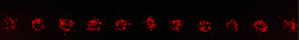
Example of a coordinated print of clusters of red fluorescent protein (RFP) labeled MCF12a cells at distances of 200µm in a linear array. Image taken 24 hours post-print. Scale bar = 200µm.

For most researchers, common types of substrates used in tissue engineering are membrane rich extracellular matrix (ECM) and collagen extracted from rattails.

“The processes of 3D culture in these two substrates has remained unchanged for nearly half a century: cells are either mixed with unpolymerized matrix to disperse them randomly throughout the substrate upon polymerization or overlaid randomly on top of a preformed hydrogel. While effective in generating organoid/tumoroid structures, the random nature of these processes has many drawbacks that limit the reproducibility and tunability of the experimental design,” explain the authors.

And while one of the greatest aspects in 3D printing today may be the sudden accessibility and affordability of 3D printers (a great example of this would be the ubiquity of such technology in schools around the world, as well as DIY workshops), that has not yet trickled down to bioprinting–with the exception of specialty labs graced with substantial funds to purchase necessary equipment and bionics.

With the inception of this project, the authors developed their own affordable, open access 3D bioprinting system. Meant to be used in scientific applications, the printer is extremely versatile, and adaptable to many different applications—from printing cells to guiding electrodes for direction of electrical pulsing of cells.

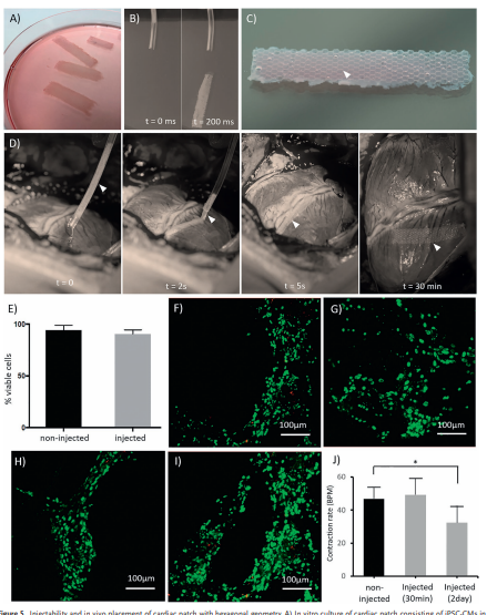
“To increase precision and maintain integrity of printed cells, we use pulled glass micropipette syringes as our cell injection ‘printhead,’” state the researchers. “Compared to standard steel needles attached to luer lock syringes, these glass micropipettes have a finer point and reduce sheer force on the cells. Combined, this minimizes disruptions to the cells and allows the hydrogel to seal behind the print. Thus, our system allows for the precise placement of cells, that then self-organize into organoids/tumoroids, making functional structures.”

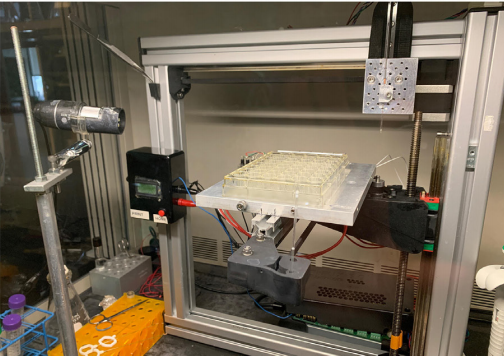
Custom benchtop 3D bioprinter and its application for printing large mammary organoids. 3D bioprinter constructed off of the Felix 3.0 (FELIXrobotics, NL) platform.

The researchers have maintained a focus on bioprinting within the realm of mammary organoids, with the potential for growing cells into predictable sizes and shapes.

“The key to the guided growth was the fact that mammary epithelial cells (MCF12a and MCF10a) would preferentially grow towards neighboring prints, forming single contiguous organoids. Using this strategy, we generated large contiguous luminal mammary organoids (> 5mm in length). This is in clear contrast to random culture where the dispersion of cells results in random organoid shape and size, with organoids never forming more than a couple of hundred microns in size,” concluded the researchers.

Our studies highlight the ease of access and the utility of the technology for basic cell and cancer biology studies. Thus, we hope to lower the bar of entry further by developing easier-to-access solutions, such as ready-built kits, and a graphic user interface (GUI) to simplify the experimental programming. The system offers a potentially revolutionary step forward for 3D culture models of development and cancer.

Bioprinting is making a huge stamp on the world, as well as the 3D printing realm—a technology that has inspired scientists in laboratories around the world to take on the challenge of tissue engineering.

And while research has already yielded so many positive results, from allowing patients to lead higher quality lives due to innovations like tunable tissue, development of microfluidic techniques, and even tailored skin grafts, the true goal—the ultimate miracle of 3D printing will be that of a human organ.

Once scientists can overcome that hurdle, many patients will rejoice in knowing that they may be able to bypass long-term waiting lists for organs made from their own cells.

What do you think of this news? Let us know your thoughts! Join the discussion of this and other 3D printing topics at 3DPrintBoard.com.

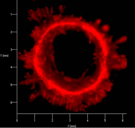
Mature organoid (21 days post-print) formed from coordinated print of clusters of RFP MCF12a cells into a circular array. Resulting organoids have been shown to have contiguous lumens stretching > 3mm in length.

[Source / Images: ‘The revolution will be open-source: how 3D bioprinting can change 3D cell culture’]

The post The Bioprinting Revolution:Accessible 3D Printing to Transform Cell Culture appeared first on 3DPrint.com | The Voice of 3D Printing / Additive Manufacturing.

University of Missouri: Thesis Student 3D Prints Collagen to Create Tissue Engineering Scaffolds

University of Missouri thesis student, Christopher John Glover, explores the use of 3D printed structures in bioprinting, outlining his findings further in ‘In Situ Polymerizing Collagen for the Development of 3D Printed Tissue Engineering Scaffolds.’ Extolling the virtues of collagen while also discussing challenges in using it, Glover explains that this natural material has been a favorite in tissue engineering, demonstrating excellent protein structures for ventures in the lab.

“Nearly all tissues in the human body contain collagen including skin, muscle, nerves, vasculature, tendons, ligaments, and even bone. Skin, for example, is 80% collagen by mass. Due to this abundance, collagen is extremely biocompatible and versatile,” states Glover. “With the proper mechanical and chemical stimuli, stem cells seeded on collagen scaffolds have the potential to differentiate down a myriad of cell lineages and become nearly any tissue in the human body.”

There can be difficulty in using collagen for some types of tissue regeneration though, and other disadvantages such as the amount of time it takes to progress from a gelatinous state to a solid.

For this project, Glover studied the manufacturing of 3D collagen-based scaffolds which he enhanced with a variety of anti-inflammatory agents such as gold nanoparticles and curcumin. Specifically, he used in situ polymerizing collagen (IPC), a unique material derived from Type 1 porcine collagen. In experimenting, he performed different post printing treatments on the test groups. Some were just left in their basic 3D printed state, which others were crosslinked without AuNP or curcumin or with either 1X or 2X AuNP or curcumin. Characterization was performed in evaluating stability of each scaffold and then noting its viability, along with which types of treatment were most successful.

Tasks for ascertaining viability were as follows:

  • To 3D print uniform and reproducible collagen-based scaffolds
  • To examine the thermal properties of the crosslinked scaffolds
  • To verify and quantify the presence of gold nanoparticles in the crosslinked scaffolds
  • To evaluate the cytotoxicity and anti-inflammatory capabilities of the gold nanoparticle and curcumin scaffolds

The six experimental groups were:

  • Uncrosslinked
  • Crosslinked
  • AuNP
  • Curcumin
  • 2X AuNP
  • 2X curcumin

“The uncrosslinked group exists to examine the effects of crosslinking alone; the AuNP and curcumin groups exist to determine the effects of each bioactive agent; the 2X AuNP and 2X curcumin groups exist to exacerbate those effects, for better or for worse,” stated Glover.

Glover customized his own 3D printer, assembled from a CNC milling machine, with translational stages manipulated by three stepper motors. Mach3 Mill software was used in design and editing. The two most common 3D prints made during the study were a grid pattern and circles used for cell assays. Glover found that resolution was not optimum with his hardware but thought it could be finer on a higher-performance printer.

The 3D printer features a 3D printed holster to house the syringe pump and is seen here printing a circular grid pattern

The Mach3 Mill software interface features many functions not utilized in
our 3D printing process, such as the tool information and spindle speed boxes.

Crosslinking with EDC or genipin proved to enhance both stability and durability of the 3D printed scaffolds.

“By comparing the application of EDC crosslinking during printing versus post printing, it was found that crosslinking post printing yielded significantly greater stabilities than crosslinking during printing,” stated the researchers.

The collagen-based scaffolds crosslinked with EDC exhibited ‘superb cell viability,’ although Glover pointed out that gold nanoparticles seemed to decrease success in viability somewhat. Genipin also decreased viability, which plummeted further with the addition of curcumin.

“As previously stated, collagen alone is a fragile material and even after crosslinking can deteriorate if over-handled. If this platform is to be utilized to produce implantable scaffolds, the durability of the collagen would need to be markedly improved. This could be accomplished by printing the IPC along with another material or by further post-print manipulation of the collagen other than simply crosslinking,” concluded Glover, who goes on to state that printer resolution would need to be improved, along with enhancing of the anti-inflammatory capabilities of the printed products.

3D printing with collagen has been of great interest to researchers lately, including uses in artistic masks, bioink, and skin grafts. Read more about collagen in tissue engineering here. What do you think of this news? Let us know your thoughts! Join the discussion of this and other 3D printing topics at 3DPrintBoard.com.

[Source / Images: In Situ Polymerizing Collagen for the Development of 3D Printed Tissue Engineering Scaffolds]

Creating Vascular Structures Using Low Cost Desktop 3D Printers

In a thesis entitled “Engineering of vascular networks within biocompatible hydrogels using 3D printing technology,” a PhD student named Juan Liu discusses the need for new technologies in wound healing. While skin flaps and grafts are the “gold standard” in clinical treatment of large skin and subcutaneous tissue defects, there are many complications that can arise from these treatments, and there is also the issue of not having enough skin to be able to harvest to cover particularly large wounds.

3D printing, however, using stem cells, allows for an unlimited amount of tissue to be created to heal large wounds. Using cells from the patient reduces the risk of rejection, as well. In order to create and maintain live tissue, however, vascularization is required, meaning that blood vessels need to be established, which is the tricky part of tissue engineering. Liu hypothesizes that open source desktop 3D printing technology can be used to design and fabricate “customized bio-artificial multicellular tissues with embedded vessel-like supply channels and corresponding bio-reactors for long-term 3D tissue culture.”

Liu outlines the following objectives:

  • Use open source software to design 3D tissues with vascular patterns and fabricate them with desktop 3D printers
  • Analyze cell survival and function over time in 3D tissues in respect to vascular patterns
  • Generate 3D printable bioreactors that allow online monitoring of the process
  • Prevent shrinking of 3D tissues
  • Generate multilayer 3D tissues with different cell types

Liu goes on to demonstrate how 3D printing technology can be used to precisely form vascular structures within cell-laden hydrogels. He also creates “customized PLA and PDMS bioreactors for continuous perfusion and real-time operation.” The vascular structures formed within the cell-laded hydrogels, which were created by 3D printing, were able to increase the viability of the cells surrounding the vascular structures.

“In addition, the final system based on PDMS has been proven to sustain long-term 3D cell culture, which is the basic for 3D cell proliferation and tissue formation in vitro,” says Liu. “Among others this approach exhibits unique advantages to fabricate a hydrogel-based multilayer vascular device in a cost-effective and fast manner, which has a huge potential for viable 3D cell culture, complex tissue engineering, disease modeling as well as drug screening.”

This model, he continues, could also be used to mimic natural tissue architecture and create bigger multilayer hydrogel constructs with corresponding layers of functional cells to study cell morphology, differentiation, and potential function of engineered tissue constructs.

“Further optimization of the hydrogel concentration in each layer, cell density and perfusion parameters may enable the preparation of 3D vascular tissues under the conditions mimicking the natural environment with better functions and matrix compositions,” he adds. “In order to overcome the tendency of shrinking in soft hydrogels, combination with eletrospun fibers are promising. The system presented in this thesis allows fabrication of vascular networks in both soft and stiff cell laden hydrogels. It may serve as a novel platform for vascularized tissue engineering, that facilitates the generation of more functional, engineered vascular tissue. It may be useful for studies of wound coverage and tissue regeneration and eventually aid the treatment of wounds.”

Discuss this and other 3D printing topics at 3DPrintBoard.com or share your thoughts below.


3D Bioprinting: Comparing Lattice Scaffolds with Traditional Rectangular Sheets

Bioprinting is not a simple endeavor – if it were, we would likely be transplanting 3D printed organs by now. It’s a delicate process that requires a number of factors to be in place, including bioinks that are both printable and biocompatible, and proper scaffolds. In a paper entitled “A Comparative Study of a 3D Bioprinted Gelatin-Based Lattice and Rectangular-Sheet Structures,” a group of researchers compares scaffolds with lattice mesh geometries to more traditional flat rectangular sheets.

“We hypothesised that the experiments performed as a part of this study would help us to observe considerable differences between the two structures, i.e., lattice and rectangle, and also open up the possibility of significantly enhancing the design of a 3D bioprinted construct for engineering cardiac tissue-on-a-chip, using bioprinting,” the researchers state.

The researchers used furfuryl gelatin (f-gelatin) as a base for their bioink, which they seeded with mouse mesenchymal stem cells. They used an ALLEVI 2 bioprinter to print the ink into two different structures – a lattice and a rectangular sheet. Rheological characterization of the bioink was conducted, and the bioprinted structures were cultured in an incubator. A live/dead cytotoxicity assay was performed, and the texture of the lattice was analyzed by scanning electron microscopy.

“The SEM cross-sectional image of the gelatin lattice revealed a highly organized, striated, patterned, and networked structure in comparison to the loosely networked and largely porous rectangular-sheet cross-section SEM, as reported in our previous study,” the researchers explain. “Porosity and pore-size are crucial to ensure cell colonization of the scaffold, deposited using bioprinting. Likewise, SEM micrographs showed a homogeneous distribution of equal sized pores within the entire area scanned and imaged….The average apparent porosity of this lattice structure was estimated to be about 50% compared to 21% for the rectangular-sheet. The results led us to conclude that although the mean pore size was significantly reduced by printing in the form of a lattice, the inherent design of the lattice allowed pores to be of a similar size and to be homogenously distributed throughout the entire structure, compared with the rectangular-sheet.”

Swelling behavior of the gels was monitored to study the hydration dynamics of the crosslinked hydrogel structure. Cell proliferation was assessed and flow cytometry was analyzed.

Results of the testing showed that the lattice structure was more porous than the flat rectangular sheet. It also exhibited a lower degradation rate.

“Further, the lattice allowed cells to proliferate to a greater extent compared to the rectangular-sheet, which initially retained a lower number of cells,” the researchers state. “All of these results collectively affirmed that the lattice poses as a superior scaffold design for tissue engineering applications.”

A scaffold is literally a foundation to build upon in bioprinting, and having an effective scaffold is key in any bioprinting application. Cells rely on a strong scaffold in order to survive and proliferate. A printable, biocompatible ink is also crucial for cells to be able to grow into tissue. The researchers find in this study that lattice structures are superior to rectangular sheets, which could mean the difference between success and failure in future applications.

Authors of the paper include Shweta Anil Kumar, Nishat Tasnim, Erick Dominguez, Shane Allen, Laura J. Suggs, Yoshihiro Ito, and Binata Joddar.

Discuss this and other 3D printing topics at 3DPrintBoard.com or share your thoughts below. 


Exclusive: 3D Printed Cardiac Patch Shows Promise for Healing Damaged Hearts

Hexagonal scaffolds induce iPSC-CM contractile properties and maturation. iPSC-CM characterization before combination with scaffold, showing A) proliferating cells, B) sarcomeric structures, C) Connexin 43 (Cx43), and D) mitochondrial localization. iPSC-CMs localize to both E) hexagonal and F) rectangular fiber scaffolds, and confocal microscopy shows G,H) Cx43 expression, and increased sarcomere density, I,J) alignment, and K) length in hexagonal scaffolds compared to rectangular scaffolds. L) Beating rate at days 2, 7, 10, 11, and 14. M) Cardiac marker and maturation-related gene expression in hexagonal and rectangular scaffolds at day 7 and day 14.

After a person suffers a heart attack, they lose about half the cells in their heart, greatly weakening the organ and increasing the odds that further attacks will occur. Doctors have begun injecting cells into the heart to grow into muscle and help contractions, but 99% of those cells get washed away. But there are alternative approaches – like 3D printing. Scientists have developed 3D printed cardiac patches that can be used to repair hearts damaged by heart attacks, but only about five have been produced worldwide.

Injectability and in vivo placement of cardiac patch with hexagonal geometry. A) In vitro culture of cardiac patch consisting of iPSC-CMs in cardiac-like ECM on large hexagonal scaffolds. B) In vitro injectability, shape recovery, and C) macro image of cardiac patch after injection. D) Application and shape recovery of cardiac patch on beating porcine heart. E) Cell viability of in vitro F,G) noninjected and H,I) injected cardiac patches. J) Spontaneous beating rate of in vitro noninjected and injected cardiac patches 30 min and 2 d after injection.

In a new study entitled “Melt Electrowriting Allows Tailored Microstructural and Mechanical Design of Scaffolds to Advance Functional Human Myocardial Tissue Formation,” a group of researchers 3D printed a world-first stretchable microfiber scaffold with a hexagonal design. They then added specialized stem cells called iPS-Cardiomyocytes, which began to contract unstimulated on the scaffold. The work was then demonstrated on the actual hearts of pigs.

We spoke with the authors of the paper to learn more about the technology used by the scientists and its implications for the future.

What are the advantages of MEW relative to other technologies? 

“Melt Electrospin Writing (MEW) has distinct advantages over other 3D Printing technologies for tissue engineering applications. MEW utilises common thermoplastics used in biomedical engineering, including Polycaprolactone (PCL). The advantage of this approach is that we are able to create microfibres with diameters often in the 10 micron range, but nano-fibres also also achievable. To put this into perspective, a single strand of hair is about 50-100 microns. Printing at such resolutions allows us to mimic the native extracellular matrix (ECM) components, and allows cells to bind onto the small fibres that at similar to collagen fibrils. Additionally, MEW allows for the control over scaffold architecture, creating scaffolds with aligned fibres and controlled fibre diameters. This specific control over architecture, allows us to tailor the mechanical properties, direct cells growth and  control the movement of nutrients. Because we can modify the scaffolds properties so well, they are often used to reinforce hydrogels (as a backbone), which are typically much weaker and less tailorable.”

What is the significance of your paper?

Representation of the workflow and fabricated microfiber scaffolds. A) Schematic illustration of the in house-built MEW device used. B) Designed hexagonal microstructure. C) 3D fiber scaffold combined with iPSC-CMs and further application in vivo through minimally invasive delivery. D) Optical images of the fabricated scaffolds: detail of microstructure with hexagonal cells (with a side length of 400 µm) composed of multiple stacked aligned microfibers. Images acquired from top and lateral perspective.

“We developed patches with controlled hexagonal micro-fibre structures that had unique flexibility and shape-recovery properties, meaning that the patch can be highly deformed without sustaining damage to its structure or cells. Moreover, such novel paths allowed the maturation of contractile human iPSC-derived cardiomyocytes, which is a breakthrough in creating a functional patch that could match an adult heart. Finally, due to the patch’s flexibility, it can be compressed and pushed through a catheter for delivery in vivo with minimally invasive laparoscopic surgery.”

Why are collagen-based hydrogels so important?

“Collagen is the most abundant protein in the human myocardial tissue and for that reason an ideal biomaterial for use in myocardial tissue engineering.”

How close are we to using 3D printing in a clinical setting?

“There are already reports of 3D printed implants used in clinics, mostly metallic or ceramic based for bone repair. However, in what concerns to our heart patch’s that include biological derived components (new cell therapies, iPSC) we believe a few more years will be required. We need first to demonstrate the efficacy and safety of such approach in animal models, which we are currently planning. Also, important challenges like integration with surrounding tissue will need to be solved: for example, currently the patches can contract autonomously, but we don’t know if they will sync with the beating of the heart once implanted.”

What work will you be doing next?

“We want to concentrate our efforts in conducting more extensive animal studies to assess feasibility and show functional effects, such as improved cardiac function. We also intend to make the patches more complex, by integrating other cell types in the patch.”

Authors of the paper include Miguel Castilho, Alain van Mil, Malachy Maher, Corina H.G. Metz, Gernot Hochleitner, Jürgen Groll, Pieter A. Doevendans, Keita Ito, Joost P.G. Sluijter, and Jos Malda.

Discuss this and other 3D printing topics at 3DPrintBoard.com or share your thoughts below.

[Images: Castilho et al, “Melt Electrowriting Allows Tailored Microstructural and Mechanical Design of Scaffolds to Advance Functional Human Myocardial Tissue Formation,” Advanced Functional Materials, 2018. Copyright Wiley – VCH Verlag GmbH & Co. KGaA. Reproduced with permission.]


New Study Shows Promise for Repairing Nerve Damage Through 3D Printing

Many injuries to the body can be healed, but nerve damage is something that can be permanent, resulting in the loss of the ability to properly feel, move or utilize the injured body part. There is a treatment for nerve damage that involves grafting healthy nerves onto the damaged parts, but the treatment isn’t perfect, according to Liqun Ning, a post-doctoral fellow in the Tissue Engineering Research Group at the University of Saskatchewan.

“Even the successful grafts only normally restore a portion of the nerve cell regional function so we are trying to find some new ways to solve this problem,” he said.

Ning is the lead author of a recent paper called “3D bioprinting of scaffolds with living Schwann cells for potential nerve tissue engineering applications,” which you can access here. In the paper, the researchers describe their goal: to 3D print scaffolds and place them within the body to try to regenerate healthy nerves. Though the scaffolds are extremely small, they are highly detailed and were created using the Canadian Light Source center at the University of Saskatchewan.

The cells seeded onto the scaffold are called Schwann cells, which are supporting cells in the nervous system that can force nerve cells to grow properly. The hope is that the scaffolds, once placed in the body, will stimulate new, healthy nerve cells to grow. The results of the study show that the 3D printed scaffolds can promote the alignment of the Schwann cells and provide cues to direct the extension of dorsal root ganglion along the printed strands.

This is only the first step, according to Ning, but it’s a step that shows great potential.

“We’re trying to test our method and the structure, the 3D printed scaffolds, with animals and to see if the structure helps to regenerate the peripheral nerve of the animal,” said Ning.

It will still be some time before any human trials can take place, but there’s a lot of promise there – like any other medical development, it will take time. The University of Saskatchewan has done some other impressive work with 3D bioprinting before, like creating 3D printed heart tissue. One of the authors of the heart tissue study, Xiongbao Chen, also worked on the recent nerve cell study.

Nerve damage can happen in the body in a number of ways – not just through injury, but due to illnesses such as diabetes. It can cause great difficulty for people who suffer from it, so any development towards restoring nerve function is an exciting one. Other work has been done advancing the bioprinting of nerve cells, so there’s hope for these patients, who number in the millions. Ning and his team believe that their work could eventually outperform grafts in restoring nerve function.

Before the first 3D printed organ is transplanted into a human being, developments such as this one may end up making the biggest difference in the lives of patients. Nerve damage is debilitating and even dangerous, and restoring the function to those damaged nerves could dramatically improve and even save lives.

Authors of the paper include Ning, Chen, Haoying Sun, Tiphanie Lelong, Romain Guilloteau, Ning Zhu, and David J. Schreyer.

Discuss this and other 3D printing topics at 3DPrintBoard.com or share your thoughts below. 

[Sources: Saskatoon Star Phoenix, CBC/Images courtesy of Liqun Ning]