Researchers around the world continually surprise us with innovation, but rarely do they reach into the roots of popular culture—and the food that accompanies it—applying it to the world of bioprinting…and with hot dogs in mind, no less. However, that’s exactly what has inspired German and Chinese scientists to build bioprinting structures, with their study outlined in the recently published ‘3D Printing of Hot Dog-Like Biomaterials with Hierarchical Architecture and Distinct Bioactivity.’
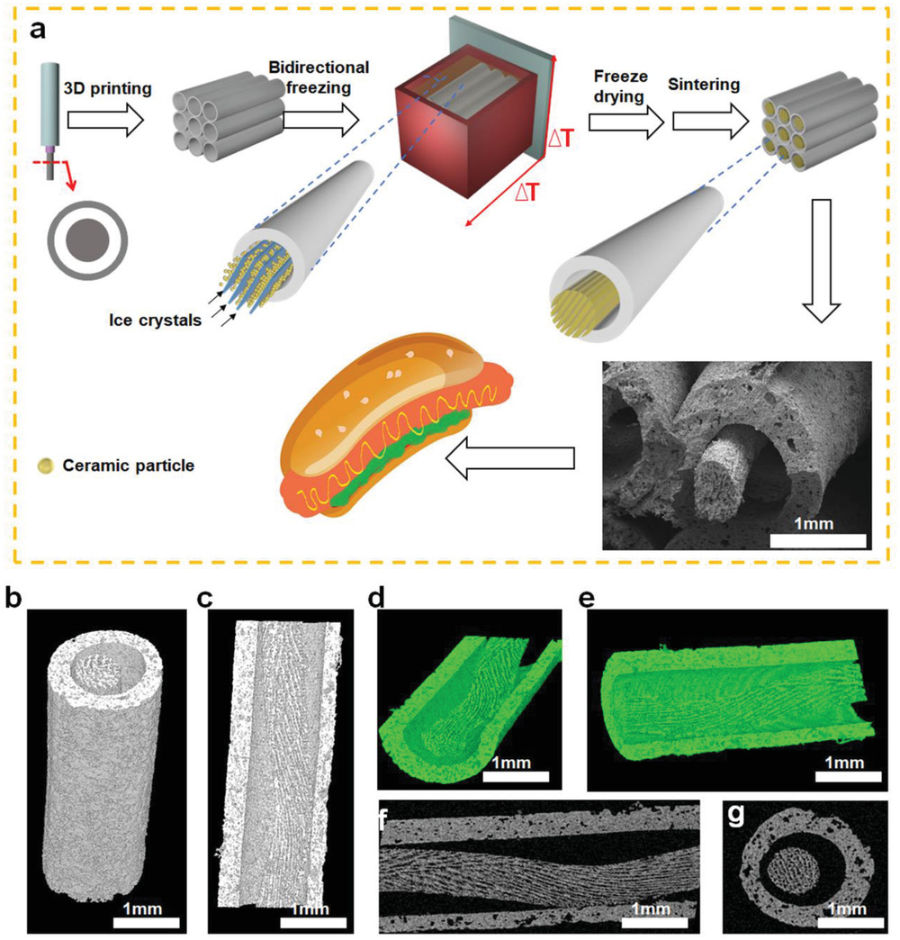
Fabrication and morphology of hot dog‐like scaffolds (HD‐AKT). a) The schemata of preparation of HD‐AKT, combining 3D printing and bidirectional freezing to realize 3D printed scaffolds containing rods with aligned lamellar microstructure. b–e) 3D micro‐CT images and f,g) 2D micro‐CT images of HD‐AKT from different views showing the scaffold structure, which is hollow tube macrospores embedded by bioceramic rods with the uniformly aligned lamellar structure of rods.
The researchers fabricated hierarchical structures using direct ink writing (DIW), with the hot dog structure figuring in with the use of tubes:
“The scaffolds are composed of hollow bioceramic tubes (mimicking the “bread” in hot dogs, pore size: ≈1 mm) embedded by bioceramic rods (mimicking the “sausage” in hot dogs, diameter: ≈500 µm) and the sausage‐like bioceramic rods possess uniformly aligned lamellar micropores (lamellar pore size: ≈30 µm),” said the researchers.
While hierarchical structures are often used in bioprinting, it can be challenging to find suitable materials in creating micro/nanostructures, and especially with DIW. And while challenges have continued in implanting 3D printed scaffolds to regenerate bone growth, there has been some success. The researchers contend, however, that hierarchy in structure is needed to promote better tissue growth, with the ‘hot-dog like structure’ lending itself to better cell adhesion and supply of nutrients, with the rods enhancing delivery of osteogenic drugs.
“By mimicking the function of nutrition supply for sausages in hot dog, the potential of the scaffolds for loading icariin (Ica, a model osteogenic drug), was investigated in this study,” explained the researchers.
“However, the Ica loading efficiency and capacity of S‐AKT were much lower than that of HD‐AKT, indicating the excellent loading capacity of the hierarchical hot dog‐like scaffolds. The thermogravimetric analysis further verified the significantly improved loading efficiency of HD‐AKT scaffolds as compared to those without hot dog microstructure.”
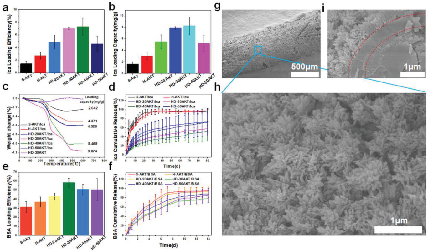
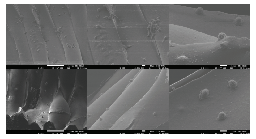
Hot dog‐like scaffolds are an excellent carrier for drug and protein. The drug Ica and protein BSA loading and release properties of the HD‐AKT. a) The Ica loading efficiency, and b) the loading capacity of traditional solid struts scaffolds(S‐AKT), H‐AKT, and different kinds of HD‐AKT. c) Thermogravimetric analysis of the scaffolds after loading the Ica. d) The Ica release of different kinds of scaffolds after loading Ica (S‐AKT/Ica, H‐AKT/Ica, HD‐20AKT/Ica, HD‐30AKT/Ica, HD‐40AKT/Ica, and HD‐50AKT/Ica). e) The BSA loading efficiency. f) The BSA release of the scaffolds. g–i) SEM images of the hot dog rod of scaffolds g) after 90 d Ica release. h) Surface images and i) the cross‐section image show lots of mineralized hierarchical structures on the surface of hot dog rod of scaffolds. Compared with the S‐AKT and H‐AKT, the HD‐AKT have higher loading efficiency and capacity. Meantime, HD‐AKT possess longer Ica/BSA release time.
The research team also discovered that other drugs could be distributed too, such as the large molecule protein bull serum albumin (BSA). Even more encouraging, the researchers found that the scaffolds exhibited ‘excellent bioactivity,’ proven through in vivo processes for bone regeneration as they implanted drugs into ‘rabbit femoral defects’ for eight weeks with no inflammation, and proof of bone tissue growth.
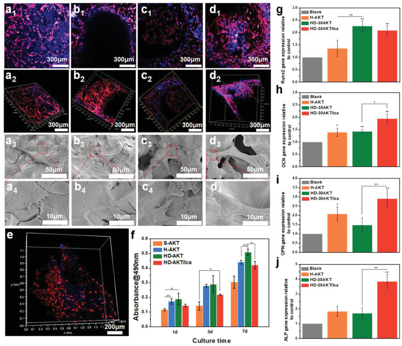
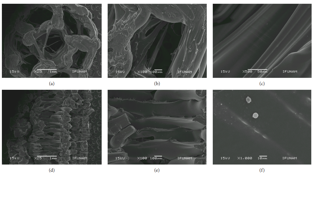
Hot dog‐like scaffolds are an excellent platform for cell delivery and differentiation. The proliferation, morphology, and relative genes expression of rBMSCs cultured on different scaffolds. a‐d) Confocal and SEM images of rBMSCs cultured on a) S‐AKT, b) H‐AKT, c) HD‐30AKT, and d) HD‐30AKT/Ica. e) The rBMSCs (red color) adhesion on the rod of HD‐AKT. f) The proliferation of rBMSCs after seeding in different kinds of scaffolds, showing hot dog‐like scaffolds are beneficial for cell proliferation. g) The relative osteogenic genes expression (OCN, Runx2, OPN, and ALP) of rBMSCs in scaffolds, indicating that the Ica release from the scaffolds promotes the relative osteogenic genes expression of rBMSCs (n = 6, *P < 0.05, and **P < 0.01.).
“Our study suggests that the hot dog‐like scaffolds can be used for the multifunctional biomaterials for drug delivery, tissue engineering, and regenerative medicine. The combined strategy of DIW 3D printing with bidirectional freezing is a promising method to prepare biomimetic and hierarchical biomaterials,” concluded the researchers.
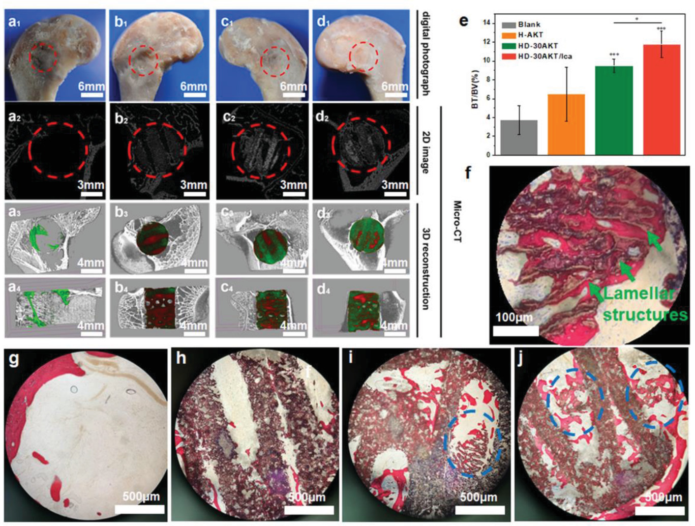
Hot dog‐like scaffolds possess excellent bone‐forming bioactivity after implanted in the femoral defects of rabbits. The characterizations of hot dog‐like scaffolds for osteogenesis in vivo. a1–d1) Digital, a2–d2) 2D micro‐CT, and 3D micro‐CT images (a3–d3: transverse view, and a4–d4: sagittal view) of the defects at week 8. In 3D micro‐CT images, green, red and white represents new bone, scaffold, and primary bone, respectively. e) Micro‐CT reconstruction analysis exhibits the volume ratio of the new bone to the original defect regions (BV/TV) at week 8. HD‐30AKT and HD‐30AKT/Ica indicate significantly. f) The newly formed bones (red) grow into the lamellar microstructures (green arrows) of hot dog‐like scaffold. g–j) Hard histological sections stained with Van Gieson’s picrofuchsin of g) Blank, h) H‐AKT, i) HD‐AKT, and j) HD‐AKT/Ica, red color stands newly formed bone and black color represents scaffolds. Newly formed bone can grow into the hierarchical rods of the scaffolds (blue circle point to the new bone in the hierarchical rods). improvement in new bone regeneration as compared to Blank control. In addition, Ica can also promote osteogenesis, suggesting that both hierarchical structure and the Ica delivery of the hot dog‐like scaffolds contribute to the bone regeneration (n = 6, *P < 0.05, **P < 0.01, and ***P < 0.001.).
While man-made processed foods are certainly inspiring to many, undoubtedly nature continues to propel scientists and designers forward from creating conductive parts to custom-made shoes, and liquid polymers for 3D printing. What do you think of this news? Let us know your thoughts! Join the discussion of this and other 3D printing topics at 3DPrintBoard.com.
[Source / Images: ‘3D Printing of Hot Dog-Like Biomaterials with Hierarchical Architecture and Distinct Bioactivity’]
The post Bioprinting Hot Dogs with Hierarchical Structures appeared first on 3DPrint.com | The Voice of 3D Printing / Additive Manufacturing.