3D Printed Esophagus Successfully Transplanted Into Rats

In the race to 3D print and transplant seemingly every part of the human body, the esophagus has been a particular challenge for scientists and doctors.

But a team of Japanese researchers has now successfully transplanted a bio 3D Printed esophagus into rats.


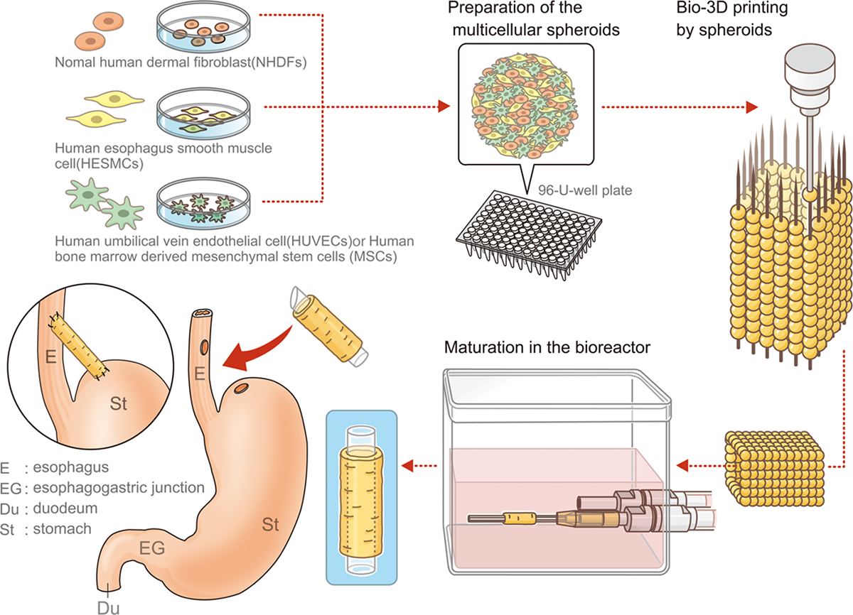
The esophagus, which was printed using Cyfuse’s bio 3D printing software Regenova, maintained its structure after the transplant and even grew to be covered by natural tissue.

These findings by the Nagasaki University-based group could mean big strides toward the first 3D-printed esophageal transplant (esophagectomy) in humans.

Why Esophageal Transplants Are So Complicated  

An esophagectomy is the main treatment for esophagus cancer. The operation involves removing all or part of the esophagus, then restructuring it using part of another organ (like the stomach or colon).

Tissue engineering/decellularization (Image: HIA)

However, this method can lead to complications including acid reflux, abnormal narrowing of the esophagus, and dumping syndrome (when food moves too quickly from your stomach to the small intestine).

The materials used in these operations typically have low biocompatibility and are done via decellularization. This is when the intracellular parts of tissue are removed so all that remains is the exterior “skeleton” which can then be used in tissue regeneration and the creation of artificial organs.

While there is nothing wrong with this method and it’s been used in several successful esophageal-related studies, it does require using existing tissue. The benefit of the 3D printing technique is that the esophageal structure can be constructed using only cells.

New Technology For New Tubes

The Japanese team opted instead to print and install a scaffold-free and more tubular structure into their rats. The tube was made with a Regenova bio 3D printer using the Kenzan method. This process first assembles human cellular spheroids into the desired 3D shape. Then, the shape cultures in a bioreactor until the spheroids fuse together to achieve the appropriate strength and structure.

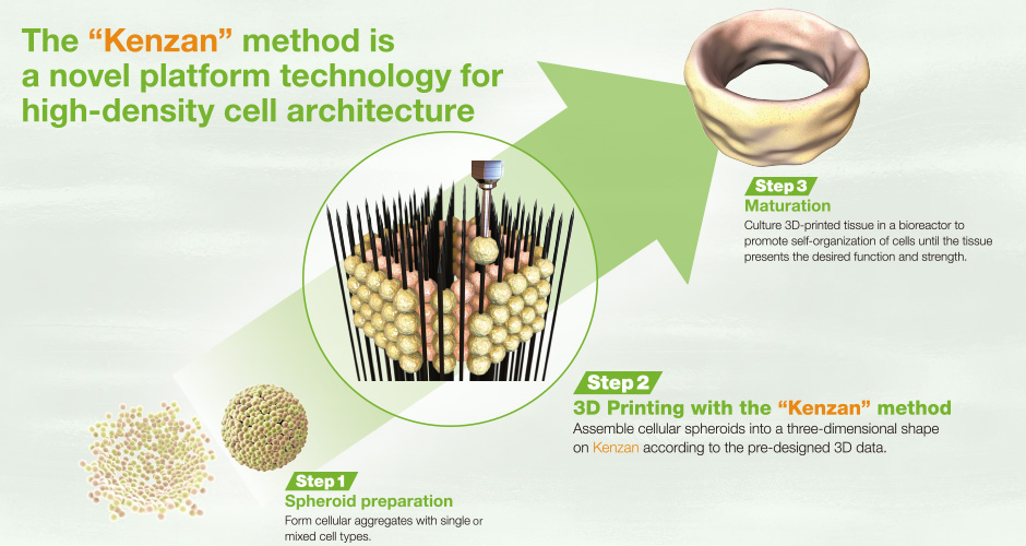
The Kenzan Method (Image: Phys.org)

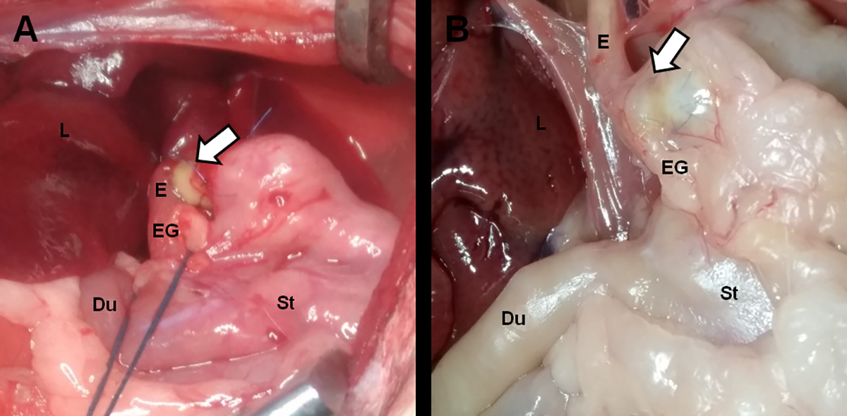
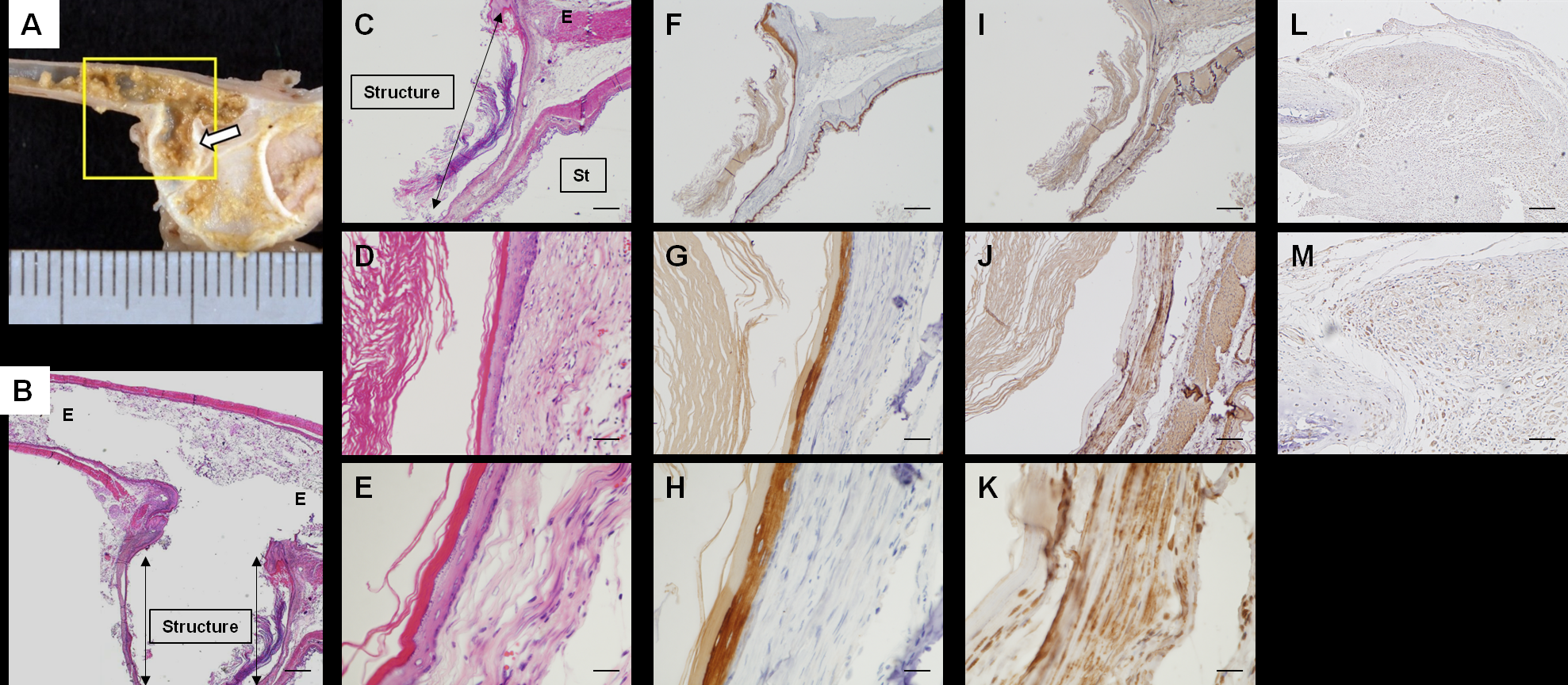
After testing for strength and flexibility, they were then transplanted into five male adult rats. Each rat survived and their esophagi maintained their shape and structure with no signs of leakage or perforation even after being exposed to gastric juices after eating. More remarkably, after 30 days scientists couldn’t detect where the rats’ natural esophageal lining and the transplanted structure began, as it became completely engrafted with natural cells.

What’s Next?

Researchers cautioned in their conclusion that despite the encouraging results, their esophagi did not perform as well as natural ones. However, the possible benefits of these results extend far beyond your food tube.

While it’s still early, using 3D printed biological and scaffold-free structures could decrease the amount of complications that come with more traditional transplant methods. No longer would patients have to sacrifice part of one of their organs (or someone else’s) to have a healthier esophagus. In times of organ shortages, this technology would obviously be vital in saving lives.

The researchers stated in their conclusion that the next step will be longer post-transplant assessment periods as well as orthotopic esophagus transplantation into larger animals.

Discuss this and other 3D printing topics at 3DPrintBoard.com or share your thoughts below.

Sources: Mayo Clinic | PLOS ONE

Japan: Researchers BioPrint & Implant Esophageal Cells Without Scaffolds

Researchers from Nagasaki University have recently made further progress in bioprinting—and without the use of scaffolds. As they become closer to their goal of 3D printing the human esophagus, the authors detail their current findings in Regeneration of esophagus using a scaffold-free biomimetic structure created with bio-three-dimensional printing.

Pointing out that they are not the first research team to attempt engineering tissue to repair defects of the esophagus, a vital tube that connects the throat to the stomach, the authors explain that their new method works because they have focused on diminishing previous issues with biological incompatibility, and challenges leading to problems like esophageal stricture.

The researchers, aware of the four cell types in the esophagus (squamous epithelial cells, fibroblasts, and smooth muscle cells), realized they had to create a structure that would not interfere with normal biological activity.

Study overview. Cells: fibroblasts,mesenchymal stem cells, smooth muscle cells, or endothelial cells were cultured respectively. Multicellular spheroids were created using mixed cell suspensions, and the artificial esophagus was then constructed with bio-3D printing using those spheroids. The artificial esophagus was matured in a bioreactor for a total of 4 weeks. Finally, esophageal transplantation of the artificial esophagus was performed.

The team began by acquiring the following:

  • Normal human dermal fibroblasts
  • Human esophageal smooth muscle cells
  • Human bone marrow-derived mesenchymal stem cells
  • Human umbilical vein endothelial cells

Esophageal-like tubes were bioprinted using a Regenova bio-3D printer and then transplanted into five male rats. They all survived the procedure, although the researchers noticed some smaller amounts of weight loss as they began their intense 30-day evaluation of the implanted structures. The rats quickly gained the weight back though, and then some. The transplanted tubes remained strong and stable, with no leaking or signs of tearing. The team noted that the rats were eating sufficient amounts, and even while exposed to gastric juices, the esophageal tube retained its integrity.

Transplantation of the structures. A. The surgical site of transplantation. B. The transplanted site at 30 days after transplantation. Arrows: transplanted site, Du: duodenum, E: esophagus, EG: esophagogastric junction, L: liver, St: stomach

Assessment of the structures at 30 days after transplantation.
A: The transplanted site. B: Hematoxylin and eosin (HE) staining at the site shown in A (yellow box). Scale bar = 500 μm. C-E: HE staining of the transplanted structure. Scale bar = 500 μm (C), 100 μm (D), and 50 μm (E). F-H: Immunohistochemical staining of the transplanted structure with anti-pan-cytokeratin. Scale bar = 500 μm (F), 100 μm (G), and 50 μm (H). I-K: Immunohistochemical staining of transplanted structure with anti-αSMA. Scale bar = 500 μm (I), 100 μm(J), and 50 μm (K). Arrows: transplanted structure, E: esophagus, St: stomach. Immunohistochemical staining of the transplanted structure with anti-HLA class 1 ABC. Scale bar = 200 μm (L), 100 μm (M).

Evaluations continued to be positive, with the 30-day mark reflecting structures ‘completely engrafted.’

“The esophageal mucosal epithelium extended into the lumen of the structure and covered its inner surface completely. Cells on the surface of the lumen expressed pan-cytokeratin. The expression of αSMA was also observed in the structure,” stated the researchers. “Expression of human HLA class I ABC was found only in the part of the transplanted structure, but there were no positive cells in the epithelial layer.”

“This means that the structure made of human cells was maintained in the rats’ body and the native rat epithelium extended on the transplanted structure after transplantation.”

Despite ultimate success in the research study, the scientists were met with some challenges and limitations; for instance, they were not able to use orthotopic transplantation successfully—and they believe it is a requirement for using this type of innovation in clinical applications. They also were unable to get data regarding neurogenesis and peristalsis of the artificial esophagus. The team realizes further studies and longer follow-up times are required—and eventually, an orthotopic transplantation model in large animals must occur.

“Although this work is our initial step of the study for the esophageal structure made with bio-3D printing system, it may contribute to the development of treatment for esophageal diseases that require transplantations,” concluded the researchers. “The structures created with bio-3D printing technology using a scaffold-free approach showed promise as a potential substitute for esophageal transplantation.”

3D printing has been an enormous boon to the field of medicine, and especially as continued strides in manufacturing new hardware, software, and materials offer so many options for improving (and sometimes even saving) the quality of life for so many patients. Researchers have recently created and 3D printed tracheas, a variety of facial prosthetics, and even skin. Find out more about the bioprinting of the esophagus for patients in need due to health problems or congenital defects here. What do you think of this news? Let us know your thoughts! Join the discussion of this and other 3D printing topics at 3DPrintBoard.com.

[Source / Images: Regeneration of esophagus using a scaffold-free biomimetic structure created with bio-three-dimensional printing]