3D Printed Medical Models May Assist in Better Monitoring of Ventricular Devices & Blood Flow

In ‘Innovative Modeling Techniques and 3D Printing in Patients with Left Ventricular Assist Devices: A Bridge from Bench to Clinical Practice,’ US researchers examine new techniques for better monitoring of left ventricular assist devices (LVAD) and troubleshooting alarms. The need is there due to altered flow dynamics caused by LVADs, which have been a developing technology for the last 30 years.

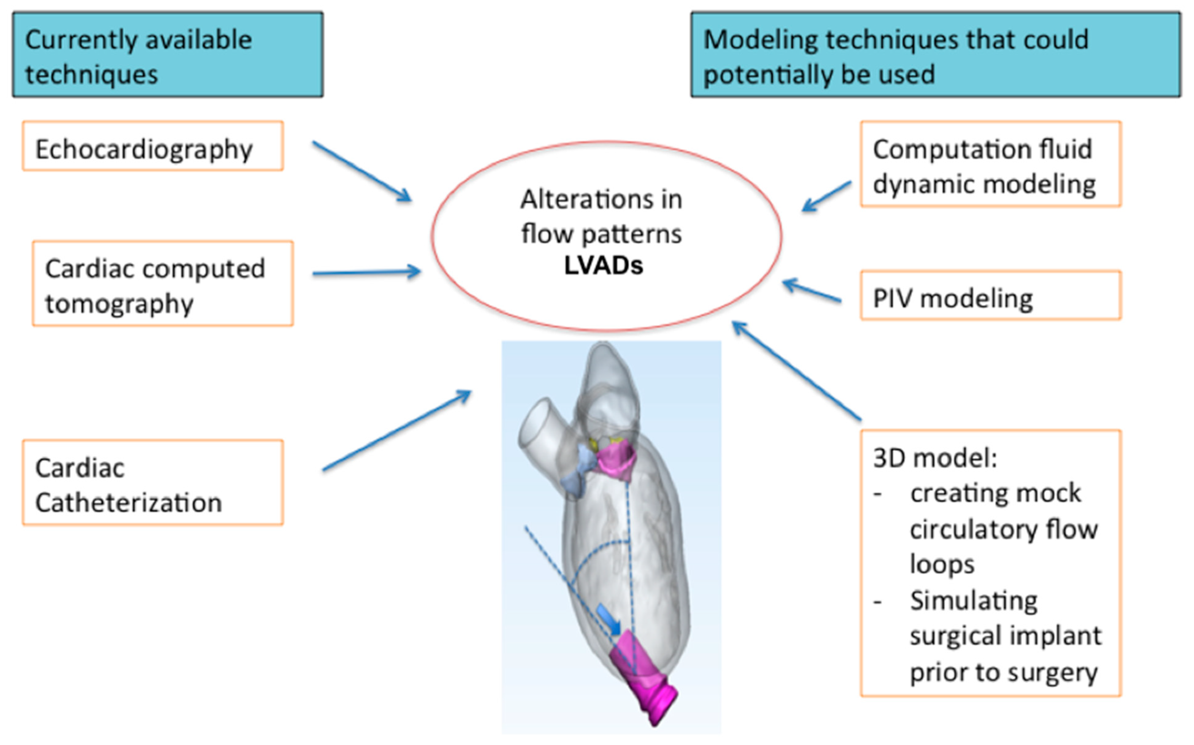
The authors examine new development methods that are not yet in use clinically but may have significant bearing in the future as they are able to improve treatment of cardiac patients who may have low cardiac output or even be in end-stage heart failure. As LVADs offer a ‘bridge’ to transplants or further therapy, some models may cause issues such as:

  • High thrombosis
  • Valve failure rates
  • Flow augmentation
  • Bleeding complications
  • Stroke

Even with superior design, issues may arise due to how the LVAD is placed in the body. Here, the researchers look further into techniques predicting LVAD flow. Not only can these techniques lead to better troubleshooting, but also better surgical planning, often with the help of 3D printed guides.

Computational fluid dynamics are used to create LVAD prototypes as well as test them:


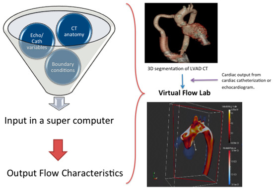
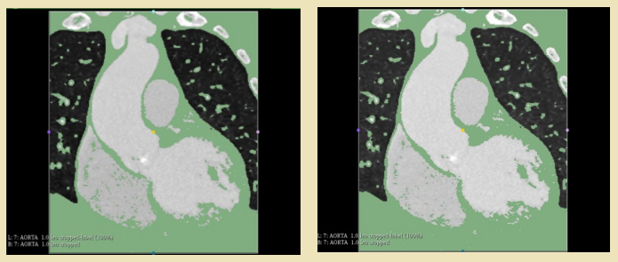
The workflow for computational fluid dynamics (CFD) analysis in a patient with LVAD.

“Prior to running a computer simulation, the accurate 3D geometry of the object needs to be defined, usually from cardiac CT or 3D echocardiography,” state the researchers. “The physical characteristics of the fluid, in this case blood, and the surrounding boundaries are defined. In these computer simulations, blood is assumed to act as an ideal fluid.”

“The quantity of blood flow through the ‘inlet’ (the left ventricle) and the ‘outlet’ (the aorta) can be derived from cardiac catheterization or echocardiography. These geometric and boundary conditions are inputs to a computer simulation that essentially functions as a virtual flow lab. The information can then be used to generate information on shear stress, heat dissipation, and wall pressure. Models for hemolysis and platelet activation can also be generated to test a device for hemocompatibility.”

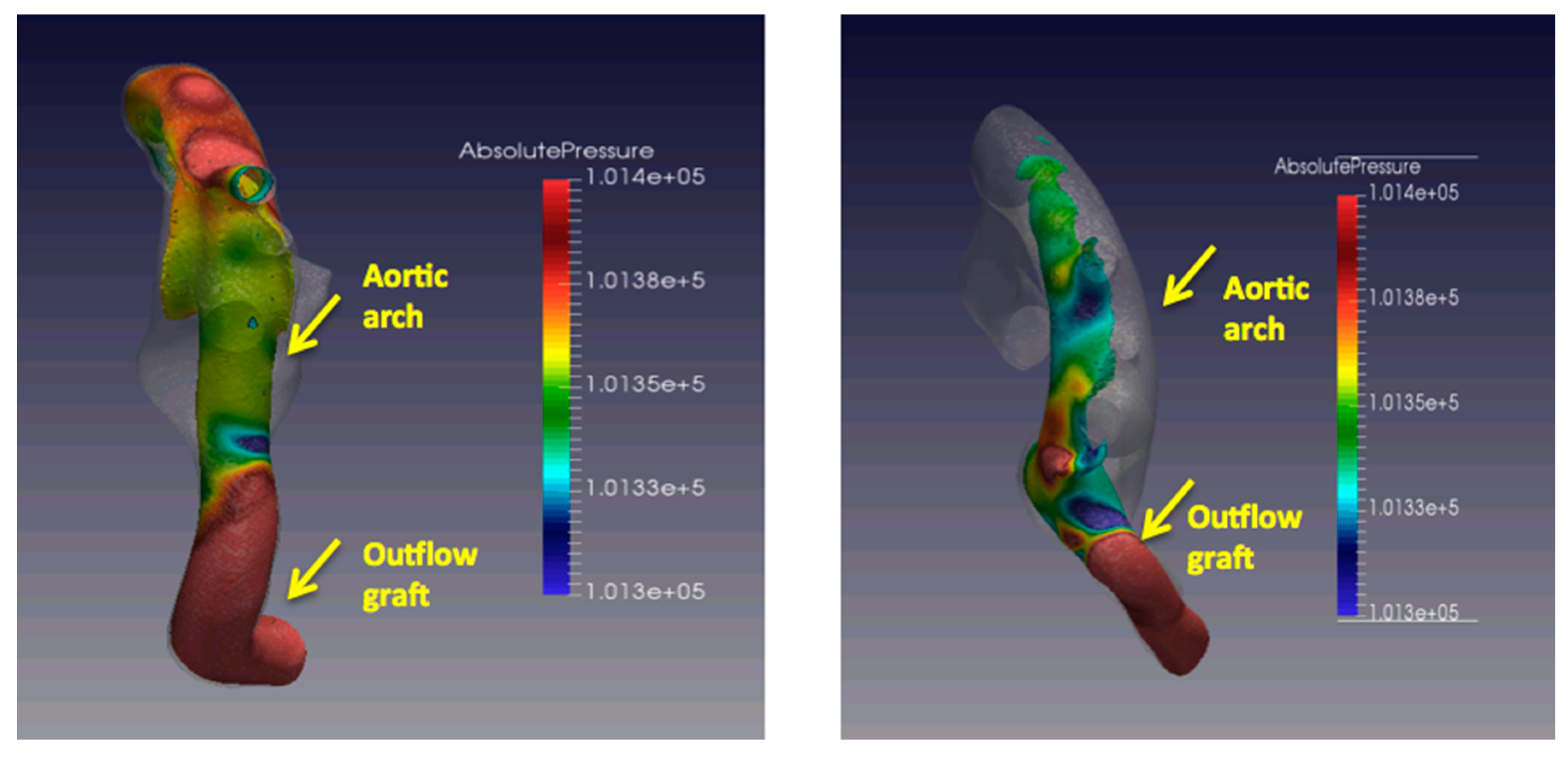
CFD simulations with varying outflow cannula angles relative to the aortic arch and variations in absolute pressure (in Pascals) and distribution on the aortic arch and great vessels.

Fluid structure interaction (FSI) allows researchers to examine fluid flow around a ‘deformable structure,’ with CFD simulations allowing assessment of the following hemodynamic parameters:

  • Pressure
  • Velocity
  • Wall shear stress
  • Displacement

Simulations allow researchers to observe disturbed flow, which may be in connection to issues with atherosclerosis and thrombogenesis. The authors point out that most CFD studies are deficient in the specific patient geometry required, along with long-term outcomes.

“CFD can prove to be a useful technique that could potentially be used clinically for LVAD implant planning, surveillance and troubleshooting of complications of patient with LVAD,” state the researchers. “Assessment of adverse fluid characteristics in relation to adverse long-term clinical outcomes may provide important insight into surgical implant techniques.”

In particle image velocimetry (PIV), researchers look at rapid sequential imaging for computing velocity vectors as well as fluid dynamics. PIV modeling involves setting up:

  • A transparent phantom organ
  • Laser
  • Camera
  • Seeding particles
  • Image processing software

The authors see potential for PIV in understanding LVAD placement and fluid dynamics.

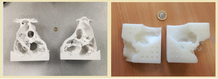
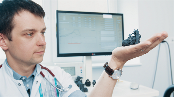
3D printed models are also being used now so that researchers can understand more about heart disease. They may be helpful in surgical planning, as well helping in the placement of ventricular assist devices (VADs). Heart models have also helped with virtual implantations, along with VADS in children too. The researchers point out that 3D printed heart models do improve anatomical positioning and surgical techniques, they are inferior to continuous flow pump simulation. They recommend better simulation models with either CFD or PIV modeling.

“As 3D printing technology evolves, there is hope to develop patient-specific models that replicate the anatomic and physiological features to be used in LVAD surgical planning,” conclude the researchers.

“In the long run, improved fluid dynamics may help improve implant techniques and lessen the burden of significant adverse events.”

An example of a flow circulatory flow loop with LVAD connected to compliance chambers and flow meters

Because there are so many heart conditions affecting so many around the world—and they can be deadly—researchers are constantly looking for better ways to treat patients. With 3D printed models and devices, patient-specific care is much more accessible, meaning that medical professionals have an easier time diagnosing heart conditions, treating them, and explaining what is going on to not only patients, but their families too. Medical students also have much more expanded training mechanisms with advanced 3D printed heart models and surgical guides.

[Source / Images: Innovative Modeling Techniques and 3D Printing in Patients with Left Ventricular Assist Devices: A Bridge from Bench to Clinical Practice]

UK Researchers Inspired by Astrophysics to Improve Imaging for 3D Printed Models

Like cancer, heart disease and the many conditions surrounding it (along with other related systems in the human body) can often cause fatal complications, leading researchers around the world to continually look for better methods in diagnosing, treatment, and surgical procedures. UK researchers, I. Brewis and J.A. McLaughlin, at Northumbria University have recently explored 3D imaging in reference to cardiovascular health care, publishing their findings in ‘Improved Visualisation of Patient-Specific Heart Structure Using Three-Dimensional Printing Coupled with Image-Processing Techniques Inspired by Astrophysical Methods.’

Brewis and McLaughlin are working in the realm of astrophysics in creating new image-processing techniques for viewing the human heart, transferring the data to an .stl file and then 3D printing a medical model. These new techniques allow for better modeling from scans, especially improving on the clarity of smaller features.

Traditionally, CT and MRI scans have supplied data for 3D printed models. As the authors of the study point out, these are ‘relatively accurate’ but still produce errors. Not only that, image processing can be a high-maintenance venture, consuming both time and money. Currently there are a range of techniques in use, including:

  • Vignetting
  • Boxcar smoothing function
  • Dilation
  • Edge detection
  • Pixel plate scale development for charge-coupled devices on spacecraft

Brewis and McLaughlin examined whether using techniques generally used in astrophysics could reduce the margin of error, along with faster turnaround time in rendering. The basic steps in arriving at a 3D model must include acquiring data, segmenting, converting files, fixing and design, and 3D printing. For the purposes of this study, the scientists used data from one anonymous patient, obtaining 856 CT images. In segmentation they were able to identify the ‘region of interest’ to be 3D printed: the heart tissue.

Segmented heart data following first thresholding of right heart region (left) and segmented heart data following second thresholding of right heart region (right). The right ventricle (bottom left) shows improved clarity of internal chamber segmentation following second thresholding

The team used Slicer to view the chest cavity from multiple angles and cut all extraneous data for a better view.

“Initial thresholding highlighted all heart tissue, with the inclusion of deoxygenated blood in both the right atrium and right ventricle,” stated the researchers in their paper. “Areas of deoxygenated blood were found to be removed by applying a second threshold in the range [-600, 70] to the chambers of the right heart.”

A second pass of thresholding resulted in converting data from 2D segments into a 3D rendering.

“This method, whilst modelling the majority of the heart’s internal structure, omitted small-scale features such as valves. In order to extract these small-scale features from the DICOM data available, an alternative approach was introduced,” stated the researchers, upon using vignetting to eliminated added interference in the background.

Netfabb image processing stages from left to right initial and divided models

Further highlighting and enhancement allowed the research team to see areas of aortic valve cusp occlusion and areas of bad pixels more clearly.

“For both the full heart and the aortal valve, the total time taken for image segmentation was on the order of tens of minutes, where some parts of the segmentation process were, naturally, more time-consuming than others,” stated the researchers. “For the method presented here, the main time-consuming step was whilst utilizing Slicer 4.8.1’s in-built eraser and draw tools.”

Once the researchers had the files for both the heart and aortic valve models ready, they used Netfabb to prep for production with an SLA 3D printer.

“The SLA printer produced more sturdy 3D models yet could not produce models which did not require additional support in order to maintain the desired structure of the internal heart chambers (i.e. free standing models) during printing,” stated the researchers. “For a simple and relatively flat structure however, such as the aortic valve, the free standing issue was not a concern and the stronger resin-based model proved to be ideal for producing thin and delicate structures such as the tricuspid valve.”

Ultimately, the team produced a 3D printed heart model with four clearly defined heart chambers, along with the aorta, superior vena cava and the pulmonary vein and artery. The patient whose data the researchers were using as an example had an aortic aneurysm that is accurately depicted in the model, seen as an area of depression in an aortic side wall. Calcification can be clearly observed too, and this type of accuracy means surgeons can operate more precisely and efficiently, saving on time in the operating room.

“The use of 3D renderings of patient data improves on traditional imaging techniques where surgeons are required to visualise a three-dimensional picture of heart defects based on a series of 2D scans by reproducing exact, real three-dimensional cardiovascular anatomy,” state the researchers. “The use of 3D modelling can also improve the physician’s understanding of individual patient anatomy such as in the case of valve replacement16 or in procedural planning for the treatment of congenital heart disease.”

What do you think of this news? Let us know your thoughts! Join the discussion of this and other 3D printing topics at 3DPrintBoard.com.

[Source / Images: ‘Improved Visualisation of Patient-Specific Heart Structure Using Three-Dimensional Printing Coupled with Image-Processing Techniques Inspired by Astrophysical Methods’]

3D printed heart models. (left) complete heart model, (right) aortic valve
model). UK one pound coin for scale.


3D Printing News Briefs: July 17, 2018

In Today’s 3D Printing News Briefs, we’re covering a lot of business and a little medical news. AMFG is partnering with a top UK bearings manufacturer to help automate its digital manufacturing workflows, while Segula Technologies has begun an industrial 3D printing partnership with digital manufacturing company Multistation. Techniplas has completed a deployment of Sharebot 3D printers to its 14 manufacturing facilities around the world, and the winners of the SkillsUSA Additive Manufacturing Competition have been announced. Finally, a pediatric cardiologist used the Sinterit Lisa to create a 3D printed model of a newborn boy’s heart to plan his risky surgery.

Bowman International Announces Partnership with AMFG

Bowman’s bearings

Automation software specialist AMFG, which recently launched a new AI software platform, has partnered with Bowman International, one of the top bearings manufacturers in the UK, as it works to grow its 3D printing capabilities through its Bowman Additive Production (AP) division. Bowman AP has several MJF and SLS 3D printers available for its use, and uses 3D printing to design and produce its end-part bearings, which has helped increase their load bearing capacity by up to 70%.

In the meantime, Bowman International’s goal is to use AMFG’s AI-powered production automation software to oversee production of said bearings, by automating production job scheduling, optimizing digital CAD files for production with printability analyses, and creating a custom digital part catalog.

“We’re very pleased to be partnering with AMFG and using their automation software to scale our already expanding AM facility,” said Jacob Turner, the Head of Additive Production at Bowman International. “Additive manufacturing is transforming the way bearings are manufactured, and we aim to continue to be at the forefront of innovating the production of bearings using AM. AMFG’s automation software will enable us to achieve this by significantly increasing the efficiency of our production processes.”

Multistation Partners with Segula Technologies

Another newly announced 3D printing partnership is the one between international engineering group Segula Technologies and Paris-based 3D printing company Multistation. The two are working together to further develop the potential of 3D printing in the industrial sector, which will allow both companies to increase their offerings and provide customers with excellent services along the AM value chain. Segula will bring its design, product-process qualification, and technology integration in industrial environments to the table, while Multistation will share and apply its expertise in AM design and simulation by determining any potential parts that could be 3D printed instead of fabricated with a more traditional method of manufacturing.

“Additive manufacturing is an integral part of a value chain within which Multistation provides a comprehensive offering; Segula Technologies was an obvious partner of choice to enable our Additive Consulting division to address manufacturers’ concerns more effectively,” said Yannick Loisance, the CEO of Multistation. “We will thus be able to supply them not just with software packages, machines and materials, but also with a more comprehensive range of high-quality engineering services that are suited to a host of different business sectors.”

Techniplas Adds Sharebot 3D Printers to Its Manufacturing Facilities

This fall, Italian professional-grade 3D printer manufacturer Sharebot joined the open innovation program at Techniplas, a top automotive design and manufacturing provider. Now, as part of its own continuing digital transformation, Techniplas has deployed Sharebot 3D printers to all of its 14 manufacturing facilities across five continents. This move will allow the company to 3D print the majority of the manufacturing products it uses every day on-site, which will equal major cost and time savings as Techniplas previously used only third-party providers for this task.

With Sharebot 3D printers installed in all of our manufacturing facilities worldwide, we are taking decisive steps toward fabricating the majority of our manufacturing line assembly tools, jigs, fixtures, gauges and even robotic arm attachments in-house. Based on our experience with Sharebot printers thus far, we expect to significantly reduce our development time and annual assembly line tooling costs in each manufacturing facility over time,” said Techniplas COO Manfred Kwade.

Winners of the SkillsUSA Additive Manufacturing Competition Announced

For the fourth year running, advanced manufacturing technology industry organization SME and Stratasys have co-sponsored the SkillsUSA Additive Manufacturing Contest, held during the annual SkillsUSA National Leadership and Skills Conference in Louisville. The winners of this year’s student contest, which asks contestants to solve real world problems with 3D printing, were just announced. This year, entrants had to design an adaptive device for a veteran, who had endured a traumatic thumb amputation, so he could keep playing his PlayStation 3. Prizes include RAPID + TCT conference passes, SOLIDWORKS’ 3D-CAD design software, SME Education Foundation scholarships (for high school participants), a one-year Tooling U-SME subscription, and a MakerBot Mini 3D printer.

“The SkillsUSA contest is designed to help students and educators realize the power of additive manufacturing to drive innovation. This year’s competition was particularly meaningful as it directly resulted in enhancing a veteran’s life with a custom solution not possible without additive manufacturing,” said Gina Scala, the Director of Marketing, Global Education at Stratasys.

The high school winners include:

  • Gold medal: Getty George and Sam Green, Martin Luther King High School, Riverside, California
  • Silver medal: Noah Logan and Johnathan Urbani, Stafford Tech Center, Rutland, Vermont
  • Bronze medal: Andrew Daddone and Layke Martin, Frederick County Career & Tech Center, Frederick, Maryland

The college winners include:

  • Gold medal: Adolfo Vargas and Alexander Kemnitz, Central Community College-Hastings, Hastings, Nebraska
  • Silver medal: Deema Al Namee and Aric Donerkiel, Vermont Technical College, Randolph Center, Vermont
  • Bronze medal: William Swaner and Ashton DeZwarte, Tenneseee College of Applied Tech-Nashville, Nashville, Tennessee

Watch a video about the 2018 competition here, and check out the winning designs here; you can also view SME’s Flickr album for more competition photos.

Surgeon 3D Prints Pediatric Heart Model with Sinterit Lisa

Desktop SLS 3D printer manufacturing Sinterit has seen its flagship Lisa 3D printer, which went through a recent upgrade, used to save lives in multiple ways, from fighting wildfires and protecting the faces of children to providing assistance in a tough pediatric cardiac surgery.

“Delivering desktop SLS 3D printer for more than three years caused that our clients send us tonnes of useful and exciting cases. Writing about all of them is hard, if not impossible, but when 3D printing helps saving lives, especially those most fragile, we feel proud, and also a duty to share it with you,” Michał Krzak, Sinterit’s Marketing Communication Manager, told 3DPrint.com.

A newborn’s heart can weigh barely 20 grams, and fits in the palm of an adult’s hand, so you can imagine that surgeries on such a delicate organ are exceedingly difficult. Jarosław Meyer-Szary, MD, from the Department of Pediatric Cardiology and Congenital Heart Defects at the University Clinical Center in Poland recently turned to Sinterit’s Lisa 3D printer to save the life of Kordian, an infant less than one month old suffering from a potentially fatal heart disease called interrupted aortic arch.

Meyer-Szary created 3D printed, life-size model of Kordian’s tiny heart, and SLS technology was able to recreate each intricate artery and vein. The model not only helped him plan the surgery ahead of time, but also helped Kordian’s mother gain a more thorough understanding of her son’s condition. Kordian is now a thriving and happy 18 month-old, thanks to Sinterit’s SLS technology.

Discuss these stories and other 3D printing topics at 3DPrintBoard.com or share your thoughts in the comments below.