CSIRO 3D Prints First Self-Expandable Stents from Shape-Memory Alloy Nitinol

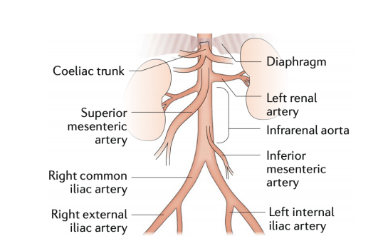
Peripheral Arterial Disease (PAD) is a condition which sees fatty deposits collect and lower the blood flow in arteries outside of the heart, most commonly in the legs. Those suffering from PAD will often experience pain while walking, and could even develop gangrene if the case is serious enough. Over 10 percent of people in Australia are afflicted with this painful condition. To treat it, a stent can be temporarily inserted inside the blood vessel to keep it open.

We’ve seen 3D printing used to fabricate stents before, which can help improve sizing options and allow for patient-specific diameters and shapes. But ,until now, there hasn’t been a way to print a self-expandable stent made of shape-memory nickel and titanium alloy nitinol. The material is superelastic, and metallurgists have had a difficult time trying to figure out a way to 3D print a self-expandable nitinol stent without compromising the unique properties of the metal alloy.

But researchers from Australia’s national science agency, the Commonwealth Scientific and Industrial Research Organisation (CSIRO), together with its Wollongong-based partner, the Medical Innovation Hub, have finally made it possible.

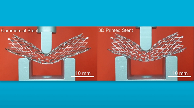
Vascular surgeon Dr. Arthur Stanton, the Chief Executive of Medical Innovation Hub, explained, “Currently, surgeons use off-the-shelf stents, and although they come in various shapes and sizes, overall there are limitations to the range of stents available. We believe our new 3D-printed self-expanding nitinol stents offer an improved patient experience through better fitting devices, better conformity to blood vessel and improved recovery times. There is also the opportunity for the technology to be used for mass production of stents, potentially at lower cost.”
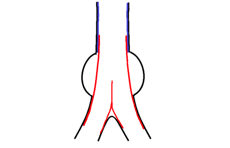
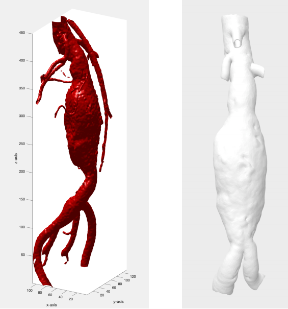
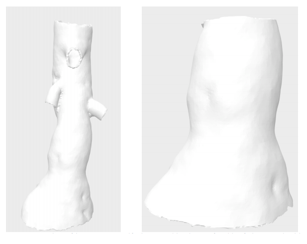
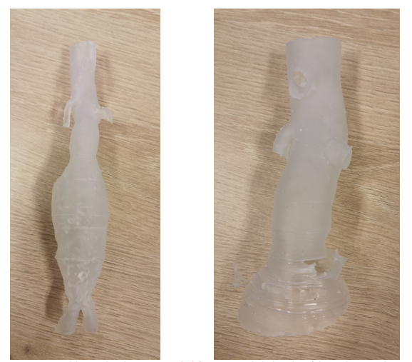
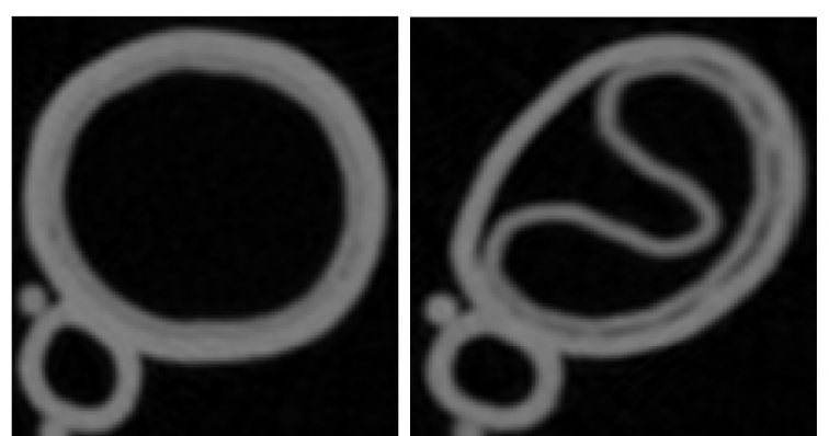
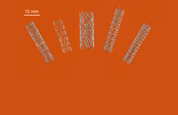
Stent model

The first 3D-printed nitinol stent is a major medical breakthrough for PAD patients, as surgeons have had to use off-the-shelf, non-custom stents for these procedures in the past. But with 3D printing, individual nitinol stents can be made right at the hospital, with the surgeon there to offer instructions—saving time and money, and reducing inventory, as well.

According to Australia’s Minister for Industry, Science and Technology, Karen Andrews, 3D printing could mark a major paradigm shift in the $16 billion worldwide stent manufacturing industry:

“This is a great example of industry working with our researchers to develop an innovative product that addresses a global need and builds on our sovereign capability.”

The proof-of-concept stents offer the potential for customization to individual patient requirements, but are equally as suitable for mass production.

Back in 2015, CSIRO opened the Lab22 Innovation Center. The specialist researchers there are focused on creating value for Australia’s manufacturing industry by developing future developments in metal additive manufacturing. CSIRO’s Lab22 collaborates with industry partners, like the Medical Innovation Hub, to build important biomedical parts, like the first 3D-printed sternum and titanium heel, and now the first 3D-printed nitinol stent.

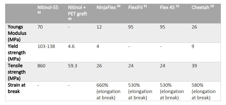
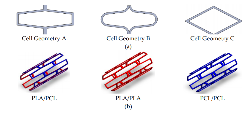
CSIRO Principal Research Scientist Dr Sri Lathabai said, “Nitinol is a shape-memory alloy with superelastic properties. It’s a tricky alloy to work with in 3D printing conditions, due to its sensitivity to stress and heat. We had to select the right 3D-printing parameters to get the ultra-fine mesh structure needed for an endovascular stent, as well as carefully manage heat treatments so the finished product can expand as needed, once inside the body.”

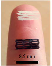
The team used selective laser melting (SLM) technology to successfully fabricate the complex mesh stent structures. Due to the level of geometric accuracy that 3D printing achieves, the stents can be made for specific patients, and nitinol allows them to expand once inside the body. CSIRO has established a new technology company, Flex Memory Ventures (FMV), to help commercialize the technology.

Discuss this story and other 3D printing topics at 3DPrintBoard.com or share your thoughts in the Facebook comments below.

The post CSIRO 3D Prints First Self-Expandable Stents from Shape-Memory Alloy Nitinol appeared first on 3DPrint.com | The Voice of 3D Printing / Additive Manufacturing.

3D Printing Models & Stents for Treatment of Abdominal Aortic Aneurysms

Joyce van Loon recently presented a Master’s thesis, ‘Personalized Endovascular Stent Grafts: Developing a Phantom Aneurysm Model to Test Personalized Stent Grafts,’ to the University of Twente. This work is centered around the use of 3D printed medical models and the potential for viable devices.

Schematic of a normal abdominal aorta and the major branches

Classification of AAA concerning the renal arteries. Suprarenal AAA includes the origin of renal arteries without the involvement of the superior mesenteric artery. A pararenal or juxtarenal AAA is when an aneurysm originates at the level of the renal arteries. A pararenal aneurysm is when the renal arteries originate from an aneurysmal aorta, and a juxtarenal aneurysm is when the aorta at the level of the renal arteries is normal. An infrarenal AAA originates below the renal arteries with a segment of non-dilated aorta between the renal arteries and the aneurysmal sac.

Caused by vascular weakness, abdominal aortic aneurysms (AAA) are irreversible conditions involving the intima, media, and adventitia. They tend to enlarge as time passes, and diagnosis is critical before a rupture. Afterward, close monitoring is required up until surgery.

“Rupture of an AAA usually leads to sudden onset abdominal or back pain and hypotension or shock. A ruptured AAA is a life-threatening situation with an overall mortality of 90% and even with prompt surgical intervention around 25% of the patients die,” stated van Loon.

“Before surgery, preprocedural imaging and planning is critical. CTA (computed tomography angiography) is superior as imaging modality and more accurate than US in estimating the diameters and lengths. During planning the anatomy, the involvement of the visceral arteries, the morphology of the aortic neck (proximal part of the aorta), the aortic angulation, existence of thrombosis, calcifications and stenosis are important factors that should be considered. This indicates the importance of a useful measurement tool in combination with the obtained images to evaluate the condition of the aneurysm and to decide which surgery should be performed and which stent graft is suitable.”

EVAR procedure. The introduction and positioning of the modular stent graft. The stent graft consists of a main device and extensions. The two femoral arteries are the access sites and incisions are made as shown.

More serious complications can arise for the 30 percent of patients not eligible for endovascular aneurysm repair (EVAR); however, customized endovascular stent grafts can be inserted. Challenges arise in using traditional methods though as there could be as much as a two-month wait.

Sketch of the combined custom-made stent graft. The arterial wall shown in black, the personalized stent graft in blue and off-the-shelf stent graft in red

During this study, data from CT scans of 65 patients functioned to create 3D models to be evaluated—and then further used to 3D print personalized stent grafts. The materials of choice for creating the aorta required flexibility and elasticity, high compliance, and material strong and durable enough to endure pressure from the stent graft. Materials must be able to fabricate complex geometries and being resistant to fluids.

“The stent graft material should have a high compliance and needs to mimic the native artery. Nitinol stents have proven to have high compliance due to its super-elasticity, it is biocompatible, hemodynamically stable, fatigue resistant, and non-corrosive,” stated van Loon. “However, we were not able yet to 3D print nitinol and we needed a comparable material to study the behavior of the stent grafts.”

CT-scan of one of the included subjects. The aneurysm is visible within the red circle. Left: Coronal slice. Right: Sagittal slice.

For the stent, van Loon stated that it required not only strength and flexibility, but it was necessary for the materials to border on the 4D level as well, able to deform with ‘self-expanding properties’ in the aorta. The material had to be soft enough to avoid any damage to the body and allow for printing of more complicated structures. The personalized stent required customization, as well as proper sealing between the aortic wall and the stent graft.

Segmentation abdominal aorta aneurysm of one of the included subjects. Left: the MATLAB segmentation results. Right: the Meshmixer results, where a large part of the renal arteries, the mesenteric artery and the iliac arteries are removed.

Aortic neck part of the segmentation, used for the aorta models and stent graft models. Left: the aortic neck and the branches of the renal arteries and superior mesenteric artery. Right: the aortic neck (infra-renal part). This part is important for measurements and sealing.

Aorta models were printed using elastic resin with a Form 2 3D printer. In testing two 3D printed models, however, van Loon stated that they both ‘showed problems,’ mainly with tearing. They were not able to finish printing of the models, in fact.

Aorta models printed in elastic resin. On the left, an aorta with the branches of the renal arteries, the complete aneurysm and bifurcation to the iliac arteries are shown. On the right, an upper part of the aorta and aneurysm can be seen, including the branches of the renal arteries and superior mesenteric artery. In the aortic neck and upper part of the aneurysm, tears are visible.

“Secondly, we printed the aortas with Agilus30 and the Stratasys ObJet260 Connex3 printer. Agilus30 is a flexible material and has a tensile strength of 2.4 – 3.1 MPa and a Young’s Modulus of 0.6 MPa,” explained van Loon. “Printing the aorta models in Agilus30 is a time-consuming (1-2 days) process but results in usable aorta models.”

“After stenting the aorta models with a 1 mm and 1.5 mm wall thickness, they rupture and are considered not suitable for further measurements. We only used the 2 mm wall thickness for further research.”

Aortic neck model, one of the first prints.

Material properties of nitinol and available and tested 3D print materials

They 3D printed stents on an Ultimaker 3, using NinjaFlex. The material was not as flexible as required for the project, and van Loon noted that the stents were ‘difficult to position’ in the sample printed aorta.

CT-scan of the printed stent grafts in NinjaFlex placed in the aorta model. Left: transversal slice of the aorta model with the personalized stent graft and a good sealing. Right: transversal slice of the aorta model with the straight stent graft, which could not unfold and bad sealing is seen at the unfolded side of the stent graft.

“The second tested material described, Cheetah, was not as flexible as thought and the solid stent grafts were not easily foldable which make it difficult to place the stent graft in the aorta model. When printed in a different geometry (meshed), the stent is more flexible and showed similar behavior as the nitinol stent graft. The stent grafts printed in NinjaFlex or Cheetah are in solid form too stiff and in mesh structure too flexible. We were not able to measure any distention differences of the aortic wall between the personalized stent grafts and the straight stent grafts,” concluded van Loon.

“The results showed that a better sealing could be obtained by using personalized stent grafts in our aorta models, however the used materials for the 3D printing of the stent grafts are not comparable with the nitinol stent graft and therefore more research is required.”

While van Loon documented obvious challenges in this study, 3D printed models are becoming indispensable in many medical applications, for assisting in surgery, diagnosing, treating and educating patients, while a variety of implants and devices are improving the quality of life for individuals around the world.

What do you think of this news? Let us know your thoughts! Join the discussion of this and other 3D printing topics at 3DPrintBoard.com.

[Source / Images: ‘Personalized Endovascular Stent Grafts: Developing a Phantom Aneurysm Model to Test Personalized Stent Grafts’]

The post 3D Printing Models & Stents for Treatment of Abdominal Aortic Aneurysms appeared first on 3DPrint.com | The Voice of 3D Printing / Additive Manufacturing.

3D Printed PLA and PCL Composite Biodegradable Stents Show Promise

PCL (white) and PLA (black) stents

Biodegradable stents have shown great potential in reducing complications in patients, but they require further study, according to the authors of a paper entitled “3D-Printed PCL/PLA Composite Stents: Towards a New Solution to Cardiovascular Problems.” The researchers outline five main requirements that a biodegradable stent must meet:

  • Their manufacturing process should be precise
  • Degradation should have minimal toxicity
  • The rate of degradation should match the recovery rate of vascular tissue
  • They should induce rapid endothelialization to restore the functions of vascular tissue but should at the same time reduce the risk of restenosis
  • Their mechanical behavior should comply with medical requirements, particularly the flexibility required to facilitate placement but also sufficient radial rigidity to support the vessel

Although the first three requirements have been thoroughly studied, according to the researchers, the last two have been overlooked. A possible way of addressing these issues would be to create composite stents using materials that have different mechanical, biological or medical properties, such as PLA or PCL. Fabricating stents with these materials using laser cutting, however – the traditional method of manufacturing stents – would not be possible. The researchers, therefore, decided to produce them using 3D printing.

They 3D printed the stents using a tubular 3D printer. The stents were then seeded with cells and left for three days, and then tests were performed to assess the morphological features, cell proliferation, cell adhesion, degradation rate and radial behavior.

“The results prove the materials’ biological compatibility and encourage us to believe that PCL/PLA composite stents would comply with the fourth requirement, i.e., rapid endothelization without risk of restenosis,” the researchers state. “PCL’s better cell proliferation may be useful to increase the proliferation of endothelial vessel cells in the external wall of the stents, while an internal PLA wall may help to reduce the proliferation of cells that produce restenosis. However, further studies with other kinds of cells or substances need to be performed to confirm this. The results here show low cell proliferation because of the small amount of material that the stents have. Additional studies that use longer culture times may be beneficial to obtain better proliferation results.”

The researchers’ initial hypothesis was confirmed: the smaller the cell area of the stent, the better the cell proliferation rate. The cell shape of the stent, however, did not show any significant influence. Because of their different molecular weights, PCL showed better cell proliferation than PLA. PLA showed a much faster degradation rate, which limits its use for biodegradable stents. Radial behavior results show that composite PLA/PCL stents could be used to improve each material’s separate limitations, with PCL offering elasticity in the expansion stem and PLA providing rigidity in the recoil step.

Overall, 3D printing proved itself to be a promising method for producing stents. Both PCL and PLA showed themselves to be biocompatible, and the composite stents showed the most promise, with medium levels of degradation rates and mechanical modulus.

“Based on the results presented here, we believe that polymer composite stents manufactured with 3D-printing processes could be a highly effective solution to the current problems that stents made of polymers have,” the researchers conclude. However, FDA rules currently limit the use of 3D-printed stents in real clinical applications and, although PCL and PLA are FDA-approved materials, there are still open challenges to be met before approval for 3D-printed implantable medical devices can be obtained.”

Authors of the paper include Antonio J. Guerra, Paula Cano, Marc Rabionet, Teresa Puig and Joaquim Ciurana.

Discuss this and other 3D printing topics at 3DPrintBoard.com or share your thoughts below.