Lung Cancer Treatment: 3D Printing Molds for Personalized Airway Stents

Australian scientists are working to improve medical devices for lung cancer treatment, sharing the outcome of their recent study in ‘Incorporating Chemotherapeutic Drug into a Personalizable Silicone Airway Stent for the Treatment of Lung Cancer and Tracheobronchomalacia.’

With a focus on relieving serious symptoms like central airway obstruction (CAO), the research team experimented with 3D printing molds to produce drug-eluting personalized airway stents, incorporated with chemotherapy drugs like Paclitaxel that inhibit the growth of cancer cells.

Interior view of current Y-stents used today, including the metallic Wallstent™ [A] and the Novatech® Dumon™ silicone stent [B] used in many CAO treatments, which do not correlate well with unique patient airway geometries [C].

Because diseases like lung cancer may leave patients struggling to breathe, pharmaceutical treatments and the use of effective devices can be critical to the quality of their lives—and even saving them in some cases. The researchers note that there are challenges with airway stents being used today due to a lack of personalization for patients, resulting in airway stent therapy that is often not effective. There may be other issues too, such as stent migration cased by improper fit.

“Unfortunately, airway stents have not developed, in large due to low relative prevalence of surgery and poor outcomes, since the release of Montgomery and Dumon stents during 1965 and 1989 respectively, despite leaps in 3D imaging and drug release technologies,” explain the researchers.

Drug-eluting stents offer potential in eliminating toxicity in delivery, as well as offering much-needed customizations for patients for better fit—reaping the rewards of one of the greatest benefits of 3D printing for the medical arena today with patient-specific treatment rather than a ‘one-size-fits-all’ premise for everyone. These benefits are heavily evidenced today in areas like prosthetics, heart valves, bio-active patches, and more.

Concentrations used during testing of drug elution from silicone stent materials.

Paclitaxel was added to the silicone molds, leaving the team of researchers to then perform a detailed assay on the Beas-2B cells derived from healthy patients and H23 adenocarcinoma cells derived from nonsmall cell lung cancer patients. The drug was insoluble in PBS, while ‘highly soluble in ethanol.’

Difference (f1) and Similarity (f2) factors used to determine the significance of the difference between release rates of paclitaxel concentrations and formulation methods in cured silicone coupons.

Variances in release rates of drugs demonstrate the potential for further manipulation, with adjustments to the paclitaxel in silicone coupons or via other techniques. The authors reported that there has been similar success with other stents.

Percentage of drug, paclitaxel, released from 250 mg silicone coupons in ethanol at 37°C, over 72 hours (n=3). Table 1 denotes A, B, C, D, E, and F silicone coupon conditions.

Cell viability for Beas-2B and H23 immortalised cell lines, grown on paclitaxel eluting silicone coupons, over 72 hours (n=6). An 80% cut-off was used to determine cellular viability.

“The implications of characterizing a successful controlled release of paclitaxel from cured liquid silicone rubber will allow clinicians to personalize treatment depending on airway geometry and control for the targeted dose of paclitaxel to the area of interest, thereby reducing the side effect profile of paclitaxel and its excipients (i.e. ethanol and polyoxyethylated castor oil) in systemic circulation,” stated the researchers.

“Further assessment in the comparability of paclitaxel release into lung-like environment is needed to characterize the effectiveness of drug release.”

What do you think of this news? Let us know your thoughts; join the discussion of this and other 3D printing topics at 3DPrintBoard.com.

[Source / Images: ‘Incorporating Chemotherapeutic Drug into a Personalizable Silicone Airway Stent for the Treatment of Lung Cancer and Tracheobronchomalacia’]

The post Lung Cancer Treatment: 3D Printing Molds for Personalized Airway Stents appeared first on 3DPrint.com | The Voice of 3D Printing / Additive Manufacturing.

An Inside Look into the ACES Lab (Part I)

A leading scientist in the field of electromaterials and one of Australia’s visionary bioprinting enthusiasts Gordon Wallace took audiences through a virtual tour into the cutting edge research labs at the ARC Centre of Excellence for Electromaterials Science (ACES), where next-generation materials research and advanced engineering for the development of customized bioinks and bioprinters take place. Located within the heart of the Intelligent Polymer Research Institute (IPRI) at Australia’s University of Wollongong (UOW) Innovation Campus, ACES turns fundamental knowledge into the next generation of smart devices to improve people’s lives and deal with some of the great challenges of the century.

With his usual enthusiasm, Wallace engaged audiences as he presented fellow researchers at work and some of the new innovations, discoveries and development of new materials for use in the field of biofabrication. During the first part of the tour, he explores the development of Graphene, 3D printed stents, and cell preparation for bioprinting. For the second part of the tour (found in a separate article), Wallace walks into another building at UOW where the recently inaugurated Translational Research Initiative for Cell Engineering and Printing (TRICEP) is leading the initiative for 3D bioprinting encompassing bioink, bioprinter, and bioprinting process developments, including the manufacturing of medical devices and the integration of living cells delivered using customized bioprinters to address specific medical challenges.

“Here at ACES we are known for our fundamental work into the discovery and the development of new materials, that can be used in energy and medical bionics,” said Wallace. “We are using the most advanced methods of fabrication to develop protocols that will enable structures and devices to be created so that we can take those fundamental advances and use them in important areas.”

Starting with the basics, Wallace first explores a lab setting where Sanjeev Gambhir, a Senior Research Fellow at the Australian National Fabrication Facility (ANFF) of the University of Wollongong, develops graphene, a material he refers to as “wondrous”, with “amazing properties for the nanoworld that we have been able to extricate into the micro and macroscopic realms to realize applications.” 

“To create a graphene-polymer composite synthesis, we modify the chemistries of graphene (which is derived from graphite, a naturally occurring mineral) so that we retain all the amazing mechanical, electric and biological properties and yet make it processible, that is, to turn it into structures and devices, using 3D printing, and eventually making it scalable,” said Gambhir.

Wallace added that “it is important that all the chemistries we use are actually scalable.” He claims that it is very different doing chemistry on a bench from processing graphene into tens of grams and managing to retain the same properties and quality as they were getting on the laboratory scale. It is all part of his vision to really make the process ready for industrial-scale manufacturing.

To show how graphene is turned into fibers for easier handling, Wallace takes audiences to the Fibre Spinning Electrodes area, where researcher Javad Foroughi, “weaves the magic” to create graphene fibers, that can even be combined with biomaterials to coat the surface of the fiber.

Working on customized 3D printed stents was Ali Jeirani, a Product Designer Development Specialist at UOW. It is one of the many processes where he uses 3D printing and takes advantage of all of the advances in material synthesis and processability at ACES, by turning them into real structures. 

“One of the important parts about the properties of a stent for applications is the design. We use G-code to create different designs and then send them to our machine to print different structures and properties,” explained Jeirani. “One of the problems of commercial stents is that they cannot be personalized for the patient, so by using 3D printing, we can customize it according to the scan of the patient. We understand that there can be very complicated stent shapes that are readily realized with 3D printing.”

According to Wallace, the graphene is often blended with other materials to improve the properties of the part, and by using small amounts of graphene and blending it with a polymer, they can create the stent. The innovative material gives the stent extra mechanical properties and could even impart electrical properties into it, which the two experts consider “one of the most interesting properties of graphene for electro stimulation”. 

“This is all made possible thanks to additive fabrication and advances in 3D printing, so it is an exciting time, since we can turn fundamental discoveries into really practical and useful structures almost immediately by working together, us at the 3D fabrication lab and our colleagues at materials processes,” continued Jeirani.

Gordon Wallace and Ali Jeirani looking into how to fabricate 3D printed stents

After delving into advancements in biomaterials and graphene, Wallace headed upstairs to the cell lab where Research Fellow at ACES, Eva Tomaskovic-Crook, revealed another important part of their work: the integration of living cells into printing protocols, which basically entails how scientists prepare the cells for printing.

They have several environments ready for the cells, from storing them in liquid nitrogen sample storage tanks–they have at least two Taylor Wharton LS750– to incubating them, which offer an environment where they nurture cells and provide the right growth conditions to expand. Incubators have a warm 37-degree environment ideal for maintaining cell growth. 

“Quality control of our cells is very important. We need to be sure that the cells maintain the ability to be pluripotent (pluripotent stem cells have the ability to undergo self-renewal and to give rise to all cells of the tissues of the body). We want to scale up the number of cells and to encapsulate them in the biomaterial.” suggested Tomaskovic-Crook.

Scaling up the number of cells is crucial because when they go into the bioprinting process they want to create a three-dimensional tissue with a high cell to biomaterial mass, not just have a few cells. According to the specialist, “it involves a process of going back and forth: scaling up the cells at the lab, then printing them, and bringing them back to the lab to interrogate the cells and see if they are still living, proliferating and turning into the cells we want them to.” 

Gordon Wallace and Eva Tomaskovic-Crook talking about preparing cells for bioprinting 

Known for their expertise in advanced materials and device fabrication, ACES incorporates collaborators from across Australia and the world. ACES is generating options for the future, so being able to peek into some of the advanced materials and device fabrication for game-changing health and energy solutions is a privilege. Not only did Wallace explain some of the most breakthrough research in biomedicine, but he also showed viewers the machines that researchers work with on a daily basis. Wallace tends to emphasize that a big part of the Australian bioprinting community is about sharing research, insights, and knowledge to advance the field. The unique landscape of the country, with its cultural and linguistic diversity as well as residence to scientists from around the globe, makes it ideal for ideas and creativity to emerge.

You can tune in to see the first part of the virtual lab tour here.

The post An Inside Look into the ACES Lab (Part I) appeared first on 3DPrint.com | The Voice of 3D Printing / Additive Manufacturing.

Florida Atlantic University Researcher Reviews Different Esophageal Stents, Including the 3D Printed Variety

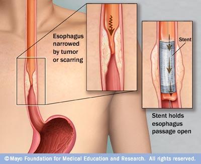
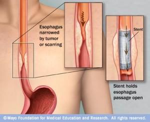
Ranking sixth among all types of cancer in mortality, esophageal cancer kills about 16,000 people in the US annually, with about 500,000 new cases diagnosed around the world last year (according to the American Cancer Society). More than half of these patients are unfortunately diagnosed when the cancer is advanced enough that a tumor blocks the esophagus, which is typically when doctors insert a palliative, self-expanding esophageal stent to allow them to eat and drink. According to a paper, titled “A Review of Self-Expanding Esophageal Stents for the Palliation Therapy of Inoperable Esophageal Malignancies,” that was published by Florida Atlantic University researcher Dr. Yunqing Kang, researchers have been looking into other therapeutic applications for these stents.

The abstract reads, “Esophageal stents have evolved in stages over the years. Current clinically used stents commonly include stainless steel or nitinol self-expandable metallic stent (SEMS) and self-expandable plastic stent (SEPS). There are many choices of different types of stents and sizes, with fierce competition among manufacturers. However, current stent technology, whether uncovered, partially covered, fully covered SEMS or SEPS, has their own advantages to solve the dysphagia, stricture, and fistula problems, but they also cause some clinical complications. The ideal stent remains elusive. New 3D printing technique may bring new promising potential to manufacturing personalized esophageal stents. Drug-eluting stents could be the new avenue to do more than just pry open a stricture or cover a defect in the esophageal lumen, a possibility of proving local anticancer therapy simultaneously. Additionally, the lack of esophageal cancer animal models also hinders the progress of stent development. This paper reviews these topics for a comprehensive understanding of this field. In a conclusion, the ultimate goal of the future esophageal stent would have multifunction to treat the underlying conditions and restore esophageal function to near normal.”

Dr. Kang was actually the recipient of a $141,743 grant three years ago to develop a biodegradable, 3D printed polymer stent that could avoid the complications of metal stents, while at the same time serve as a drug delivery system. He has obviously continued his work, and in this study, he reviewed the applications of a variety of stents, including self-expanding, biodegradable, 3D printed, and drug-eluting.

Self-expanding metal stents, or SEMS, are the most widely used kind for malignant esophageal cancer, and come in covered, uncovered, and partially uncovered. However, use of SEMS has led to complications like recurrence of tissue growth, bleeding, fistulas, and stent migration. Because they don’t have the painful, rigid metal ends of SEMS, self-expanding plastic stents (SEPS) don’t cause these issues, and can also be easily removed. In addition, they can improve quality of life and dysphagia, and reduce how many dilatation sessions patients with benign structures require. But, while studies show that SEPS have a much higher rate of migration than SEMS, they are often used to treat perforations, fistulas, and esophageal leaks.

“Although SEPS causes high rate of migration, it appears to be safer than metallic stent as the plastic material may not cause significant tissue trauma,” Dr. Kang wrote.

Biodegradable (BD) stents were developed to reduce the number of complications from migration and growth of hyperplastic and tumor growth tissue, as well as to avoid removal.

“Two types of biodegradable polymer stents are available currently,” Dr. Kang wrote. “One is the ELLA-BD stent (ELLA-CS, Hradec Kralove, Czech Republic), which is composed of polydioxanone, a surgical suture material (Figure 2(b)) [35], and the other one is the poly-L-lactic acid (PLLA)-BDstent (Marui Textile Machinery, Osaka, Japan), which consists of knitted PLLA monofilaments [23].”

Biodegradable stents don’t need to be removed once they’re implanted, which means less time on the operating table for patients, and less chance of recurrent dysphagia. But, studies show they’re still likely to migrate, which causes the patient pain.

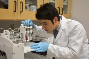
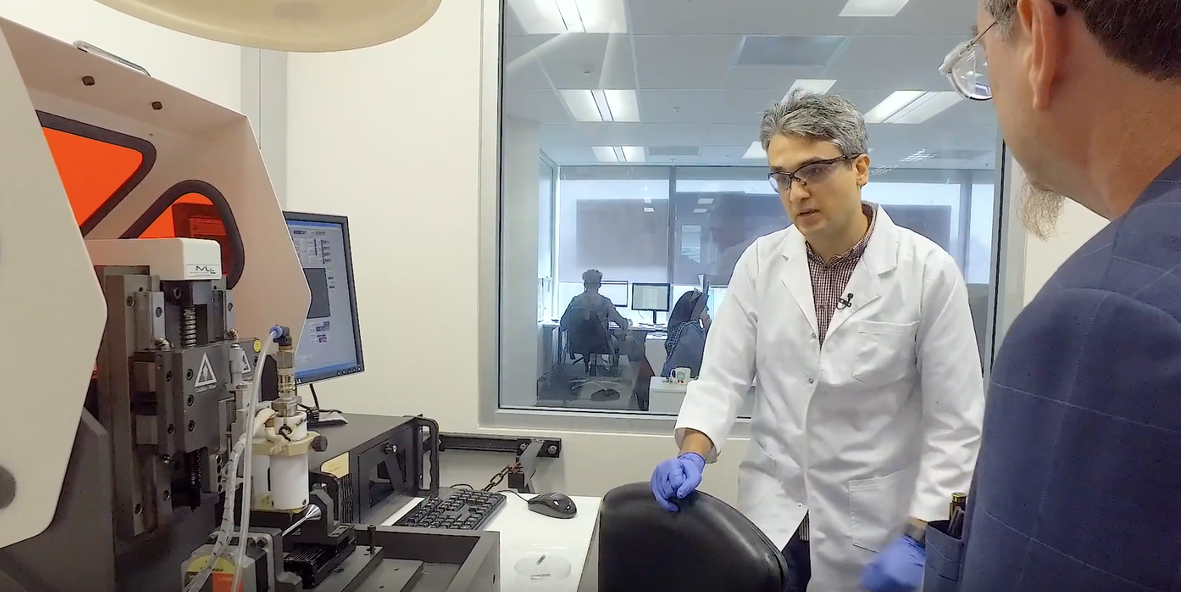
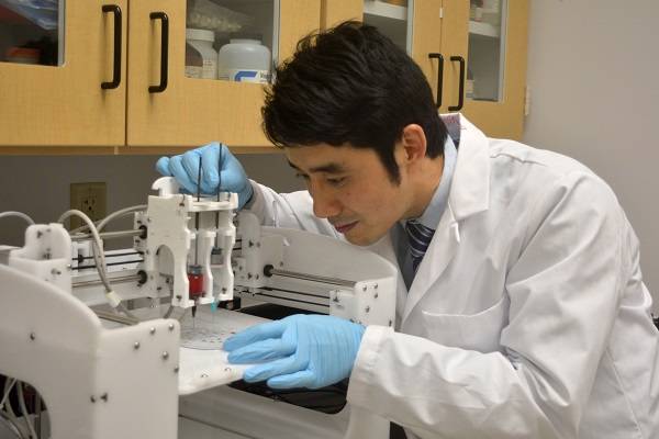
Dr. Yunqing Kang. [Image: Florida Atlantic University]

“From these results, it can be seen that although biodegradable stents may provide a valuable alternative to SEPS and SEMS, and also may eliminate the need for repeat esophageal dilations, biodegradable stents still presented some complications of migration and tissue regrowth. Also, biodegradation may lead to the collapse of stents after placement due to the collapsed degradation of the stent, quickly losing the mechanical strength,” Dr. Kang explained. “From these studies, it can be seen that the degradation properties of a BD stent determine its mechanical integrity. Studies showed that both ELLA-BD stent and PLLA-BD stents, the two currently available BD stents, can be degraded by hydrolysis, which is accelerated at low ambient pH. The stents began to degrade after 4 to 5 weeks and dissolved during a period of 2 to 3 months.”

Dr. Kang concluded that longer term studies are still needed to investigate “the relative efficacy or safety of esophageal biodegradable stents.”

“Therefore, the question of which type of stent should be recommended for the effective treatment of complex and refractory benign strictures, also malignant tumor remains unclear.”

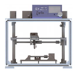
3D printing is also used to create a variety of stents, such as airway stents, vascular stents, and biodegradable stents as well. The technology offers advantages over conventional fabrication of polymer stents, and is our best shot for personalization.

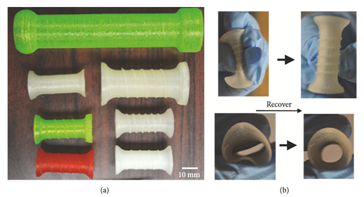
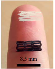
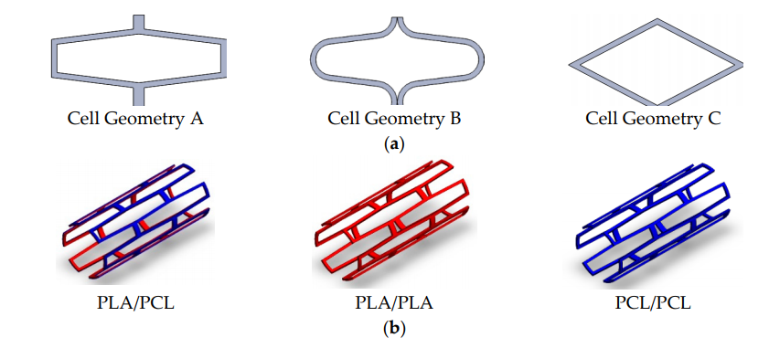
Photographs show the different types of 3D printed stents with different structures and material ratios (a). The stent was compressed and then recovered to the original shape (b).

“In our study, we used a 3D printing technology to produce a flexible polymer esophageal stent (Figure 3) [49]. We found that our 3D printing technique can print an esophageal stent with different size and shape,” Dr. Kang explained. “This is the first study using 3D printing technique to produce a polymer esophageal stent. Although the function of the 3D-printed flexible polymer stent has not been proved in vivo, the in vitro study showed that the 3D-printed esophageal stent has promising potential to treat malignant esophageal diseases. It can self-expand, and 3D printing technique can design and print different sizes and shapes of the stent easily.”

Again, the researcher noted that more study was required.

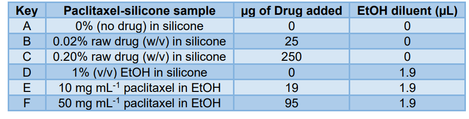
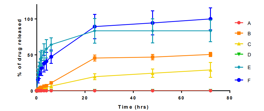
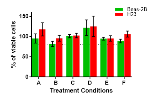
Dr. Kang also looked at drug-eluting stents (DESs), which are good for vascular applications but not clinically available yet to help treat esophageal cancer. Studies continues on this, and researchers have tried loading anticancer drugs, like 5-fluorouracil and paclitaxel, and whether sustained release of a stent with a bilayer film structure was possible.

“This kind of localized sustained delivery system in combination with the stent appeared to be a promising strategy to treat malignant esophagus cancer,” Dr. Kang wrote.

Esophageal cancer cells [Image: Reuters]

Finally, many types of models, like 3D cell spheroid models and animal xenograft/orthotopic ones, are used to evaluate the functionality and efficacy of a new drug delivery or treatment in cancer studies. But it’s not as easy for esophageal cancers with strictures, or benign esophageal diseases, due to the diameter and length of most FDA-approved stents not being applicable in these models. Dr. Kang stated that we need larger-sized animal models, like pigs or dogs, to study esophageal cancer and stents.

“The challenge is not only because of the complex surgery on a large animal, but also the potential difficulty of inoculating tumor cells or tissue in the orthotopic esophagus for tumor formation,” Dr. Kang explained. “In the future, new animal models or alternative animal modelling technologies still need to be developed and established for esophageal cancer stenting.”

I don’t enjoy thinking about testing medical theories out on animals, but I admit I see the point of needing larger animals for a necessary evil such as this.

Currently, popular palliation therapies for patients with inoperable esophageal malignancies suffering from dysphagia who use esophageal stents includes SEMS, SEPS, and biodegradable stents. But, as Dr. Kang’s research shows, novel stents that can get around some of the existing complications are desperately needed.

“In addition to improving the functionality of the drug-loaded stent with markedly reduced adverse effects, new ideal stents will allow to be tailored to individual needs at much lower cost,” Dr. Kang concluded. “Additionally, there is an unmet need to develop a large animal esophageal cancer model in vivo and establish a functional esophageal cancer model in vitro to test stents and study esophageal cancers.”

Discuss this research, and other 3D printing topics, at 3DPrintBoard.com or share your thoughts below. 
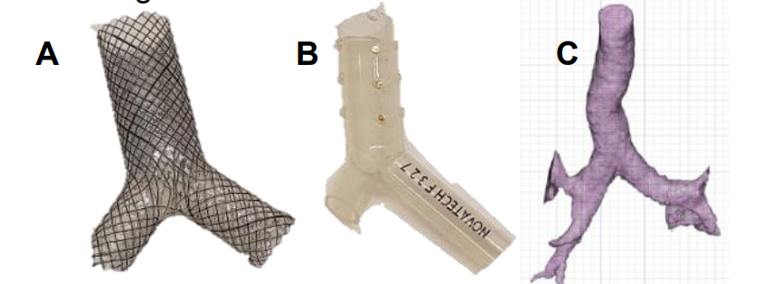
3D Printed PLA and PCL Composite Biodegradable Stents Show Promise

PCL (white) and PLA (black) stents

Biodegradable stents have shown great potential in reducing complications in patients, but they require further study, according to the authors of a paper entitled “3D-Printed PCL/PLA Composite Stents: Towards a New Solution to Cardiovascular Problems.” The researchers outline five main requirements that a biodegradable stent must meet:

  • Their manufacturing process should be precise
  • Degradation should have minimal toxicity
  • The rate of degradation should match the recovery rate of vascular tissue
  • They should induce rapid endothelialization to restore the functions of vascular tissue but should at the same time reduce the risk of restenosis
  • Their mechanical behavior should comply with medical requirements, particularly the flexibility required to facilitate placement but also sufficient radial rigidity to support the vessel

Although the first three requirements have been thoroughly studied, according to the researchers, the last two have been overlooked. A possible way of addressing these issues would be to create composite stents using materials that have different mechanical, biological or medical properties, such as PLA or PCL. Fabricating stents with these materials using laser cutting, however – the traditional method of manufacturing stents – would not be possible. The researchers, therefore, decided to produce them using 3D printing.

They 3D printed the stents using a tubular 3D printer. The stents were then seeded with cells and left for three days, and then tests were performed to assess the morphological features, cell proliferation, cell adhesion, degradation rate and radial behavior.

“The results prove the materials’ biological compatibility and encourage us to believe that PCL/PLA composite stents would comply with the fourth requirement, i.e., rapid endothelization without risk of restenosis,” the researchers state. “PCL’s better cell proliferation may be useful to increase the proliferation of endothelial vessel cells in the external wall of the stents, while an internal PLA wall may help to reduce the proliferation of cells that produce restenosis. However, further studies with other kinds of cells or substances need to be performed to confirm this. The results here show low cell proliferation because of the small amount of material that the stents have. Additional studies that use longer culture times may be beneficial to obtain better proliferation results.”

The researchers’ initial hypothesis was confirmed: the smaller the cell area of the stent, the better the cell proliferation rate. The cell shape of the stent, however, did not show any significant influence. Because of their different molecular weights, PCL showed better cell proliferation than PLA. PLA showed a much faster degradation rate, which limits its use for biodegradable stents. Radial behavior results show that composite PLA/PCL stents could be used to improve each material’s separate limitations, with PCL offering elasticity in the expansion stem and PLA providing rigidity in the recoil step.

Overall, 3D printing proved itself to be a promising method for producing stents. Both PCL and PLA showed themselves to be biocompatible, and the composite stents showed the most promise, with medium levels of degradation rates and mechanical modulus.

“Based on the results presented here, we believe that polymer composite stents manufactured with 3D-printing processes could be a highly effective solution to the current problems that stents made of polymers have,” the researchers conclude. However, FDA rules currently limit the use of 3D-printed stents in real clinical applications and, although PCL and PLA are FDA-approved materials, there are still open challenges to be met before approval for 3D-printed implantable medical devices can be obtained.”

Authors of the paper include Antonio J. Guerra, Paula Cano, Marc Rabionet, Teresa Puig and Joaquim Ciurana.

Discuss this and other 3D printing topics at 3DPrintBoard.com or share your thoughts below.