University of Hong Kong: Sterilization & Infection Rates in 3D Printed Models & Guides Used Intra-operatively

Hong Kong researchers explore the use of 3D printed medical models but advance in a more unique direction with concerns over infection. Detailing their findings in the recently published ‘A review of the manufacturing process and infection rate of 3D-printed models and guides sterilized by hydrogen peroxide plasma and utilized intra-operatively,’ the authors discuss the use of innovative bespoke devices for surgical planning.

As 3D printing has made impacts within the medical field, medical professionals, patients, and their families benefit due to better avenues for diagnosis, treatment, and education for everyone involved. But there are even more specific uses for 3D printed medical models today in promoting patient-specific treatment with improved pre-operative planning procedures and even intra-operative processes.

“In the specialty of orthopedics, 3D models can allow for visualization of bony anatomy and implant contouring, whilst guides can be created to direct osteotomies as well as screw entry sites,” explained the researchers.

Much attention is paid to the strides being made due to 3D printing, but outside issues such as sterility are critical to the health of patients also. Typical methods include ethylene oxide (EtO) gas and hydrogen peroxide plasma, which is a result of excitation beyond the gaseous phase—with free-radical formation allowing for sterility.

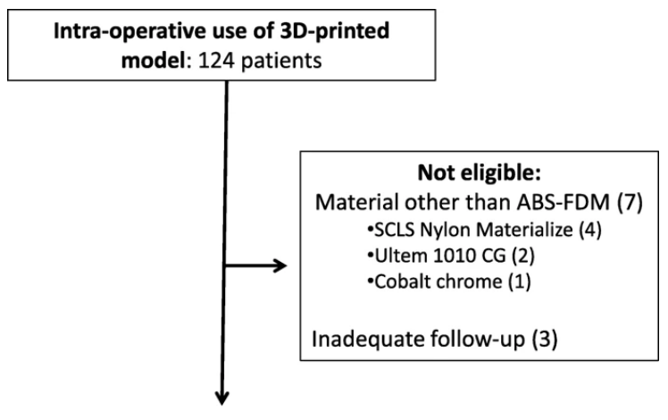
Patient eligibility and exclusion. More than 300 models were rendered by computer software from 2015 – 2019. The numbers of models proceeding to manufacturing, sterilization and intraoperative use amounted to 124. A further ten patients were excluded from analysis due to use of materials other than ABS (7 patients) as well as failure to reach 3-months of follow-up subsequent to surgery (3 patients), leaving a total of 114 patients eligible for analysis.

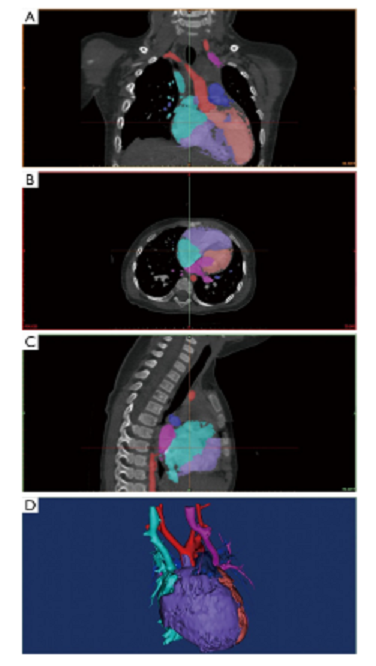
To date, the University of Hong Kong has produced over 300 3D models and guides. Beginning in 2015, their orthopedic academic unit began 3D printing models and guides on-site; and while they were at first fabricating models exclusively for orthopedics, over time they also began 3D printing for ‘other surgical contexts’ too. The authors confirm that out of the 300 models produced, 114 have been used for intra-operative purposes. Their review goes on to cite details regarding cases using models and guides, identifying those in which infections occurred, and highlighting risk factors.

3D printed models and guides were designed using Meshmixer and printed on a Fortus 450mc 3D printer with ABS-M30i.

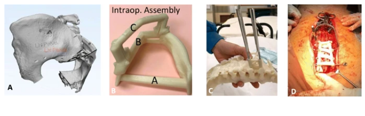
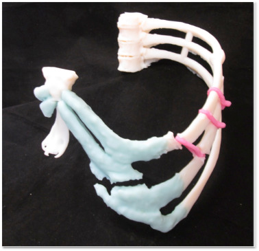
Aspects of model/guide manufacturing unique to intra-operative usage. a 3D-rendering of pelvis model with initials engraved upon the left ilium to allow for correct patient identification. b Photos taken by instrument nurse demonstrating proper grouping and assembly of a surgical guide for pedicle screw placement so corresponding components may be packaged and sterilized together. The assembled guide was contoured to fit upon bony surface landmarks of the posterior spinal vertebra, as demonstrated during testing upon a 3D-model of the same patient c and during the definitive surgery (d)

3D prints were sterilized with hydrogen peroxide plasma, and low temperatures prevented deformation in material.

“A surgical time-out procedure ensured that the printout was used for the correct patient, anatomical region and procedure, in accordance to initials upon the surface,” explained the authors. “Post-operatively, printouts were similarly subject to low temperature disinfection then returned to the surgeon in charge.”

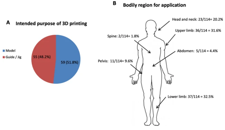
Application of 3D printouts. a Intended purpose of 3D printout showing 59/114 (51.8%) of printouts being utilized as anatomical models and 55/114 (48.2%) as guides/jigs intra-operatively. b The 124 cases utilized intraoperatively spanned different regions of the body as well as surgical specialties. The numbers relevant to each region and their percentages in relation to the whole patient cohort is shown

The researchers examined 3D prints from 124 patients with models used intra-operatively during surgical planning and management.

“Seven cases were excluded as printouts were not constructed from ABS, of which four cases utilized nylon, two case utilized polyetherimide (Ultem1010 CG), and one case utilized cobalt chrome,” explained the authors.

“Three cases were excluded because of inadequate follow-up. A total of 114 models remained for subsequent analysis. Fifty nine out of 114 (51.8%) were anatomical models utilized on-table for planning and / or implant contouring. The remaining 55/114 (48.2%) were utilized as guides or jigs specific to patient anatomy to facilitate corrective osteotomies, screw insertion or pin placement.”

Ultimately, 10.9 percent of the guides or jigs developed infections, while 3.3 percent of the models developed infections at the surgical sites.

“All six cases of guides/jigs with infection were utilized to facilitate osteotomies. Both models with infection were utilized for implant contouring, one during fixation of a pilon fracture and the other for an orbital floor blowout fracture,” stated the researchers.

Pointing out that while the infection rate of 7 percent was comparable to previous literature published regarding traditional techniques, the authors realize the importance of users to ‘be aware of potential caveats,’ despite the overall safety of the application. There are also intrinsic challenges in the fabrication of patient-specific devices and ensuring the safety of tissue biocompatibility.

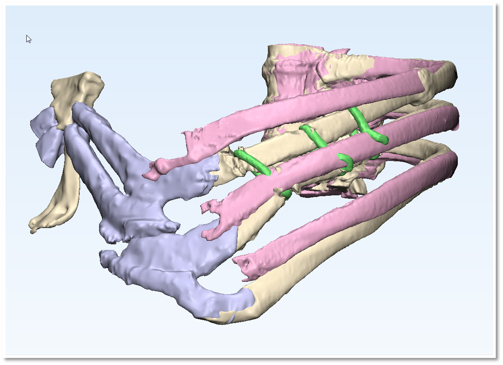
Cross-section of pelvis 3D model demonstrating irregular luminal spaces. a Arrows indicate surface openings upon the posterior ilium on a 3D model of the pelvis. The dotted line and arrowhead demonstrates the level of transverse sectioning subsequently performed. Cross-sectional appearance following software rendering b and physical sectioning c of the same pelvis model demonstrating irregular trabecular spaces contained within

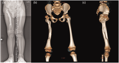
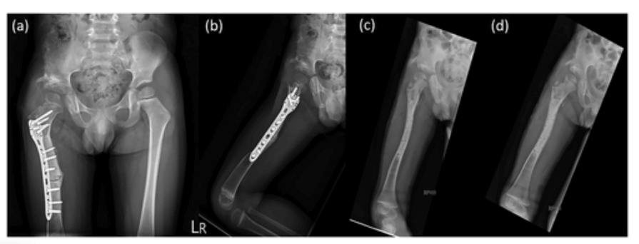
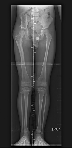
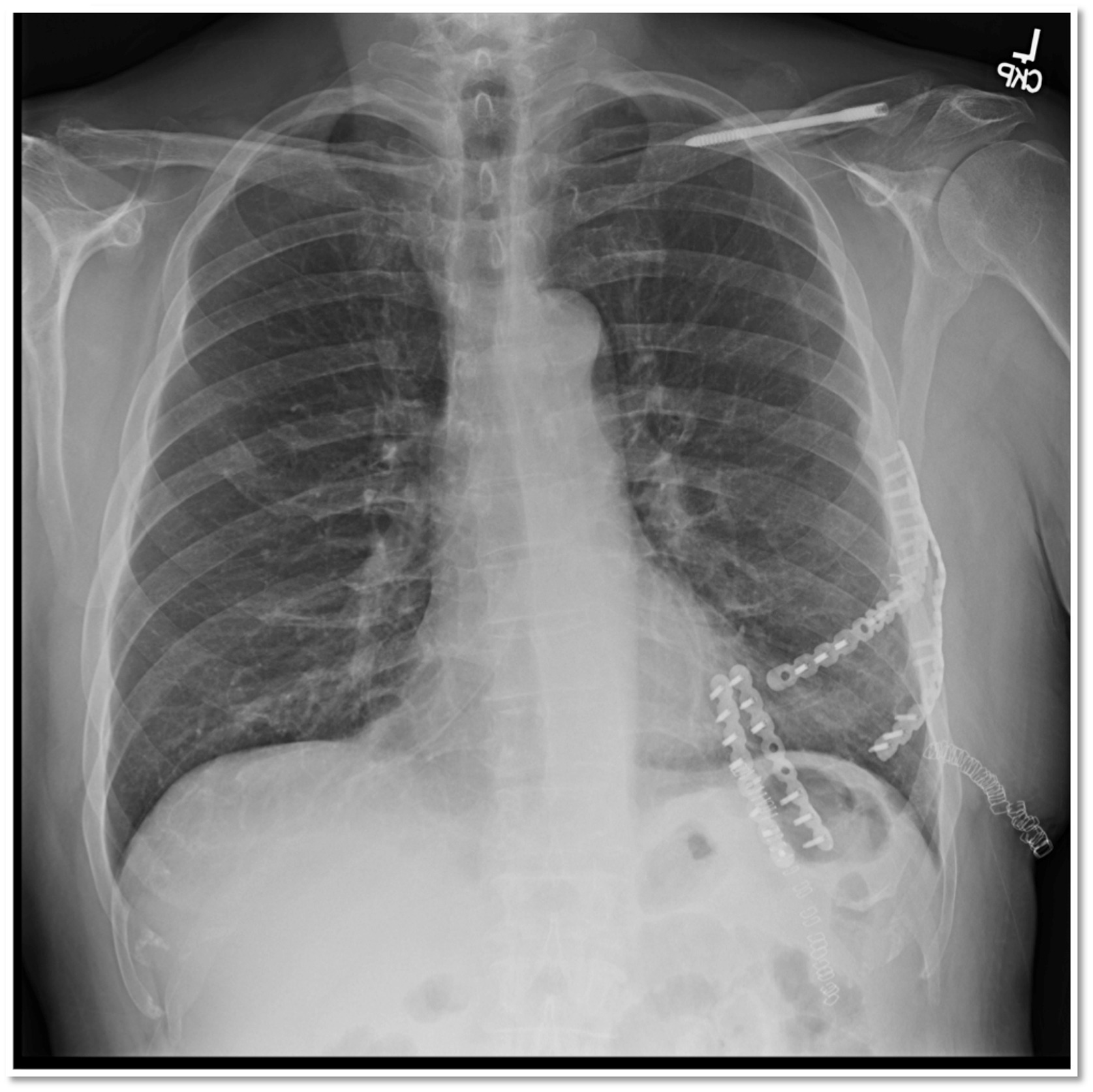
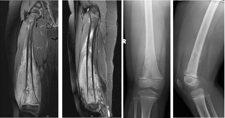
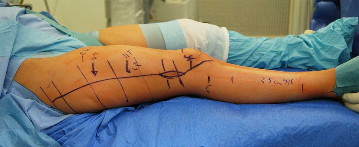
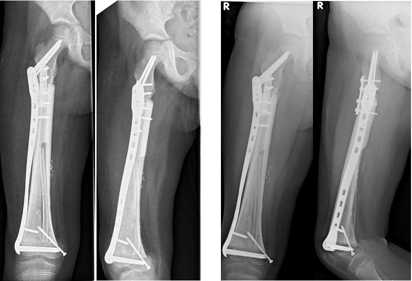
Intraoperative use of osteotomy guide. a Software rendered image of guide intended for corrective osteotomy and shown upon the tibial shaft (b) with the osteotomy site marked in teal. c Intra-operative photo with the guide secured and oscillating saw engaged in preparation for osteotomy and (d) upon completion. e Intra-operative x-ray demonstrating reduction and fixation of the tibial shaft following corrective osteotomy. f Similar guide retrieved post-op demonstrating damage to ABS over the osteotomy slit with the potential to release debris

“It is worth noting that prior studies detailing infection-related outcomes of 3D printouts have not explicitly utilized them intra-operatively, and this is one of the first studies to have done as such. Our overall impression was that our process of sterilization and on-table usage is safe, and that surgical complexity and tissue manipulation as reflected by increased operating time were the main culprits for infection,” concluded the authors.

“In detailing the design, printing, and sterilization of 3D printouts as well as infection-related outcomes amongst this sizable cohort, we demonstrate that our production process is safe for continuation and may be adopted elsewhere.”

What do you think of this news? Let us know your thoughts! Join the discussion of this and other 3D printing topics at 3DPrintBoard.com.

[‘A review of the manufacturing process and infection rate of 3D-printed models and guides sterilized by hydrogen peroxide plasma and utilized intra-operatively’]

The post University of Hong Kong: Sterilization & Infection Rates in 3D Printed Models & Guides Used Intra-operatively appeared first on 3DPrint.com | The Voice of 3D Printing / Additive Manufacturing.

Zhejiang University Sheds Light on APVC with 3D Printed Surgical Models

Researchers from China’s Zhejiang University are looking for new ways to improve preoperative planning for procedures for children, with their findings outlined in the recently published ‘Utility of three-dimensional printing in preoperative planning for children with anomalous pulmonary venous connection: a singer center experience.’ Many of the greatest benefits were put into action for this study as the 3D printed models could be used for completing diagnosis, assessing treatment options, and planning for surgery.

In this study, Chinese medical scientists studied 17 children diagnosed with anomalous pulmonary venous connection (APVC) from November 2017 to January 2019. Ages ranged from only two days old to twenty months old, with the following variations:

  • Ten children suffering from total supracardiac APVC
  • One child suffering from intracardiac APVC
  • Mixed type APVC in one child
  • Partial APVC in three children

Data from CT scans was imported into Mimics 19.0 software for 3D modeling and design of the heart model to display elements of the heart such as papillary muscles, muscle bundles, and outflow tracts. While very little research has been performed for APVC citing the assistance of 3D printing, it is clear from this study that the use of models allows for much greater light to be shed on the condition.

“We manually labelled each area according to the left ventricle (LV), right ventricle (RV), LA, RA, aorta (AO), and pulmonary artery (PA) area modules of the CT heart module and distinguished them with different colors. Special attention was paid to labeling the boundaries of each part,” stated the researchers.

Image segmentation and postprocessing in Mimics 19.0 software. The colored masks were segmented for 3D modeling. (A) Coronal plane; (B) transverse plane; (C) Sagittal plane; (D) 3D modeling. Green, superior vena cava and right atrium; purplish red, pulmonary veins and left atrium; purple, right ventricle; orange, left ventricle; red, aorta; dark blue, pulmonary artery.

Four cases diagnosed with supracardiac type TAPVC. (A) Refers to patient 1, view from posterior; (B) refers to patient 5, view from posterior; (C) refers to patient 9, view from posterior; (D) refers to patient 10, view from posterior. *, obstruction exists at the junction of the common pulmonary venous confluence to the left-sided vertical vein (VV). Ao, aorta; PA, pulmonary artery; PV, pulmonary vein; SVC, superior vena cava. Orientation labels: I, inferior; L, left; R, right; S, superior.

Two cases diagnosed with intracardiac type TAPVC. Part of the hollowed heart model was segmented to emphasize the PV and RA we focused on. Pulmonary vein through the coronary sinus opening in the right atrium. (A) Refers to patient 11, view from inferior; (B) refers to patient 12, view from the right. CS, coronary sinus; IVC, inferior vena cava; PV, pulmonary veins; RA, right atrium. Orientation labels: A, anterior; I, inferior; L, left; P, posterior; R, right; S, superior.

One case (patient 13) diagnosed with infracardiac type TAPVC, view from posterior. The red arrow indicates the junction of the pulmonary vein and the inferior vena cava. Ao, aorta; IVC, inferior vena cava; PV, pulmonary vein; VV, vertical vein; TAPVC, total anomalous pulmonary venous connection. Orientation labels: I, inferior; L, left; R, right; S, superior.

Three cases diagnosed with PAPVC. Images (A,B) both represent patient 15. (A) The blue arrow points to RPV flowing into the right atrium. View from posterior. (B) The blue arrow represents the outlet of RPV, flowing into the right atrium. View from the right. (C) Patient 16 diagnosed with PAPVC; the blue arrow represents RPV flowing into the right atrium. View from posterior. (D) Patient 17. The blue arrow represents RSPV flowing into the SVC. View from posterior. Ao, aorta; CoA, coarctation of aorta; IVC, inferior vena cava; LIPV, left inferior pulmonary vein; LSPV, left superior pulmonary vein; RA, right atrium; RIPV, right inferior pulmonary vein; RPV, right pulmonary vein; RSPV, right superior pulmonary vein; SVC, superior vena cava. Orientation labels: A, anterior; I, inferior; L, left; P, posterior; R, right; S, superior.

Two cases underwent cardiac CT examination during follow-up. (A,D) Blood volume; (B,C) hollowed models. Images (A,B,C) represent patient 3 after repaired supracardiac TAPVC. (A,B) view from posterior; (C) view from anterior. (C) The blue and yellow arrows represent the opening of the right pulmonary vein and left pulmonary vein. There is no anastomotic stenosis. (D) Superior view of patient 4 after repaired supracardiac TAPVC. Stenosis of LSPV (blue arrow) is shown compared to other pulmonary vein branches. Ao, aorta; LA, left atrium; LIPV, left inferior pulmonary vein; LSPV, left superior pulmonary vein; PA, pulmonary artery; PV, pulmonary vein; RIPV, right inferior pulmonary vein; RSPV, right superior pulmonary vein. Orientation labels: A, anterior; I, inferior; L, left; P, posterior; R, right; S, superior.

3D printing of the personalized heart models was completed via an ISLA 650 3D printer (Shining 3D, China). Preoperative planning was then based on the models, along with medical history of the patients, and imaging data. The models were also used as surgical guides in the operating room upon being sterilized. Each patient-specific heart model took around half an hour to two hours to model, with 3D printing requiring anywhere from two to five hours. Surgeries were performed on all 17 patients, and each procedure was successful.

“The malformations demonstrated by the 3D models were consistent with intraoperative observations, and presurgical planning was in line with real surgery programs. These heart models could be sterilized and brought into the operating room for surgery navigation. These 3D models greatly assisted the presurgical planning for APVC surgery and were of great clinical value from our experience.”

“After surgeries, these heart models were evaluated on whether they were of high quality, and whether they could help presurgical planning, reduce unforeseen circumstances, and benefit medical education. An evaluation pertaining to the issues above was conducted via questionnaire by our cardiac surgeons and cardiologists.”

3D printed heart models and medical devices such as implants have been used in connection with cardiac issues and defects, with guides and models used in everything from complex medical training to pediatric surgeries, methods for creating heart patches, and more.

What do you think of this news? Let us know your thoughts! Join the discussion of this and other 3D printing topics at 3DPrintBoard.com.

[Source / Images: ‘Utility of three-dimensional printing in preoperative planning for children with anomalous pulmonary venous connection: a singer center experience’]

The post Zhejiang University Sheds Light on APVC with 3D Printed Surgical Models appeared first on 3DPrint.com | The Voice of 3D Printing / Additive Manufacturing.

Taiwan: Researchers Rely on 3D Printed Models & Surgical Guides for Pediatric Orthopedic Surgery

Medical researchers and orthopedic surgeons in Taiwan at Kaohsiung Veterans General Hospital continue to explore better ways to heal bones and manage defects, with their findings outlined in the recently published ‘Anatomic three-dimensional model-assisted surgical planning for treatment of pediatric hip dislocation due to osteomyelitis.’

While bone defects are already a challenge to manage, obviously the problem is compounded in children, with smaller bones being even more difficult to repair in surgery. Currently, there are few options for a good device meant for small bone repair during pediatric osteotomies—making it difficult for surgeons around the world to correct both subluxated hip joints and deformed femurs in children.

The authors (and surgeons) performed corrective surgery on a four-year-old boy with a post-osteomyelitis deformity. In preparing for the surgery, they relied on a 3D printed model of the bone for studying the condition, surgery and preparing the site for the appropriate implant. Because this type of surgery requires ‘meticulous planning,’ the doctors required both 2D and 3D assistance, in the respective forms of axial images and 3D virtual models of patient anatomies.

Radiographs taken before corrective surgery. (a) Triple film showing the proximal femur deformity with osseous recovery. Three-dimensional computed tomography image: (b) anteroposterior and (c) lateral views

As the surgeons examined the patient and reviewed the CT, they noticed a genu valgus deformity (more commonly known as a ‘knock-knee’ condition). Another corrective surgery was scheduled, with 3D CT imaging examined for bone tissue analysis. The surgeons realized, however, that the procedure would be more successful overall with a life-size 3D model. They were able to outline a patient-specific plan, also bringing in additional assistance from an orthopedic consulting firm focused around 3D orthopedics and ‘patient-specific instrumentation.’

Customized-to-patient three-dimensionally–printed guide. (a) The patient-specific guide for our patient. (b) Two resecting osteotomies can achieve optimal joint congruency and varus angle correction. (c) Correcting the femoral rotation would result in joint translation in both the coronal and axial planes

What was also very valuable to the surgery—and the outcome for the little boy involved—was that the surgeons could use the model to practice on, exercising ‘simulations of possible osteotomy options.’

“After a few osteotomy options had been analyzed, one osteotomy cut was made vertically to the femoral shaft on the subtrochanteric area, and another was made on the middle third of the femur to correct the bowing deformity of the midshaft,” stated the researchers. “Correction of femoral rotation can result in either joint translation in the coronal and axial planes or difficulty with fixation, both of which could be prevented with the help of the 3D model in the present case.”

The results of the surgeries were successful, with the patient able to stretch and begin other mobilization activity after four months.

Postoperative (a) anteroposterior and (b) lateral views. Fifteen-month postoperative (c) anteroposterior and (d) lateral views

“The result of our case suggests that the use of 3D printing models improves the postoperative performance as shown by both physical function and radiological evidence,” stated the authors in the concluding discussion.

“The use of a 3D-printed patient-specific guide is a safe, modern, affordable, and promising method that offers advantages including a shorter surgical time, optimally positioned implant placement, acceptable alignment, and a probable lower rate of complications. The utilization of 3D-printed models for skeletal deformity surgery, especially complex and difficult pediatric surgery, provides superior precision and foreseeably better outcomes. We strongly believe that with the promotion of 3D printing methodology, models for preoperative planning may soon become the gold standard for pediatric deformity correction surgery.”

3D printing continues to make impacts in the area of healing bones, regeneration and planning for complex surgeries with a range of medical devices and models. What do you think of this news? Let us know your thoughts! Join the discussion of this and other 3D printing topics at 3DPrintBoard.com.

Triple film at 2-year postoperative follow-up showing no significant leg length discrepancy (<0.5 cm)

[Source / Images: ‘Anatomic three-dimensional model-assisted surgical planning for treatment of pediatric hip dislocation due to osteomyelitis’]

The post Taiwan: Researchers Rely on 3D Printed Models & Surgical Guides for Pediatric Orthopedic Surgery appeared first on 3DPrint.com | The Voice of 3D Printing / Additive Manufacturing.

Mayo Clinic Researchers 3D Printing Models & Surgical Guides for Chest Wall Reconstructive Surgery

In the recently published ‘3D printed modeling contributes to reconstruction of complex chest wall instability,’ researchers from the Mayo Clinic Rochester look into new methods for creating 3D printed models and surgical planning devices for the treatment of complex conditions, pointing toward the example case of a 55-year-old male who underwent resuscitative thoracotomy after developing a pulmonary hernia—a condition usually caused by part of the lung bulging or tearing into the surrounding wall.

The researchers 3D printed two different models during the case study, with one demonstrating anterior fractured cartilage, and the right side acting as a mirror for the ‘normal ribs’ to demonstrate misalignment of the other side. The models were used to plan the surgery, involving a thoracotomy incision and more.

Chest trauma often requires surgical stabilization of rib fracture (SSRF). 3D printing has been reported as useful during intricate cases in the past for creating chest wall prostheses, but not with reconstruction using standard surgical tools.

“Given the compound curvature and dynamic anatomy of the chest wall, the utilization of patient specific 3D printed models to facilitate detailed operative planning prior to undertaking complex non-union or re-operative chest wall repair with SSRF presents a substantial opportunity,” stated the researchers.

The patient studied during this research had actually been stabbed, causing the trauma to his left chest area—also resulting in cardiac arrest. Surgery was performed, but the pulmonary hernia developed later and was repaired in a second procedure. He continued to be in pain, however, especially when laughing or coughing or stretching. As surgeons prepared to restore his chest wall with plates and bicortical screws, they used 3D printed models consisting of:

  • Vertebral bodies
  • Ribs
  • Cerclage wires
  • Cartilage

Model photos demonstrate non-union at the costal cartilage.

The models offered a clear perspective for the surgeons regarding areas like the disruption at the costosternal joint which was causing left chest distortion. As they continued to study the 3D printed models, the doctors were able to decide on their surgical approach. The authors point out, however, that reconstruction of the chest wall is a complicated procedure, ‘fraught with difficulties,’ to include:

  • Malalignment
  • Altered geometry
  • Patient compensatory muscular hypertrophy
  • Spinal changes

With 3D modeling, however, surgeons can optimize the entire surgical and possible implantation process too.

“Without 3D models, each individual case requires the surgeon, using two-dimensional non-life-size images, to create a 3D model mentally and then translate that to a life size plan. The patient-specific life size 3D printed model allows the surgeon to more easily comprehend and understand the anatomy without mental gymnastics,” state the authors. “These models offer a life size, multimaterial, tactile approach that was heretofore inaccessible. Secondarily, it allows the surgeon to pre-operatively bend plates to the patient’s specific anatomy, preprocedurally practice the osteotomies, and preoperatively have the plates sterilized, thus saving operative time.”

Overlaid model demonstrates the degree of mal-alignment based on mirror image data from the contra-lateral side.

As 3D printed models can be printed from the medical facility, all the benefits of 3D printing technology are recognized from affordability in production to speed to customization—and best of all, patient-specific treatment.

“At our institution, we have built an in-hospital centralized 3D printing facility to serve the surgical theater when complex cases arise. This allows point-of-care multidisciplinary collaboration and 3-D printing additive manufacturing expertise to the surgeons involved which accelerates innovative personalized care,” stated the researchers in their final discussion and conclusion.

“We believe that the early adoption of this technology by surgeons can help improve surgical quality and provide more individualized patient care. Centers should pursue the integration of 3D printed models into their practice and active collaborations between surgeons and modeling experts should be sought at every available opportunity.”

3D printing holds a special place in the world of medicine as the technology has made such sweeping changes in the way medical professionals are able to diagnose and treat other medical issues too like brain tumors, examine heart conditions with cardiac patches, and even create devices like 3D printed protheses for children with undeveloped eyes—and countless other issues making life much more tolerable for patients around the world. What do you think of this news? Let us know your thoughts! Join the discussion of this and other 3D printing topics at 3DPrintBoard.com.

Post-operative film showing reduced and plated ribs and costosternal articulation.

[Source / Images: 3D printed modeling contributes to reconstruction of complex chest wall instability]

Turkey: Researchers Innovate Further in Creating Titanium Hip Implants

In ‘Design, manufacture, and fatigue analysis of lightweight hip implants,’ Turkish researchers Yunus E. Delikanli and Mehmet C. Kayacan explore and test better ways to fabricate hip implants for total hip arthroplasty (THA). Most humans are aware of the critical importance of their hip joints—especially when something goes wrong and a major health issue arises, or deterioration from age becomes an apparent, and painful.

The ball-and-socket joint allows humans to perform most of the required actions for mobility—from taking a seat, to walking or running—or more athletic activities requiring jumping. This joint is expected to handle a lot of wear and tear over a lifetime, and fractures due to trauma can occur at any age but are much more expected in the elderly. According to the research team, ‘heritage, nutrition, and lifestyle’ can play a role too.

“In cases involving high body weight and physical activity, the load on the femur increases, which in turn results in bending and torsional stresses in the femoral component of an implant,” stated the researchers. “If these stresses are repetitive and variable, fatigue fractures or deformations may arise in hip implants.

(a) Small pore (0.3 mm, KG) and (b) large pore (0.6 mm, BG) implants.

Striving to innovate further in creating implants, the researchers used titanium metal powder (Ti6Al4V alloy) for 3D printing. Nine samples were created, from .03 mm to solid, using Kubisch Raumzentrierten (KRZ) geometries in a lattice structure, with a porosity of 78.3 percent, 3D printed on an EOS M280 direct metal laser sintering (DMLS) machine. Upon fabrication of the samples, the researchers realized a reduction in weight of up to 17 percent—due to the ability to not only make complex structures but also with hollowed out interiors.

In testing, the research team found that ‘maximum equivalent stresses’ were exhibited in what is called the ‘neck region’ of each implant. Lightened implants exhibited greater stresses even with the same loading—attributed to less of a cross-sectional area, and a more complex one. Each implant was deemed successful after five million load cycles—with an infinite fatigue life.

Lightening process of the implants.

“All the implants produced with DMLS have been shown to exhibit enough fatigue performance according to the requirements of the ISO 7206 standard. In addition, FEA findings are highly consistent with fatigue test results,” concluded the researchers. “Thus, the displacements outside of the investigated pore size range can be predicted with sufficient accuracy by FEA. This enables us to save production costs and obtain an idea about the implant performance without carrying out any building process.”

Upon its inception decades ago, very few could have realized the impacts 3D printing and surrounding technologies would have on medical patients today, who are now able to enjoy a better quality of life due to a range of different implants and devices brought forward by innovative researchers who have created everything from surgical guides for replacement surgeries to knee implants to spinal implants.

Find out more about hip arthroplasty and the role of 3D printing here. What do you think of this news? Let us know your thoughts! Join the discussion of this and other 3D printing topics at 3DPrintBoard.com.

Cell development channels of KG (a) and BG (b) hip implants.

[Source / Images: ‘Design, manufacture, and fatigue analysis of lightweight hip implants’]

Swiss Hospital Will Use axial3D’s Software Platform to Improve Patient Care with 3D Printed Medical Models

[Image: axial3D]

The award-winning, Belfast-based medical 3D printing and healthcare technology firm axial3D is focused on helping the global healthcare industry adopt 3D printing by using its patient-specific medical models to improve surgical outcomes, assist patients and doctors in better understanding ailments and treatments, and facilitate pre-operative planning.

Now, on the heels of a new partnership with Tallahassee Memorial HealthCare, the company has announced that it is collaborating with top Swiss medical center University Hospital Basel (USB) in order to improve process management and patient care and outcomes at the hospital’s interdisciplinary 3D Print Lab.

“3D printed models have been shown to help surgeons complete complex life-saving surgeries that would be otherwise impossible,” axial3D’s Ryan Kyle told 3DPrint.com. “University Hospital Basel’s new collaboration with axial3D will help to deliver high-quality 3D printed models much quicker than before.”

The hospital, which has about 7,000 people on staff, is northwest Switzerland’s biggest healthcare facility. Its 3D Print Lab uses patient image data to fabricate realistic anatomical models, and other objects, using a variety of different materials and 3D printing methods. Now it will be using axial3D’s new cloud-based platform, axial3Dassure, to support its 3D printing program.

[Image: University Hospital Basel]

By using axial3Dassure, USB will optimize its 3D Print Lab in order to provide a greater level of performance and patient care. The software, which has an end to end workflow, provides features like processing and quality management, so that hospitals and medical centers can meet their expanding business needs through its powerful analytics. The new axial3Dassure platform will also help support collaboration within the hospital’s 3D Print Lab with such features as email notifications and task-driven workflows.

Daniel Crawford, axial3D

“We are very excited to be working with the team at University Hospital Basel. They are a leading force in medical 3D printing, not just in Europe, but globally, and this alliance will ensure the expertise they have developed can support our company’s growth by informing the ongoing development of axial3D’s software solutions,” said axial3D’s CEO and Founder Daniel Crawford. “With a growing requirement for 3D printing within healthcare, a centralized management platform is necessary for any 3D print lab, which plans to scale and grow in the coming years. University Hospital Basel has taken strides in its commitment to improving outcomes for patients through technology advances in the form of this collaboration.

“Our software will help the hospital gain insight into the statistics and figures usually hidden within data, ultimately allowing them to measure clinical impact and value 3D printing is having for patients. The workflow management capability will allow the hospital to speed up the creation, processing, and delivery of 3D printed models, while ensuring auditability, reliability and standardization.”

By using axial3Dassure software, USB will be able to increase efficiency and improve compliance and productivity. The hospital’s 3D Print Lab, which includes over 20 desktop and industrial 3D printers, will now be better equipped to manage communication, quality control, tracking, and workflow management.

In addition, USB will benefit from the company’s orthopaedic auto-segmentation software module, which is embedded within the axial3Dassure platform. This module will help lower the amount of time that is typically required during pre-production of 3D printing orthopaedic models.

Finally, by partnering with axial3D, USB will be able to speed up the creation, processing, and delivery of its 3D printed surgical guides.

“Our initial focus for the use of 3D printed surgical guides was within the Department of Cranio-Maxillofacial Surgery where 3D printing has now become routine,” explained Philipp Brantner, Senior Physician of Radiology and the Co-Director of the 3D Print Lab at University Hospital Basel. “Having access to onsite printing has revolutionized how we treat those patients, some who arrive with life-threatening injuries that require immediate action. The functionality that we now get provided will allow us to speed up production and treat patients more effectively and efficiently.”

What do you think about this news? Discuss this story and other 3D printing topics at 3DPrintBoard.com or share your thoughts in the Facebook comments below.

3D Printing Helps a Five-Year-Old Girl to Dance Again

Five-year-old Daria loves dancing, but that was imperiled when she was diagnosed with Ewing’s Sarcoma, a rare malignant bone tumor, in her right leg. The tumor was large, extending almost to her distal femur, so removing it while still allowing her to walk – and dance – normally was a special challenge. Careful planning was required, but thankfully, Daria’s doctors had the tools to do just that.

“With all the soft tissues and skin in the way, what surgeons experience in surgery can be completely different to what they see on a 2D scan,”  said Mieke Motmans, Clinical Engineer at Materialise. “Using 3D digital and physical models gives an extra level of information and to work out exactly how best to proceed – it’s like the difference between outlining a journey on a paper map, and using a GPS that tells you exactly where you are going and when you will get there.”

Dr. A.H. Krieg and colleagues at the University Children’s Hospital in Basel, Switzerland had to remove the tumor and reconstruct Daria’s femur using a combination of her own live fibula and a donor bone.

“Endoprostheses (artificial bones) are the usual limb-salvage treatment if the tumor is close to the knee joint or extends to the epiphysis, but this is rarely possible for children as young as Daria – there are high complication rates,” said Dr. Krieg. “Making the reconstruction a success meant using 3D printing to its full potential and working with the right experts.”

Materialise has worked with surgeons to plan for more than 2,000 osteotomy cases, or corrective surgery where a bone is cut to allow realignment. The company has also designed and 3D printed more than 700 custom implants or endoprostheses.

“I’d actually worked with Mieke and her team before, so I knew they could help,” said Dr. Krieg. “The first step was uploading our MRI and CT scans of Daria’s leg to Materialise’s online portal for evaluation.”

Motmans specializes in oncology cases. Her team began by aligning meshes from Daria’s CT and MR scans, checking them in the Materialise Mimics medical imaging processing software and verifying the tumor’s position.

“Finding the tumor outline is where we start,”  she said. “Sometimes tumors are only visible in MRI, so Mimics helps us transfer its boundaries to the more accurate CT scans. Then we combine the CT scan ‘slices’ to build the 3D bone geometry model that lets surgeons see the overall picture and zoom in to the detail. Some surgeons have a very clear idea of what they want to do, while others will simply send us the scans and ask us for a solution. Either way, getting to this stage – seeing the 3D model – is often a critical moment. The model’s accuracy lets us verify the planned approach and sometimes it can be very obvious that the initial plan just isn’t going to work.”

Materialise and the hospital team worked together to map out the surgical procedure and position the resection plains that would be used to show exactly where saw cuts would be needed around the tumor. They had to retain as much bone as possible to keep Daria’s knee joint intact to support the screws for the titanium bridge that would hold the remaining bone, the allograft and fibula as they grew together during osteosynthesis. The margins were tight because the tumor was so big and so close to Daria’s knee.

“Where to cut the tumor influenced the size of the hole, which in turn governed the required shape of the donor bone,” said Motmans. “We couldn’t print anything until the whole planning stage – where to resect the tumor and allograft, the plate size, the position of the screws – was complete.”

The surgeons had to trim their normal safety margins and sacrifice the lower epiphyseal plate which contains live growing bone tissue, but they were finally able to set their resection planes. Materialise then 3D printed the models and the three cutting guides. Slots in these templates, two for the femur and one for the allograft, would be used to precisely position the surgeon’s saw blade, with carefully positioned holes directing the drill to ensure correct screw positions and insertion angles. The allograft guide defined the channel where the fibula would be inserted. Motmans used models showing pre- and post-operative bone geometry to check that each cutting guide and plate fitted precisely.

“The operation lasted ten hours, involved seven surgeons and was a great success,” said Dr. Krieg. “Although Daria still has a long way to go in rehabilitation, she now has the chance to make a nearly full recovery and use her leg without major restrictions.”

“The skilled teams that perform these operations are amazing,” said Motmans. “To be able to help them is a privilege. 3D printing means surgeons can plan ahead, allowing them to deal with problems before they even enter the operating room, and so avoid surprises during surgery. That’s an outcome that’s great for everyone.”

X-ray 6 weeks and 3 months after the surgery

Discuss this and other 3D printing topics at 3DPrintBoard.com or share your thoughts below.

[Source/Images: Materialise]


Natural Plant-Derived Resins Used to Make Antibacterial 3D Printing Filament

Hospital-acquired infections are a growing problem everywhere. The CDC calls them a “major, yet preventable threat to patient safety,” and the key to preventing them lies in keeping bacteria from spreading in a setting where bacteria is rampant. As 3D printing becomes more and more prevalent in the medical field, it is vital to make sure that 3D printed implants and tools do not play a role in spreading disease. Certain companies have been working on creating antibacterial 3D printing filament, and a group of researchers has conducted a study on bioactive filaments with antimicrobial and antifungal properties. You can access the paper, entitled Bioactive Potential of 3D-Printed Oleo-Gum-Resin Disks: B. papyrifera , C.myrrha , and S. benzoin Loading Nanooxides—TiO2 , P25, Cu2O, and MoO3, here.

The researchers point out that bacteria have managed to develop resistance to many antibiotics, but that there are many natural antibiotics to which resistance has not yet been developed. They extracted oleo-gum-resins from benzoin, myrrha, and olibanum plants and combined some of them with 10% of metal nano oxide particles. 3D printer filament was created from the resins and metals, then 3D printed into disks which were subject to a number of tests.

“Due to their intrinsic properties, disks containing resins in pure state mostly prevent surface-associated growth; meanwhile, disks loaded with 10% oxides prevent planktonic growth of microorganisms in the susceptibility assay,” the researchers explain. “The microscopy analysis showed that part of nanoparticles was encapsulated by the biopolymeric matrix of resins, in most cases remaining disorderly dispersed over the surface of resins. Thermal analysis shows that plant resins have peculiar characteristics, with a thermal behavior similar to commercial available semicrystalline polymers, although their structure consists of a mix of organic compoundsThe disks 3D printed from the natural materials, in most cases, inhibited the growth of the clinical pathogens being studied, and when nano oxide particles were added, the materials were even more effective.

Whats more these materials behaved just like some polymers do. The resins,

showed thermal behavior inherent to semicrystalline polymers such as polyester and polyurea; at some point, the molecules disposed in amorphous matrix obtain enough freedom of motion to spontaneously rearrange themselves into crystalline forms. This transition from amor-phous solid to crystalline solid was evidenced by distinct exothermic peaks, as the temperature increases to 500samples, eventually reaching its melting point.

In short this is a promising study. Polymeric behavior from these Oleo-Gum-Resin may make it easy to process them just as many other 3D printing materials. Furthermore, as 3D printing is being increasingly used to create things such as surgical instruments, surgical guides and implants, special consideration should be given to the materials that are used to 3D print these tools. Of course, all surgical instruments and implants are made to be sterile before being used, but what if they could be made from materials that actively prevented infection? There’s a big difference between tools that are free of pathogens and those that actively repel pathogens. Surgeries could be made safer and recoveries quicker, without the complications and extended hospital stays that happen when infections are acquired. 3D printing surgical tools from these materials will not eliminate all hospital-acquired infections; there are a number of causes for these diseases that go beyond surgeries themselves and threaten anyone who has to stay in a hospital. But if the use of these materials could cut down even a little bit on surgical complications, that would be progress.

Authors of the paper include Diogo José Horst, Sergio Mazurek Tebcerhani, Evaldo Toniolo Kubaski, and Rogério de Almeida Vieira.

Discuss this and other 3D printing topics at 3DPrintBoard.com or share your thoughts below. 


Using Patient-Specific 3D Printed Surgical Guides for Total Knee Replacement

While surgery has always, ultimately, been about the patient, it hasn’t always been patient-centered. Historically, patients have not had an easy time of understanding exactly what their surgery entails and have often been treated as if they were ancillary to the surgical problem presented. This can’t all be blamed on uncaring medical staff, as most people involved in medicine do care and care profoundly. Instead, it has largely been a result of resources and standing custom. Surgical procedures are complicated and difficult to understand, hence the reason why experts are the ones who address them, and the pressure and stress involved in going into a procedure largely blind has made it difficult for surgeons to relax and broaden their focus to include the patient beyond the problem.

3D technology is making great contributions to medicine, from aiding in research to assisting in the preparation of students to practice medicine to producing the tools necessary to perform better operations. It is being integrated into the surgical theater and changing the face of surgery as we know it. One of the ways it is doing this is through the provision of a greatly improved ability to plan for the procedure. 3D technology not only allows the medical team a sneak preview, 3D printing can create models of the particular areas to be addressed and allow surgeons to study them in advance. This helps minimize surprises and therefore reduces the stress on both the patient and the medical staff.

The staff at Orthoparc in the Netherlands has figured out another way to help create and deliver the best in patient centered care. Using 3D technology, they have developed a method of patient-centered total knee replacement that allows a patient to walk in, in the morning, and walk out that same day. Such a possibility requires a highly interconnected team of specialists working together to ensure that not only does the patient get the knee replacement they need, but their psychological, nutritional, and whole health needs are met as well.

Dr. Saskia Boekhorst

One component of this is the integration of 3D printed, patient-specific surgical guides that take the uncertainty out of the procedure itself. These surgical guides are produced using data gathered about an individual patient’s knee and are fabricated in-house on a 3D printer. When placed upon the patient during surgery, they guide the surgeon to exactly where cuts need to be made in relationship to where the knee is resting. Dr. Saskia Boekhorst is an orthopedic surgeon at Orthoparc, and she described the impact these guides have had in her experience:

“I’m a big fan of the patient-specific knee guides because this technology allows me to place the components of the knee arthroplasty exactly in the right axis of the leg in all dimensions. The first one hundred cases I’ve double checked by manual measurements, because it was a different approach and I am very careful. But after seeing very nice and consistent results, I became convinced.”

This increase in precision means that the surgery is more exactly what the patient needs and reduces the risk of unnecessary or erroneous incisions. The surgical guides also make the surgery less invasive, removing the need to drill into the femur canal as in traditional knee replacement surgery procedures. They also help decrease the amount of time the patient has to spend in surgery, as Dr. Boekhorst explained:

“The positioning and alignment are already done by me within an interactive planning software in which I can rotate the knee in all directions and see how the prosthesis could be placed for a particular patient. You will never be able to see this in all these dimensions and directions with a real patient because of soft tissue.”

All of this adds up to mean that there is less anesthesia necessary and less powerful post surgery painkillers required. Not only does that reduce the risks associated with such powerful medicines but allows the patient to be more fully cognizant and mentally focused on an increased timeline, something which is necessary for the implementation of a physical therapy regimen.

It’s not just good for the patients, it also makes sense for the surgical team in terms of reducing stress and ensuring they are better prepared for the operation. In addition, using these guides means that fewer surgical tools need to be prepped and sterilized as the knee guides come in a comprehensive ‘knee in the box’ package that includes all the necessary instrumentation and two sizes of preselected implants to treat a single patient. This means less overhead and a reduction in logistics cost, something which allows the medical practice to reinvest its resources elsewhere, such as in its staff and patients.

What do you think of this news? Let us know your thoughts; join the discussion of this and other 3D printing topics at 3DPrintBoard.com.

[Source/Images: Materialise]