Dental Students Compare Conventional and 3D Printed Surgical Training Models

There are few things I hate more than going to the dentist. That’s why I’m always glad to hear stories of dental students using 3D printed training models to learn on – if they have to work in my mouth, then I want them to know what they’re doing. A group of researchers from University Hospital Münster in Germany published a paper on this topic, relaying the results of their work using real patient data to create 3D printed surgical training models for root tip resection. Then, they compared them against a commercial typodont model, which is a common simulation model used at university dental clinics with replaceable gingiva masks and teeth that often “show idealized eugnathic situations, which are rarely encountered in everyday practice.”

“Furthermore, the ready-made standard models do not usually depict special pathological or anatomic situations,” they wrote.

A root tip resection, or apicoectomy, removes inflammation around the tip of the tooth’s root. The researchers explained that the typodont model at their university features teeth “in direct contact with the hard plastic that simulates the jawbone,” and simulates the inflammation (apical granuloma) with wax, though it’s missing a sensitive periodontal ligament.

“The teeth used are idealized stereotypes. Anatomical variations, such as extremely long or even curved roots, cannot be simulated with these industrially produced models. Therefore, we have developed a method to create more realistic, individualized training models,” the researchers explain.

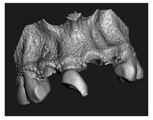
The model they created is of a real patient’s upper jaw with three anterior root apices, periodontal ligament, and the apical granuloma, along with a gingival mask.

“We also present an evaluation of the model by dental students and compare it with their evaluation of the conventional typodont model,” the team wrote. “Our intention was to evaluate whether dental students accept the 3D-printed surgical training model just as well as the popular typodont model.”

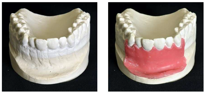
L-R: Modified plaster cast, modified plaster cast with wax layer.

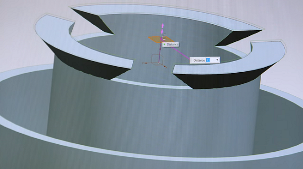
They used CAD/CAM technology to design the training model, which allowed them to add the simulated inflamed tissue, and took a conventional impression of the area in question in order to make a plaster cast. The gingiva was modeled with a 1 mm thick layer of wax, and an industrial 3D scanner was used to attain the shape of the modified cast with and without the wax gingival mask.

L-R: Scanned surface of the plaster cast without wax layer and meshes of the three teeth aligned to the upper jaw.

The cone beam computed tomography (CBCT) data of another patient was used to create 3D models/meshes of teeth 11, 12, and 21 in Materialise Mimics, and the 3D reconstruction was modified using Rhinoceros 5. To make a model of the periodontal ligament, which the typodont model doesn’t include, they deleted the upper parts of the teeth mesh and thickened the rest by 0.25 mm in Geomagic Wrap.

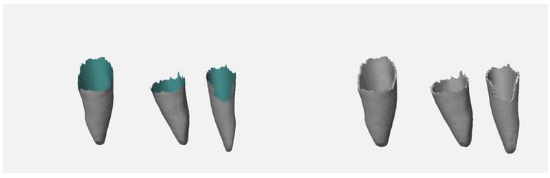
L-R: Meshes of the roots (rear faces of mesh in blue-green), extruded root surfaces representing periodontal ligament.

They constructed a 6 mm sphere around the root apex of tooth 11 to simulate an apical granuloma.

“The material used to represent the periodontal ligament and the apical granuloma is softer than the material used for the other parts of the model. This allows a more realistic representation than in the typodont model,” they explained.

Meshes of the granuloma on tooth 11 and the periodontal ligament on teeth 11, 12 and 21, 3D printed in soft support material (red).

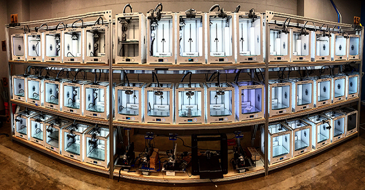
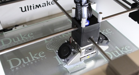
The 3D printed model also includes a silicone gingival mask so students can practice the surgical incision. A 3D printed matrix technique was used to fabricate the mask directly onto the model, and the model was 3D printed out of liquid photopolymer on an Objet Eden 260V PolyJet 3D printer. The undercut areas and the cavities in the model that simulated apical granuloma and periodontal ligament were filled with a soft support material. It took roughly six hours to 3D print 12 models in a single build.

Silicone gingival mask.

“Dental students, about one year before their final examinations, acted as test persons and evaluated the simulation models on a visual analogue scale (VAS) with four questions (Q1–Q4),” the researchers wrote.

35 students evaluated the typodont model, while 33 students used the 3D printed simulation model. Participants watched a video of the root tip resection exercise, and then completed the procedure once. They were given a questionnaire about the simulation model and the difficulty of the exercise, rated on a visual analogue scale (VAS). There was also an optional free-text section if a participant wanted to express their opinion in their own words.

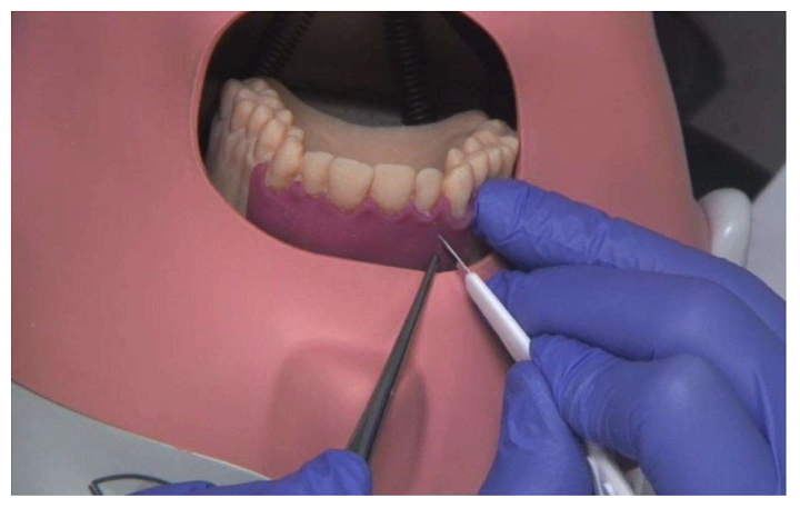
Surgical incision guidance on the 3D printed model in the phantom.

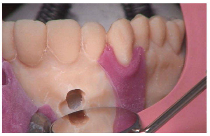
Osteotomy of the root tip.

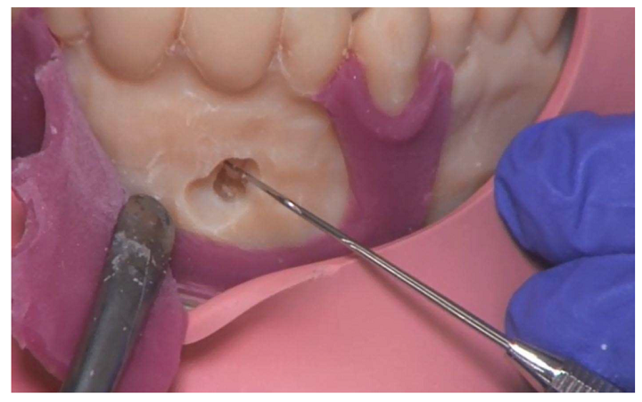
Presentation of the root tip. Note: torn gingiva mask.

Resected root tip with demarcation to the bone.

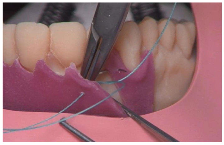
Suture exercise on the gingiva mask.

54.5% of the Group 2 participants said in the free-text section that the gingiva mask in the 3D printed model tore during the procedure, while 20% in Group 1 said that it detached from the typodont model.

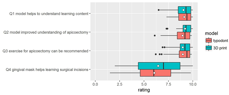
Questionnaire results; white dots denote the mean values.

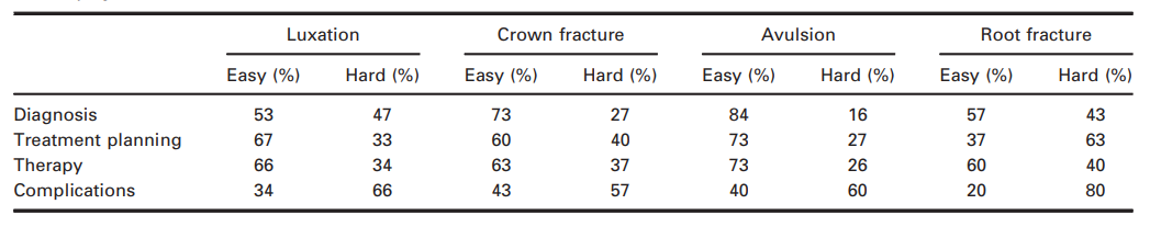
“Shapiro–Wilk normality tests revealed that, with the exception of Q4, normality cannot be assumed,” they explained. “Wilcoxon rank sum tests were therefore carried out to identify differences in the assessments of the two model types. The alternative hypothesis for each test was “The rating for the typodont model is higher than that for the 3D printed”. As the p-values presented in Table 1 reveal, the alternative hypothesis has to be rejected in all cases.”

Table 1.

The researchers determined that their 3D printed training models were “not inferior to the industrially manufactured typodont models,” and that the approach is very flexible – the models can be easily redesigned and adapted for different learning scenarios, and it’s much faster to fix them when necessary. While the 3D printers weren’t cheap, the material costs for a 3D printed single-use model were only about €10, compared to €300 for the multi-use hypodont model.

“A shortcoming of our study is that the exercises were performed by students without surgical experience. As a result, there is a lack of professional evaluation of the models in terms of how well they reflect the reality. Thus, we were not able to check an important quality aspect of the models,” the researchers noted.

“Future studies with experienced surgeons could provide more information about the realism of the 3D-printed models.”

Other issues include the missing color difference between anatomical structures or cortical and cancellous bone structures, and the gingiva mask needs improvement, either through alternative technologies or materials.

“Individual 3D-printed surgical training models based on real patient data offer a realistic alternative to industrially manufactured typodont models. However, there is still room for improvement with respect to the gingiva mask for learning surgical incision and flap formation,” they concluded.

Discuss this and other 3D printing topics at 3DPrintBoard.com or share your thoughts below.

The post Dental Students Compare Conventional and 3D Printed Surgical Training Models appeared first on 3DPrint.com | The Voice of 3D Printing / Additive Manufacturing.

Germany: Research Shows Good Response from Students Using 3D Printed Dental Traumatology Training

Authors M. Reymus , C. Fotiadou, R. Hickel, and C. Diegritz explore the uses of 3D printed models in dental traumatology training, with their findings outlined in the recently published ‘3D printed model for hands-on training in dental traumatology.’ For their study, they used an SLA printer to create a 3D printed model of a dental patient’s maxilla, mimicking several different traumatic dental injuries.

Being able to create accurate models exhibiting significant trauma offers a host of benefits to dental students who can take their time in a deliberate learning mode rather than waiting to rush in to see what could be a relatively small number of injured patients on-site. This also accentuates the enormous amount of learning gained from lectures. The hope is that more knowledge can be gained about dental traumatology, as the researchers point out that dental accidents are often treated by general dentists who may not have an adequate education or experience to deal with such cases overall.

The researchers wanted to make a model that was not only realistic but would allow for students to practice both diagnosis and treatment too. They also wanted to design a product that would translate from educational settings to dental clinics. With these hands-on tools available, the authors also created another level to their study regarding the use of dentaltraumaguide.org, offering the resource to only half of the students participating in the study—and comparing their knowledge.

The model was designed and 3D printed as follows to show dental trauma for a 16-year-old boy:

“The data generated were exported as single DICOM files and imported to Invesalius for Mac (Centre for Information Technology Renato Archer, Amarais, Brazil) to convert it into one .stl file. This file was subsequently imported to Meshmixer for Mac 11.0 (Autodesk, San Rafael, CA, USA) and trimmed to a region extending from the right first premolar to the left premolar. The right lateral incisor, the left first incisor as well as the left second incisor were cut out of the STL-mesh and exported as single STL-files.

Using the function ‘Boolean difference’, these teeth were cut out, leaving imitation tooth sockets in their original position. Additionally, the right lateral incisor was positioned at a 30° angle towards the palatal from its original position, and again, the function ‘Boolean difference’ was used to imitate a lateral luxation of the tooth perforating the buccal bone. The left lateral incisor was separated into two parts at its apical third imitating a horizontal root fracture. The extracted left incisor was not changed, imitating an avulsion. The mesial edge of the right incisor was removed, exposing the pulp chamber to imitate a complex crown fracture.”

Computer-aided designed model with empty tooth sockets, buccal perforation and complicated crown fracture.

The 32 undergraduate students were tasked to work on the case, even simulating a conversation with the mother of the injured boy as they practiced asking the correct questions about the accident, as well as advising on post-traumatic behavior. Upon examining the 3D printed model, they were given information about every tooth, and asked to offer the following:

  • Diagnosis
  • Treatment plan
  • Recall regime
  • Prognosis of each injured tooth

The assessment was considered in these areas:

  • Pre-treatment
  • Therapy
  • Post-treatment
  • Recall
  • Complications

“The presented workflow allowed the manufacturing of a radiopaque model that imitated a luxation injury, a complicated crown fracture, an avulsion, and a horizontal root fracture in a realistic way,” stated the authors.

Radiograph of the right lateral incisor with a luxation injury (left) and Radiograph of the empty tooth socket of the left incisor and the left lateral incisor with a horizontal root fracture (right).

And while their goal was for such a workflow to be easily transferred to another dental school, they would need to own a CBCT and a stereolithographic printer, along with software that could be offered free. The 32 students were asked to evaluate the model, with 57 percent reporting it to be ‘very realistic,’ and 43 percent choosing ‘rather realistic.’

“The diagnosis of the lateral luxation was evaluated to be the most difficult of all injuries, whereas the avulsion was the easiest injury to diagnose. Concerning treatment planning, the horizontal root fracture was rated as being the most difficult injury. When listing possible complications, the students had serious problems with the horizontal root fracture.

Students’ evaluations of difficulty in diagnosis, treatment planning, therapy and knowledge about complications for each injury.

“All participants reported to have gained new knowledge on dental traumatology, and 97 percent felt better prepared for treating traumatic dental injuries in the future.

“Students seem to focus especially on the diagnosis and treatment of traumatic injuries to teeth when dealing with dental traumatology. This is logical because these steps are of outmost importance for immediate care when confronted with a trauma case. Fortunately, both groups of students in the present study achieved their best results in these fields. The group without access to dentaltaumaguide.org, however, had only poor results when faced with developing a recall regime and knowing about possible complications,” concluded the researchers.

Many dentists and orthodontists rely on 3D printing today for digital dentistry, dentures, and even grafts for issues like alveolar augmentation. What do you think of this news? Let us know your thoughts! Join the discussion of this and other 3D printing topics at 3DPrintBoard.com.

Printed model with gingival mask.

[Source / Images: ‘3D-printed model for hands-on training in dental traumatology

The post Germany: Research Shows Good Response from Students Using 3D Printed Dental Traumatology Training appeared first on 3DPrint.com | The Voice of 3D Printing / Additive Manufacturing.

3D Printing News Briefs: October 7, 2018

We’ve got a shorter edition of 3D Printing News Briefs for you today. Siemens Corporate Technology is working on process simulation for additive manufacturing. BIOMODEX is launching a realistic, 3D printed new training product, and an orthopedic surgeon is using 3D printing to repair bone fractures. Finally, several companies are collaborating and using metal 3D printing to make a customized component for the upcoming Ironman race.

Siemens Working on 3D Printing Process Simulation

Often in metal 3D printing, all kinds of defects can occur, such as distortion and local overheating. Getting the 3D print right the first time around is the goal that experts of Siemens Corporate Technology are working to achieve. Process simulation for additive manufacturing is a pretty important step on the way to industrializing the technology, as getting complex geometries correct at the beginning of the process could save time and money down the line.

“Our vision is to develop this additive manufacturing process in such a way that we can actually print a model created in the CAD system, getting it right the first time and printing it perfectly,” said Ursus Kruger of Siemens Corporate Technology in Berlin. “We call this the first-time-right principle, which we want to achieve here.”

Learn more about Siemens’ work in the video below:

BIOMODEX Launching New 3D Printed Training Product

The Left Atrial Appendage Closure Solution (LAACS) station

With the launch of its new training product, medical technology startup BIOMODEX is officially entering the interventional cardiology space. Its new Left Atrial Appendage Closure Solution (LAACS) lets physicians work on their skills using a super realistic, 3D printed multi-material heart. The startup’s patented INVIVOTECH technology makes it possible to create 3D printed organs based on a patient’s medical imaging, like CT scans. It’s also possible to reproduce an organ’s surrounding tissue and biomechanics as well.

“Our mission is to provide as realistic an experience as possible for physician training,” said Carolyn DeVasto, the Vice President of Global Commercialization at BIOMODEX. “Our advancements in patient specific 3D printing using INVIVOTECH and ECHOTECH allow physicians to train in a clinical setting using the same techniques they use in an actual procedure.  Ultimately, we want to provide the physicians an opportunity to test drive any procedure on our solution to improve safety and clinical outcomes.”

BIOMODEX’s patented ECHOTECH also allows physicians to observe the 3D printed heart using fluoroscopy, or any TEE ultrasound system. This means that they will be training with the same techniques they’ll be using in real life procedures, which is invaluable in the operating room.

Repairing Fractures with 3D Printing

Nathan Skelley, MD, an orthopaedic surgeon and sports medicine specialist at the Missouri Orthopaedic Institute, is working on a research project about a specific issue related to trauma orthopaedics – reducing and fixing bone fractures.

“In the United States, we’re very fortunate that I have an almost endless supply of plates and screws,” Dr. Skelley said. “I’ve never been in a situation in the OR where I don’t have another screw or I don’t have another plate to fix one of these fractures. But in the developing world or in rural environments, those resources are not always the case.”

Dr. Skelley and his team are testing if they can easily replicate the plates, screws, and tools they use so often in these types of common trauma and sports procedures with 3D printing, so physicians in areas not quite as developed as the US can perform necessary orthopaedic surgery. You can learn more about his work in the video below:

Metal 3D Printing for Ironman World Championship

Next week, the Ironman World Championship, a yearly culmination of several Ironman triathlon qualification races held around the world, begins in Hawaii. For this particular race, Canyon, Swiss Side, and Sauber Engineering are working together on Project 101 for Patrick Lange, last year’s Ironman World Champion. The goal is to make the Lange, the fastest Ironman, even faster, by using metal 3D printing to fabricate a customized aero cockpit that fits Lange’s arm shape and position perfectly. CFD (Computational Fluid Dynamics) simulations were used to confirm that his tri-bar extensions were producing a decent amount of drag, so the project partners worked out a design to integrate them into Lange’s arms.

Swiss Side 3D printed the first concept and tested it back in May, and Lange’s arms were scanned at Sauber to ensure the perfect fit. Canyon and Swiss Side designed and optimized the aerodynamics for the new aero cockpit, and using FEM (Finite Element Method) structural analysis, the parts were optimized for weight and stiffness. The most recent iteration was 3D printed in plastic and tested in another wind tunnel session so Lange could approve its performance. Then, Sauber used titanium to 3D print the final parts; aluminum was used to create ultra-light shells for the elbow pads.

“While working on Project 101, we did something that has never been done before in triathlon,” Lange said. “I am very proud to be part of this project. We tested my new aero cockpit in the wind tunnel and the results confirmed a significance performance improvement. This will have a direct impact on my bike-splits in Kona. I can’t wait to show the world my new aero cockpit and deliver a strong performance on October 13th at the big race in Kona, Hawaii.”

What do you think? Discuss these stories and other 3D printing topics at 3DPrintBoard.com or share your thoughts in the Facebook comments below. 

Duke University’s 3D Printing Innovation Lab Allows Surgeons to Create Accurate 3D Printed Medical Training Models

3D printers in Duke University’s Innovation Co-Lab [Image: Innovation Co-Lab Studio]

3D printing is becoming increasingly more accessible and affordable in many industries, including the medical field. We often see the technology used for the purposes of creating accurate training models and simulators, so that medical professionals can practice surgeries and procedures ahead of time – this not only saves on costs, but can also allow surgeons to offer a better level of care.

Tawfig Khoury, MD, an otolaryngology (ear and throat) resident at Duke University, is focused on the latter, and uses 3D printing to improve patient care. He makes 3D printed medical models of the ear’s delicate temporal bones, which are later used for the purposes of medical training.

“One focus of my research has been taking CT scans of temporal bones, and printing an exact, patient-specific replica. Our residents can then practice drilling and performing other tests without having to work on an actual patient,” Dr. Khoury explained.

Tawfiq Khoury, MD, Otolaryngology
Resident

Dr. Khoury works on his 3D printed models at the university’s Innovation Co-Lab Studio, which contains a network of over 80 3D printers, ranging from MakerBot and Markforged to Ultimaker and Formlabs, that have been used for various projects since the facility began to really grow last year and explore new uses for 3D printing at the university.

“With recent renovations, we now have a state-of-the-art facility, with high-end equipment across an entire floor dedicated to the lab,” Dr. Khoury said.

“The Innovation Lab is a great example of how different departments across the hospital, as well as other healthcare groups, residents, and students, can work together to create something of value for the community.”

The lab, previously described as a “creativity incubator,” also includes 3D scanning equipment, CNC machines and laser cutters, digital modeling workstations, and a multitude of electronics.

Physicians from several of the university’s medical specialties, including cardiology, neurosurgery, and neurology, use the patient record system Epic to access an ordering system in order to have medical models 3D printed in the studio from ultrasounds and CT and MRI scans. Occasionally, the Innovation Co-Lab Studio can provide its 3D printing services at no cost if the 3D printed replica models are created specifically for patient care.

One of the 80 3D printers in Duke University’s Innovation Co-Lab Studio [Image: Cara O’Malley]

In order to receive and handle requests for 3D prints from around the world, the studio uses 3DPrinterOS, the popular online cloud management system, as a service to the university’s community. 3DPrinterOS users have access to an online, live-streaming video of the project while it’s being 3D printed.

Since the facility’s expansion, a wider community of users have been taking advantage of its services. The expansion also gives Dr. Khoury the opportunity to, according to a post by Scott Behm with Duke’s Department of Surgery, “set his sights on some short- and long-term goals.”

Dr. Khoury feels that 3D printing, even though it can already create accurate models for the purposes of medical training, can go even further at the university. Before his residency at Duke is complete, he hopes to set up an efficient system in order to assist patients with facial trauma who must have maxillofacial reconstruction surgery. His main goal in this is to enable the routine creation of 3D printed models for eventual use in implants for this type of procedure.

Someday in the future, Dr. Khoury believes that we will be able to rely on 3D printers as a way to create organic replacement organs or body parts out of bioink or hydrogel, such as an eardrum, which can then be infused with live cells and implanted in a patient’s body.

Discuss this story and other 3D printing topics at 3DPrintBoard.com or share your thoughts in the Facebook comments below.

3D Printing in Architecture, Engineering, Product Design: 3D Hubs Announces 2018 Student Grant Winners

Last year, 3D Hubs, the world’s largest online network of 3D printing services, reported that nearly 500 applicants from 300 universities around the world applied for its extremely popular Student Grant program, which encourages students to make a positive influence on the world by using 3D printing in a creative way.

Just a few months ago, 3D Hubs announced that it would be kicking off its Student Grant program for the second year in a row, and offering grants to students who were able to illustrate the best use of 3D printing in the architecture, engineering, and product design fields. The grant amount was increased this year from $500 to $1,000, to be used for project expenses and continued funding. Unsurprisingly, the reception for the grant program was great once again.

“We’ve had some amazingly innovative applicants from around the world using 3D printing for some unique applications,” George Fisher-Wilson, the Communications Manager for 3D Hubs, told 3DPrint.com.

There were applicants from more than 50 countries this time around, entering a wide array of innovative 3D printed projects that, as Fisher-Wilson told us, were diverse, including “underwater jetpacks, prosthetics for mountain biking and a 3D printed head with sensors used as a training device for robotic surgery.”

Today, 3D Hubs has announced the three awardees for this year’s 3D Hubs Student Grant program, who were chosen based on the core concept, impact, and functionality of their projects, along with how creatively 3D printing was used to make their ideas a reality. 44.6% of all entries this year were for the Product Design category, followed up by 27.5% for Architecture and 25.9% for Engineering. For the second year in a row, Loughborough University in the UK had the most entries, while New York’s Pratt Institute came in second and the Politecnico di Milano in Italy was third.

“After the success of last year it was great to see more refreshing and new ideas being submitted,” said Filemon Schoffer, the CMO of 3D Hubs. “Our goal is to give students affordable access to all manufacturing technologies so [their] ideas don’t have to be restrictive. The 3D Hubs Student Grant is always a great way to showcase the talented people we having using the platform who will be pushing the industry forward.”

The winners of this year’s Architecture category were Benedikt Boschert and Miriam Boldt from Hochschule Koblenz in Rhineland-Palatinate, with their 3D printed model of a public swimming pool. Their particular project turned the old culture of bathing on its head and into a new, more modern concept.

“With the background of a real task for our city, this concept is weaving the conditions to [an] optimal design of contemporary public swimming pools,” explained Boschert and Boldt in their project description.”

The students 3D printed over 25 pieces for the swimming pool model with an FDM 3D printer, then bonded them together, which helped them turn their original design into a physical model.

Rory Geoghegan and Dr. Mendelsohn from UCLA, who are in the bioengineering and biomedical engineering field, were awarded the 3D Hubs grant in the Engineering category for their Training Platform for Transoral Robotic Surgery.

Using an FDM 3D printer and PLA materials, the two created a 3D printed model of a human head, which also includes a synthetic oral tumor and an anatomically accurate aerodigestive tract, to be used as a training platform.

“Robotic surgery can be used to remove tumors from deep inside the mouth,” Dr. Mendelsohn and Geoghegan explained. “Currently there is no training platform to facilitate the necessary skills acquisition for surgical residents.”

The model is cost-effective, which is good if replacement parts are needed quickly during training, and also includes force sensors underneath the structures that are most often damaged by new surgeons, such as the lips and teeth.

Archie O’Brien from Loughborough University was awarded the final 3D Hubs grant, in Product Design, for his 3D printed, underwater jet pack. He calls his project, named Cuda, the “fastest underwater jetpack” in the world, and says it can be used for multiple purposes, such as underwater exploration, lifeguard duties, “and of course fun.”

The functional 3D printed prototype, designed in SOLIDWORKS, shows what O’Brien calls a “complex use of 3D printing,” as the prototype and most of its internal components were 3D printed using mostly PLA.

During this year’s program, 3D Hubs also took a close look at the most commonly used 3D modeling software – SOLIDWORKS was at the top, with Rhino following closely behind – and the most popular materials (PLA was the clear winner) used by students for their projects.

To take a look at the rest of these findings, and all of the top ten finalists in each of the three award categories, check out the 3D Hubs blog post. While dates and categories won’t be confirmed until later this year, the 3D Hubs Student Grant, open to all registered students, will definitely be returning for a third iteration in 2019.

Discuss this story, and other 3D printing topics, at 3DPrintBoard.com, or share your thoughts in the Facebook comments below.

[Images provided by 3D Hubs]