
This week’s edition of Sliced, the 3D Printing Industry news digest, answers: How is 3D printing helping to break the land speed record? Can bioprinters be used in rugged military environments? What is the largest 3D printed building in the world? And how can you create your own robot vacuum cleaner? Read on for the […]
Singapore: World’s First 3D Printed Polymer Ribcage Reconstruction
Australia based Anatomics was an early pioneer in using 3D printing for surgeries. Under Neurosurgeon Paul D’Urso the firm has done a lot of work in personalized implants, surgical planning and 3D printed medical devices. In thoracic surgery, CMF implants and orthopedics the firm has rolled out software solutions as well as implants and materials for surgeons to use. Patient-specific surgical tools, patient-specific spinal cages, sternums or software to design surgical implants.
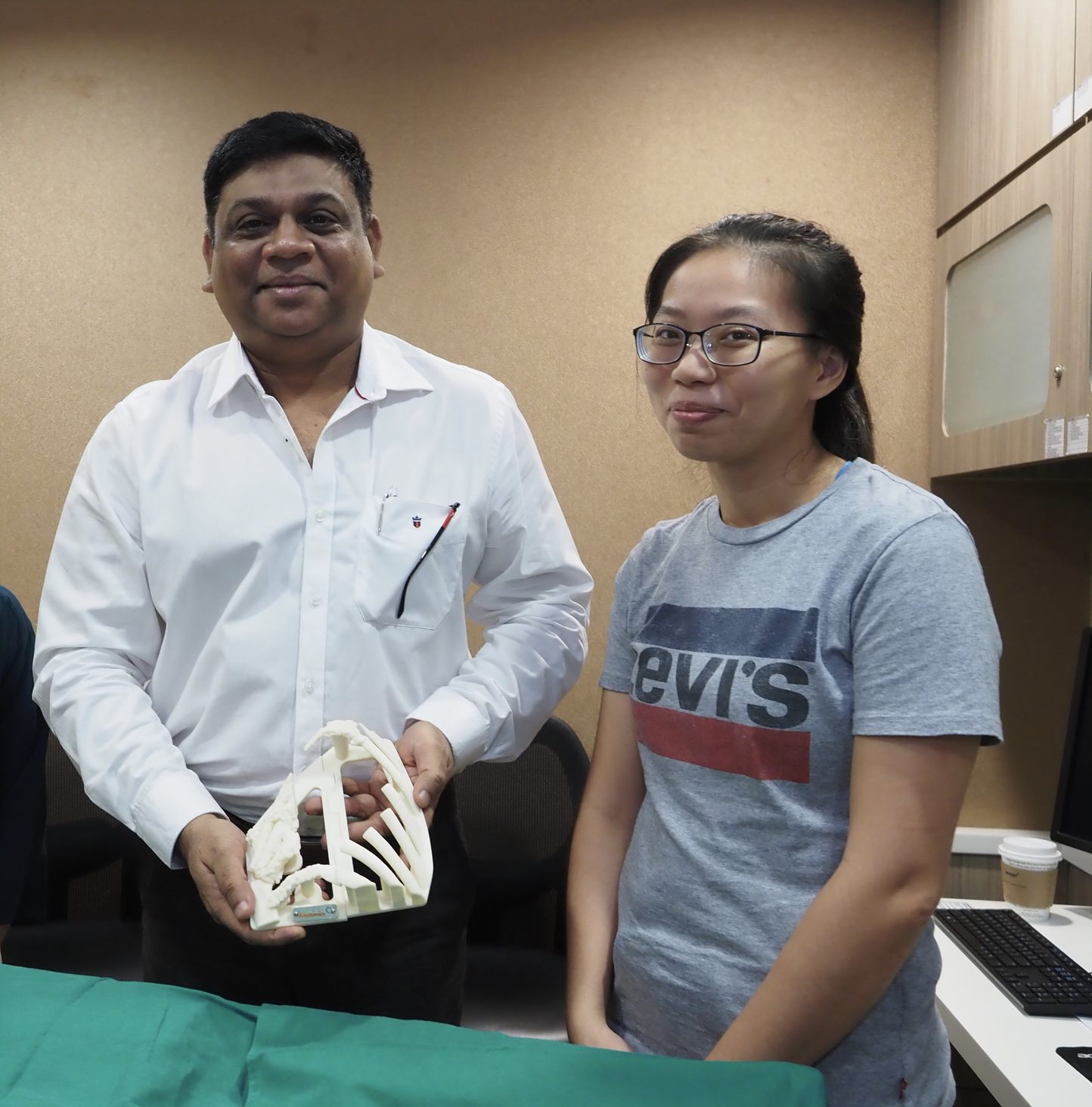
Now Anatomics worked with Singapore’s Tan Tok Seng Hospital to do a world-first, a Polymer ribcage reconstruction. Using Anatomics’ StarPore polymer tissue scaffold a 26 year old cancer patient who needed a new ribcage was helped. The surgery was a success and the patient went home after two days. In addition to the 3D printed ribcage reconstruction itself, patient-specific surgical guides, medical models and cutting guides and templates were also created to aid the surgery.
The StarPore has, “strength, flexibility and its capacity to allow cell and tissue ingrowth. StarPore scaffolding will last for a patient’s life span whilst keeping the same shape, with natural tissue holding it in place. The material provides a scaffold for human tissue to grab onto and grow into, due to biological trabecular porosity similar to that found in the body.”
The surgery was conducted by Dr Aneez Ahmed, Head of Service of Thoracic Surgery at Tan Tock Seng Hospital, and his team. Dr Ahmed says that,
“There’s nothing like a like for like comparison with the StarPore solution for this case. Thus the StarPore solution definitely improves the patient’s health outcome as there is no alternative possible…An important advantage of the Anatomics solution was the ease of use. Instead of doing one rib at a time, multiple times, in the conventional way, we cut out and detached the affected chest wall and inserted the implant easily, making the reconstruction very easy. “The Anatomics personalized solution (StarPore implant, BioModel, 3DP cutting guide and 3DP implant positioning template) helped achieve the success. The usual chest wall reconstruction takes an average of about 4-6 hours. We were able to do this case in about two and a half hours”
Patient-specific implants have long been touted as the future of implantology and orthopedics. Theoretically, they should result in quicker procedures, less blood loss and faster recovery times. In some cases, this may be true while in others a standard implant could suffice. In the case of novel procedures however 3D printing coupled with digital surgical planning is becoming a tried and tested route to the unknown and big new results. The integrated approach seen here would derisk the procedure somewhat and make planning an execution much clearer. I would assume that there would be value in something fitting right the first time but absolutely know that there is value in personalized cutting guides and specific labeling to make sure that errors are reduced; especially with lengthy, new or complex procedures.
The post Singapore: World’s First 3D Printed Polymer Ribcage Reconstruction appeared first on 3DPrint.com | The Voice of 3D Printing / Additive Manufacturing.
The Real Wolverine: Anatomics Works with Surgeon to Design 3D Printed Titanium Metacarpal Implant
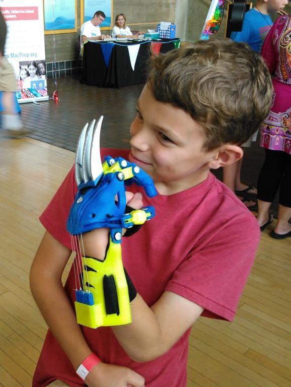
[Image: e-NABLE]
While I did not read the comic books, I was a big fan of the ’90s X-Men cartoon series growing up. In fact, the first short story I ever wrote was about a canine with superpowers named X-Dog…pretty original, right? My love for the mutant superheroes was reawakened when Bryan Singer’s blockbuster movie X-Men came out in 2000, and the world was treated to the first glimpse of Hugh Jackman as Wolverine, the moody mutant with a shadowy, unknown past.
Since then, 3D printing has given other Wolverine fans a way to embody him without having to turn to adamantium. But this has gone one step further now, with a story, as Paul D’Urso, MD, a neurosurgeon at Epworth Healthcare and the Executive Chairman of Australian medical device company Anatomics, tells us, about a “Real Life Wolverine.”
D’Urso said, “Anatomics pioneered the use of 3D printing in surgery in 1995 and has helped surgeons in 40 countries with the most difficult & complex reconstructive surgical problems that no other company could provide a solution for.”
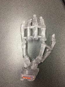
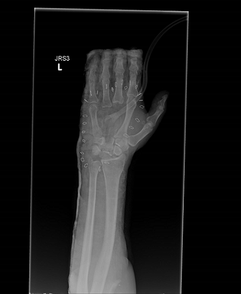 Let’s back up a little first. The five metacarpal bones, which are located between the carpal bones of the wrist and the phalanges of the fingers, make up the intermediate portion of the human hand. Equivalent to the metatarsal bones in the foot, the tops of the metacarpals form our knuckles where they join to the wrist.
We’ve seen 3D printed haptic models representing carpal and metacarpal bones during various hand movements created and studied for potential use in preoperative planning for hand surgery, a 3D printed wrist brace for stabilizing a broken metacarpal while healing, 3D printed patient-specific models and surgical guides for metacarpal deformities, and even a 3D printed titanium bone to replace a metacarpal thumb bone.
Anatomics, a not-for-profit company, has plenty of experience using 3D printing in the medical field, such as 3D printing the first titanium sternum and set of ribs and vertebrae. The company recently had to call on this experience to help with an important case.
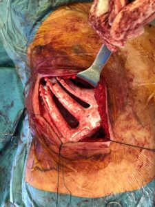
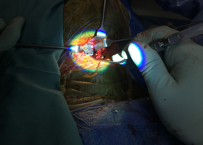
“This man had a severe workplace injury and lost the bones in the middle of his left hand,” D’Urso told 3DPrint.com. “As there was no ‘off the shelf ‘ solution his hand surgeon, Dr Dan Rowe, called upon Anatomics to reconstruct the patient’s hand with a 3D printed titanium implant.”
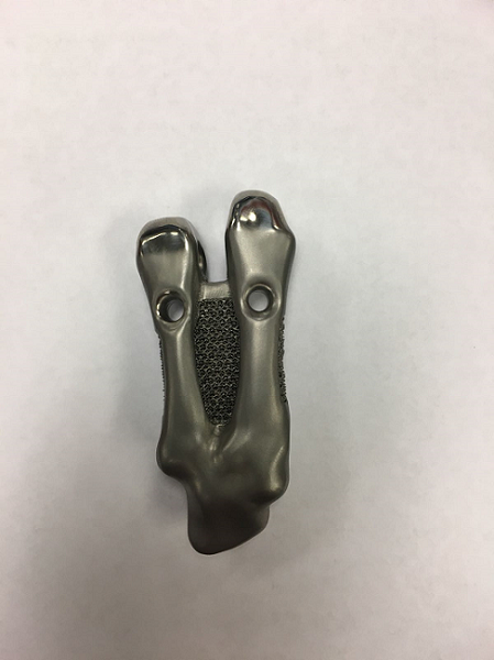
Anatomics stepped up to the plate and got to work. Together with Dr. Rowe, engineers from the medical device company designed a 3D printed, patient-specific metacarpal implant to replace the patient’s two missing metacarpals and missing capitate that had resulted from his injury.
The implant was designed with titanium mesh, which makes soft tissue ingrowth possible once it’s inserted into the patient’s body. Titanium is biocompatible, with a high resistance to corrosion, both of which combine to allow bone to grow. These qualities make it the perfect material for medical implants, once it’s combined with 3D printed lattice structures.
“The implant was designed to articulate with the other bones in the hand and had special channels in it that allowed the tendons to secure it in position,” D’Urso tells us.
“The implant contained a porous lattice to allow for tissue integration.”
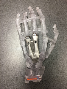
During the patient’s hand reconstruction surgery at Greenslopes Private Hospital in Queensland, Dr. Rowe implanted the 3D printed titanium metacarpal. D’Urso tells us that “Dr Rowe is very pleased with initial results!”
Okay, so the patient didn’t actually end up being a real Wolverine, as only a very small portion of his bones were replaced with metal, but he was still part of something pretty amazing. Innovations like this one are what make 3D printing so important in the medical field. While I imagine the patient still didn’t have the easiest recovery – he did lose the bones in the middle of his hand, after all – I’m willing to bet his quality of life will be better with a 3D printed implant made specifically for his hand than it would be with an off-the-shelf implant, or worse, no implant at all.
Discuss this story and other 3D printing topics at 3DPrintBoard.com or share your thoughts below.
Patient-Specific 3D Printed Spinal Solutions by Anatomics
3D printed anterior cervical cage
Australian medical device company Anatomics has a very prolific 3D printing portfolio in the healthcare industry, from assisting with a 3D printed heel bone to 3D printing the first titanium sternum and set of ribs and vertebrae. Anatomics founder Paul D’urso, MD, a neurosurgeon at Epworth Healthcare, began his medical 3D printing research way back in 1991, four years before starting the not-for-profit Anatomics, which has since helped over 5,000 patients with its custom 3D printed medical solutions.
D’Urso said, “Anatomics has lead the world in custom 3D printed spine technology for over 20 years and is proud to have developed numerous world first applications.”
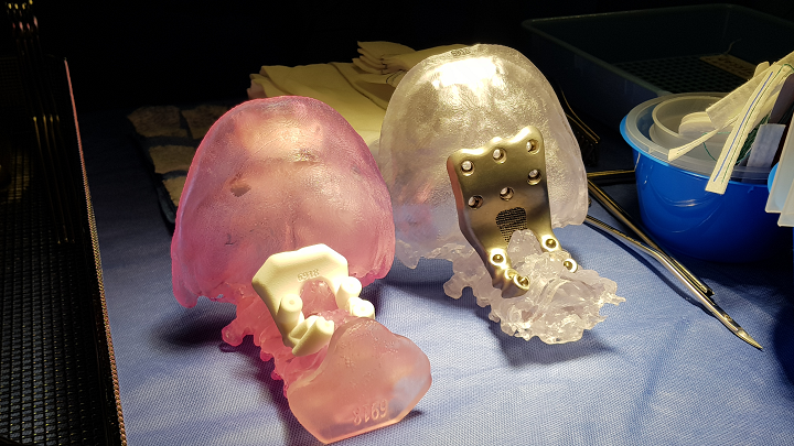
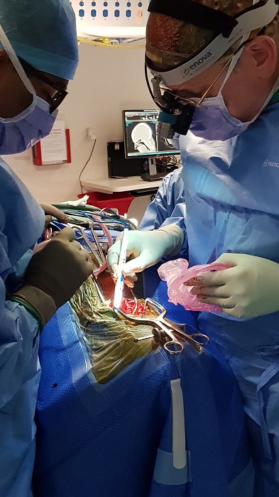
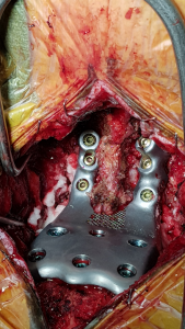
3D printed occipito-cervical plate
D’Urso has a particular interest in developing spinal applications for biomodeling, and at the recent 3DHEALS conference in San Francisco, reported that he’s used 3D printing in nearly 700 spinal fusion procedures, including what D’Urso tells us is “the world’s first custom occipito cervical plate.”
“In the future Anatomics plans to create disruptive Spinal Solution Centres that will enable Community Based Personalised Healthcare allowing surgeons and hospitals to 3D print a range of spinal implants and share designs through-out the world,” D’Urso told 3DPrint.com.
 The success of some of the innovative 3D printed solutions that D’Urso and Anatomics have developed, including 3D printed custom spinal implants, have recently been described in articles and research papers printed in various publications, such as the European Spine Journal and the Journal of Clinical Neuroscience.
The latter paper, titled “Designing patient-specific 3D printed devices for posterior atlantoaxial transarticular fixation surgery,” discussed how biomedelling and 3D printing are both useful tools for pre-surgical planning, developing titanium implants and patient-specific tools, and intraoperative stereotaxy – a minimally invasive surgical procedure which uses a 3D coordinate system to locate small targets inside the body and then perform an action, like an ablation, biopsy, injection, or implantation, on them.
The abstract reads, “Atlantoaxial transarticular screw fixation is an effective technique for arthrodesis. Surgical accuracy is critical due to the unique anatomy of the atlantoaxial region. Intraoperative aids such as computer-assisted navigation and drilling templates offer trajectory guidance but do not eliminate screw malposition. This study reports the operative and clinical performance of a novel process utilising biomodelling and 3D printing to develop patient specific solutions for posterior transarticular atlantoaxial fixation surgery. Software models and 3D printed 1:1 scale biomodels of the patient’s bony atlantoaxial spine were developed from computed tomography data for surgical planning. The surgeon collaborated with a local medical device manufacturer using AnatomicsC3D to design patient specific titanium posterior atlantoaxial fixation implants using transarticular and posterior C1 arch screws. Software enabled the surgeon to specify screw trajectories, screw sizes, and simulate corrected atlantoaxial alignment allowing patient specific stereotactic drill guides and titanium posterior fixation implants to be manufactured using 3D printing. Three female patients with unilateral atlantoaxial osteoarthritis were treated using patient specific implants. Transarticular screws were placed using a percutaneous technique with fluoroscopy and neural monitoring. No screw malposition and no neural or vascular injuries were observed. Average operating and fluoroscopy times were 126.0 ± 4.1 min and 36.7 ± 11.5 s respectively. Blood loss was <50 ml per patient and length of stay was 4–6 days. Clinical and radiographic follow up data indicate satisfactory outcomes in all patients. This study demonstrates a safe, accurate, efficient, and relatively inexpensive process to stabilise the atlantoaxial spine using transarticular screws.

The paper also explained how operative ergonomics and the placement of atlantoaxial transarticular screws can both be simplified using 3D printing. Authors include Ganesha K. Thayaparan, with Epworth’s Department of Neurosciences, and Anatomics’ Mark G. Owbridge, Robert G. Thompson, and D’Urso.
Discuss this and other 3D printing topics at 3DPrintBoard.com or share your thoughts in the comments below.
[Images provided by Anatomics]