Improvements to the BioFabrication Facility on the ISS Thanks to Lithoz

Scientific discoveries and research missions beyond Earth’s surface are quickly moving forward. Advancements in the fields of research, space medicine, life, and physical sciences, are taking advantage of the effects of microgravity to find solutions to some big problems here on Earth. Researchers in 3D printing and bioprinting have taken advantage of space facilities that are dedicated to conducting multiple experiments in orbit, such as investigating microgravity’s effects on the growth of three-dimensional, human-like tissues, creating high-quality protein crystals that will help scientists develop more effective drugs, and even growing meat with 3D printing technology.

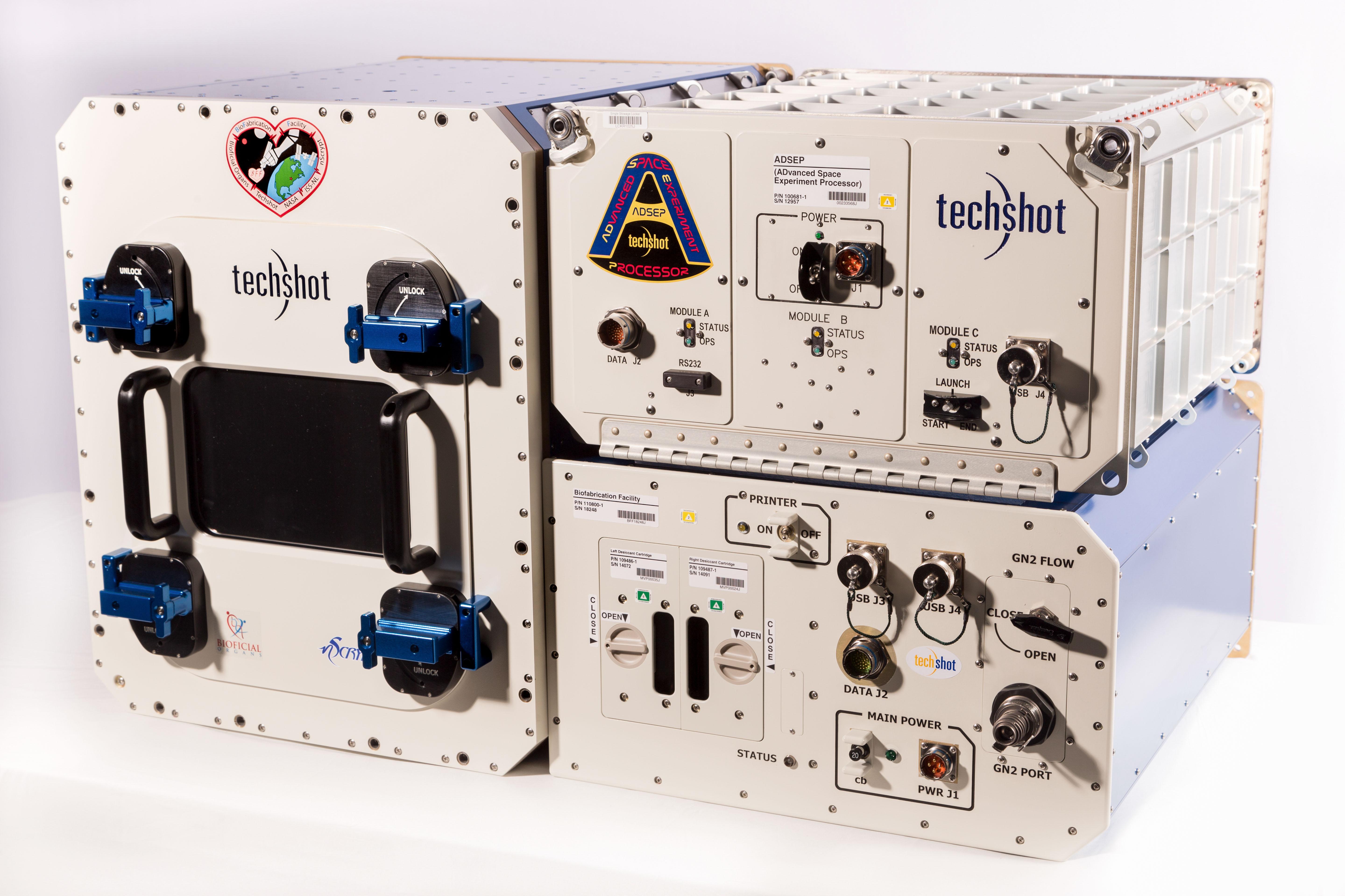
The BioFabrication Facility (BFF) by Techshot and nScrypt (Credit: Techshot)

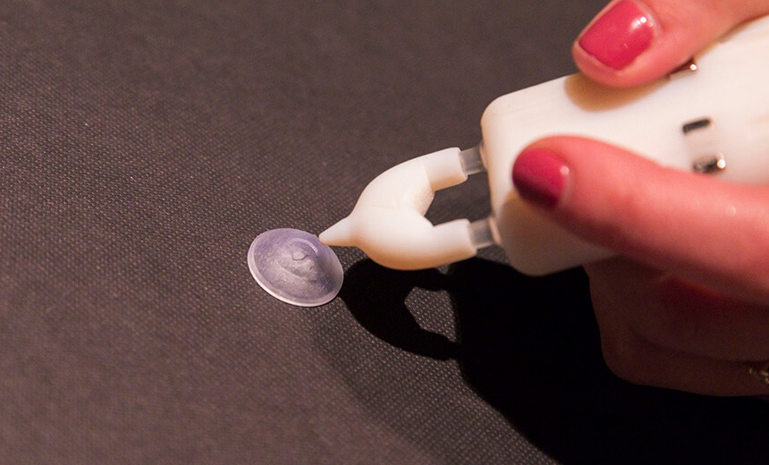
On November 2, 2019, a Northrop Grumman Antares rocket successfully launched a Cygnus cargo spacecraft on a mission to the International Space Station (ISS). The payload aboard the Cygnus included supplies for the 3D BioFabrication Facility (BFF), like human cells, bioinks, as well as new 3D printed ceramic fluid manifolds that replaced the previously used that were printed out of polymers. According to Lithoz – the company behind the 3D printed ceramic fluid manifolds – they are enabling advancements in bioprinting at the ISS.

The additive manufactured ceramics have been in service since November 2019 and Lithoz claims they have proven to provide better biocompatibility than printed polymers, resulting in larger viable structures.

Lithoz, a company specializing in the development and production of materials and AM systems for 3D printing of bone replacements and high-performance ceramics, printed the ceramic manifolds using lithography-based ceramic manufacturing (LCM) on a high-resolution CeraFab printer in collaboration with Techshot, one of the companies behind the development of the BFF. Moreover, the ceramic fluid manifolds are used inside bioreactors to provide nutrients to living materials in space by the BFF.

Testing of the ceramic 3D printed manifolds is focusing on biocompatibility, precision, durability, and overall fluid flow properties; and the latest round of microgravity bioprinting in December yielded larger biological constructs than the first BFF attempts in July.

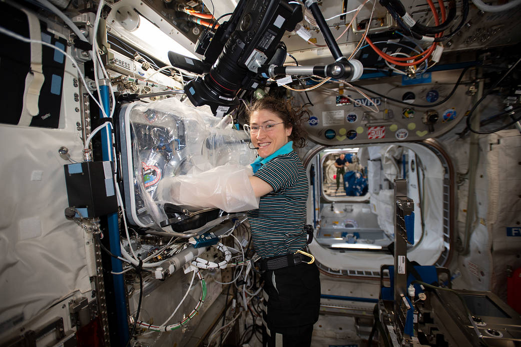
NASA engineer Christina Koch works with the BioFabrication Facility in orbit (Credit: NASA)

Techshot and Lithoz engineers and scientists worked together to optimize the design and the manufacturing processes required to make it. Techshot Senior Scientist Carlos Chang reported that “it’s been an absolute pleasure working with Lithoz.”

While Lithoz Vice President Shawn Allan suggested that “their expertise in ceramic processing really made these parts happen. The success of ceramic additive manufacturing depends on working together with design, materials, and printing. Design for ceramic additive manufacturing principles was used along with print parameter control to achieve Techshot’s complex fluid-handling design with the confidence needed to use the components on ISS.”

Headquartered in Vienna, Austria, and founded in 2011, Lithoz offers applications and material development to its customers in cooperation with renowned research institutes all over the world, benefiting from a variety of materials ranging from alumina, zirconia, silicon nitride, silica-based for casting-core applications through medical-grade bioceramics.

This work, in particular, highlighted an ideal use case for ceramic additive manufacturing to enable the production of a special compact device that could not be produced without additive manufacturing while enabling a level of bio-compatibility and strength not achievable with printable polymers. Lithoz reported that Techshot engineers were able to interface the larger bio-structures with the Lithoz-printed ceramic manifolds and that the next steps will focus on optimized integration of these components and longer culturing of the printed biological materials. While conditioned human tissues from this mission are expected to return to Earth in early 2020 for evaluation.

Back in July 2019, Gene Boland, chief scientist at Techshot, and Ken Church, chief executive officer at nScrypt, discussed the BFF at NASA’s Kennedy Space Center in Port Canaveral, Florida, how they planned to use the BFF in orbit to print cells (extracellular matrices), grow them and have them mature enough so that when they return to Earth researchers can encounter a close to full cardiac strength. Church described how a tissue of this size has never been grown here on Earth, let alone in microgravity. The 3D BFF is the first-ever 3D printer capable of manufacturing human tissue in the microgravity condition of space. Utilizing adult human cells (such as pluripotent or stem cells), the BFF can create viable tissue in space through a technology that enables it to precisely place and build ultra-fine layers of bioink – layers that may be several times smaller than the width of a human hair – involving the smallest print nozzles in existence.

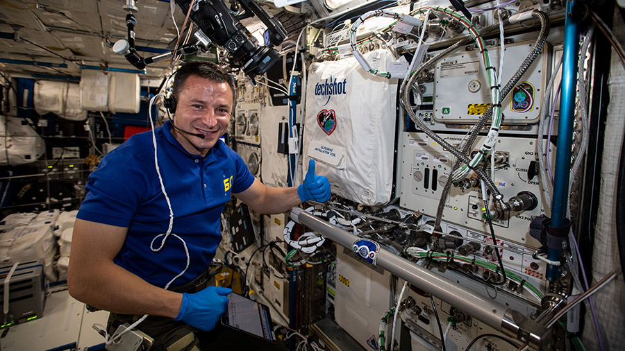
Flight engineer Andrew Morgan works with the BioFabrication Facility (Credit: NASA)

Experts suggest that bioprinting without gravity eliminates the risk of collapse, enabling organs to grow without the need for scaffolds, offering a great alternative to some of the biggest medical challenges, like supplying bioprinted organs, providing a solution to the shortage of organs.

With NASA becoming more committed to stimulating the economy in low-Earth orbit (LEO), as well as opening up the ISS research lab to scientific investigations and experiments, we can expect to learn more about some of the most interesting discoveries that could take place 220 miles above Earth. There are already quite a few bioprinting experiments taking place on the ISS, including Allevi and Made In Space’s existing Additive Manufacturing Facility on the ISS, the ZeroG bio-extruder which allow scientists on the Allevi platform to simultaneously run experiments both on the ground and in space to observe biological differences that occur with and without gravity, and CELLINK‘s collaboration with Made In Space to identify 3D bioprinting development opportunities for the ISS as well as for future off-world platforms. All of these approaches are expected to have an impact on the future of medicine on Earth.

The post Improvements to the BioFabrication Facility on the ISS Thanks to Lithoz appeared first on 3DPrint.com | The Voice of 3D Printing / Additive Manufacturing.

The Future of Bioprinting Research Has a New Road Map

Improving efficiency, optimizing technology, increasing awareness, even reducing costs and time, these are all traits that result from strategic road maps, and in the case of bioprinting, where the outcomes affect tissue engineering, bespoke outcomes for patients, regenerative therapy, and much more, having a blueprint for the entire industry seems like a bright idea. Especially when this blueprint highlights some of the challenges on the way to achieving meaningful and innovative scientific development.

Published in IOP Science’s Biofabrication Journal, “The Bioprinting Roadmap” features advances in selected applications of bioprinting and highlights the status of current developments and challenges, as well as envisioned advances in science and technology. The roadmap brings together researchers, specialists, and physicians from a myriad of universities, institutions, and hospitals around the world, combining their knowledge to focus on different aspects of bioprinting technology. These include: Jürgen Groll, professor of functional materials in medicine and dentistry at the University of Würzburg in Germany; Binil Starly, professor of mechanical engineering at North Carolina State University; Andrew Daly, an orthopedic surgeon at Emory University Hospital; Jason Burdick, a bioengineer from the University of Pennsylvania; Gregor Skeldon, a life science medical writer at Maverex, in the UK; Wenmiao Shu, professor of biomedical engineering at the University of Strathclyde in Glasgow; Dong-woo Cho, mechanical engineer at Pohang University of Science and Technology; Vladimir A. Mironov, chief scientific officer of 3D Bioprinting Solutions, and many more.

The roadmap focuses on a broad spectrum of topics, in a detailed and readable fashion that showcases broad knowledge of the field by its authors, as well as a great deal of research that went into the making of the guide. The paper is categorized into the following sections:

  1. Charting the progress from cell expansion to 3D cell printing
  2. Examining the developments and challenges in the bioinks used for bioprinting
  3. Looking into bioprinting of stem cells
  4. Presenting a strategy for bioprinting of tissue vascular systems and tissue assembly:
  5. Examining the potential for using 3D printed biohybrid tissues as in vitro biological models for studying disease
  6. Analyzing how 3D bioprinting can be used for the development of organs-on-a-chip
  7. Biomanufacturing of multi-cellular engineered living systems
  8. Exploring how researchers are pushing boundaries with bioprinting in space
  9. Investigating the developments of bioprinting technologies

The introduction of the research work was in charge of Wei Sun, chair professor of the College of Engineering from Drexel University, in Philadelphia, and Tsinghua University in Beijing, China, who said to IOP Publishing that “there are a number of challenges to overcome, including the need for a new generation of novel bioinks with multi-functional properties to better transport, protect and grow cells during and after printing; better printing processes and printers to deliver cells with high survivability and high precision; efficient and effective crosslinking techniques and crosslinkers to maintain the structure integrity and stability after printing; integration with microfluidic devices to provide a long term and a simulated physiological environment to culture printed models.”

Wei Sun (Credit: Drexel University)

“Due to the rapid advancements in bioprinting techniques and their wide-ranging applications, the direction in which the field should advance is still evolving,” went on Sun. “The roadmap aims to address this unmet need by providing a comprehensive summary and recommendations, useful to experienced researchers and newcomers to the field alike.”

The research sheds light on the main roadblocks to overcome in the future. The specialists consider that the next technologies will use “multiple modalities” in one single platform and “novel processes, such as cell aggregate bioprinting techniques” to create scalable, structurally‐stable, and perfusable tissue constructs. But in the meantime the challenges that remain are many. In his introduction, Sun talks about the need for a new generation of novel bioninks with multifunctional properties to better transport, protect, and grow cells during and after printing; better printing processes and printers to deliver cells with high survivability and high precision; efficient and effective crosslinking techniques and crosslinkers to maintain bioink structural integrity and stability after printing, and integration with microfluidic devices to provide a long-term and a simulated physiological environment in which to culture printed models.

Other researchers highlighted the importance of further improving bioreactor-based cell-expansion systems to lower barriers to the adoption of bioprinting in regenerative medicine and tissue engineering product markets. While Skeldon and Shu suggest that stem cell bioprinting can be realized if scientists find a way to reduce the shear stress of bioprinting stem cells.

Another obstruction is the high production costs and the difficulty of having large-scale production of organoids. Jinah Jang and Dong-Woo Cho from the Pohang University of Science and Technology suggest that there have been remarkable advances to the recreation of vasculatures and large organs even though challenges remain immense.

Whereas some of the highlights of the research include novel benefits from bioprinting in space, considering that microgravity conditions enable 3D bioprinting of tissue and organ constructs of more complex geometries with voids, cavities, and tunnels; cell expansion as a critical upstream process step for cell and tissue manufacturing; the great promise of stem cells in biomedical research and applications, which through bioprinting, can be particularly positioned in 3D in relation to other cell types and/or biomaterials, as well as progress in stem cell biology and in vitro culture which is opening up new doors to regenerative medicine and better physiological cell-based assays for disease models.

With so many advances in bioprinting around the world and remaining challenges to overcome, both seasoned researchers and newcomers will find an interesting and complete summary of the bioprinting industry. This game plan can help researchers come together to create new novel processes and fill in current technological gaps, as the researchers suggest.

The post The Future of Bioprinting Research Has a New Road Map appeared first on 3DPrint.com | The Voice of 3D Printing / Additive Manufacturing.

Interview with Gordon Wallace on Bioprinting Solutions to Medical Challenges

In a race to combat very specific medical challenges, Gordon Wallace has developed groundbreaking and bespoke advances in bioprinting, both in specialized 3D printing devices and customized bioinks. Along with fellow researchers at the University of Wollongong (UOW), in Australia, working with clinicians and institutions, they seek to find solutions for rare conditions, like microtia, schizophrenia, epilepsy, as well as for more common afflictions, including corneal ulcerations and damaged cartilage.

A distinguished professor at UOW, Wallace, has been working in bioprinting for the past 25 years with a background in materials science. His first foray into biotechnology was developing new electrode materials for Graeme Clark’s pioneer multi-channel Cochlear Implant for severe-to-profound deafness, and in developing those new materials Wallace realized that conventional fabrication wasn’t good enough, which quickly sparked an interest in developing new 3D printing techniques.

“We have been fortunate because all of our projects and research have been driven by our incredible clinician network throughout the country,” explained Wallace to 3DPrint.com during an interview. “This is a highly interdisciplinary field, so through collaborative efforts bioprinting has advanced a lot. Our collaboration spans from timely producers of materials, like seaweed farmers–we extract the molecules form this type of algae and use it as a source of our bioink–right through the materials processing in mechatronic engineering, designing and building applications with cell technologists and medical specialists.”

Gordon Wallace

Wallace has been identifying and customizing materials and bioprinters to deliver solutions like the BioPen for cartilage regeneration, the iFix Pen to treat corneal ulceration, bioprinter 3D Alek to reproduce the complex geometry of an external ear, leading a project to reproduce brain cells and even developed a specialist Pancreatic Islet Cell Transplantation (PICT) bioprinter. Most of the developments are taking place at the ARC Centre of Excellence for Electromaterials Science (ACES), where Wallace is Executive Director; the Translational Research Initiative for Cellular Engineering and Printing (TRICEP), which also has the expert as Director, and the Australian National Fabrication Facility (ANFF) Materials Node, based at the UOW Innovation Campus. The university and its partner institutes have quickly become some of the go-to-places for medical bioprinting advances. TRICEP is a 100% owned initiative of the UOW and can commercialize opportunities in 3D bioprinting including printer manufacturing, biomaterials, bioinks, and material-cellular combinations to address significant industry challenges that require an exclusive, tailored solution; it even houses a range of additive manufacturing technologies, including the highest resolution metal printer in Australia and the country’s leading biofabrication capability to develop biomaterials.

Back in 2014, Wallace along with researchers at the Department of Surgery at St Vincent’s Hospital Melbourne used stem cells to build 3D structures to encourage the formation of cartilage in human tissues claiming they were on the cusp of reaching their goal: “true cartilage regeneration.” Soon after, a team led by Wallace developed the BioPen, which allows surgeons to use a hand-held co-axial 3D printer pen filled with stem cell ink to ‘draw’ new cartilage into damaged knees in the middle of a surgical procedure, providing practitioners with greater control over joint repairs, and reduce surgery time. 

“The BioPen will eventually repair damaged bones, muscles and tendons, and reduce the need for joint replacements by regenerating cartilage, which we have already done in preliminary studies in animals. During this year and the next, we hope to lead some preclinical studies, and if those turn out to be successful the plan is to move into clinical trials with Professor Peter Choong, Director of Orthopaedics at St Vincent’s Hospital Melbourne. Repairing the cartilage in the knee is pretty huge in Australia because it is a sports oriented country with a big demand, especially in younger patients that might have suffered a sports-related injury, and thanks to this, they could avoid osteoarthritis later in life,” explained Wallace.

Gordon Wallace and the BioPen

Soon, other clinicians became interested in the utility of the BioPen, thinking it could be applicable to other medical disciplines, such as for treating wounds and ulcers in the eye. Gerard Sutton, Ophthalmic Surgeon and Professor of Corneal and Refractive Surgery at the Save Sight Institute, at the University of Sydney’s School of Medicine, noticed what Wallace was doing and thought that he could print a different kind of ink directly into the eye. They soon started working together to revolutionise the treatment of corneal ulceration by developing the iFix Pen, which delivers a special bioink formulation that has the capacity to facilitate healing and prevent infection in treating the disease, which causes severe eye pain, visual morbidity and visual loss, and accounts for 55,000 hospital admissions in Australia every year. The exciting new collaborative corneal bioengineering program started in 2017 and was awarded over 780 thousand US dollars for research and development. Animal testing is already underway.

The iFix Pen by Gordon Wallace and Gerard Sutton

“At the lab we use both stem cells and biomaterials. The source of the cells varies depending on the clinical application that we are pursuing, as well as the different kinds of combinations of biomaterials that constitute the bioink wich also depends upon the patient. The choice is driven by the clinical application, while optimizing performance in that specific biological environment requires a different combination of cells and materials for bioinks,” he continued.

Another very specific project is the development of a custom 3D bioprinter dedicated specifically to treating microtia, a congenital deformity that results in an underdeveloped external ear. Named 3D Alek, the machine was recently installed at the Royal Prince Alfred Hospital (RPA), in Sydney, since it was done in collaboration with associate professor and Ear, Nose and Throat surgeon at RPA, Payal Mukherjee, who treats a lot of kids who have the disease. According to Wallace, Mukherjee’s vision was to create a 3D printed external ear. Ultimately, the goal is to 3D print a living ear using a patient’s own stem cells, something the team is developing now. 

Gordon Wallace and frequent collaborator Payal Mukherjee

“But bioprinting is not without some challenges, which are still in optimizing the cells and material combinations for particular applications. Also, a general challenge in 3D printed structures for tissue regeneration is the ability to encourage vascularization of that structure so that bigger defects can be treated. I believe that it is a challenge that many groups around the world, including our own, are concerned about,” suggested Wallace. 

Challenge or no challenge, Wallace is hard at work. At the UOW Intelligent Polymer Research Institute (IPRI), which he founded and also directs, Jeremy Crook, Associate Professor at UOW and ACES, is leading a project to reproduce brain cells through bioprinting, to study conditions like schizophrenia, epilepsy, and depression. 

“Driven by clinical need, we are really interested in how diseases like epilepsy and schizophrenia develop, and one way to try and get some insights into that is to be able to 3D bioprint stem cells from the patient and try to create functional neural networks on the bench to understand how those diseases develop, as well as to gain insight into that little bit of tissue we created with bench research to test interventions, such as pharmaceutical.”

A few years back Crook and Wallace indicated that many neuropsychiatric disorders result from an imbalance of key chemicals called neurotransmitters, which are produced by specific nerve cells in the brain. Defective serotonin and GABA-producing nerve cells are implicated in schizophrenia and epilepsy while defective dopamine-producing cells are involved in Parkinson’s disease, so the team started using 3D printing and bioink to make neurones involved in producing GABA and serotonin, as well as support cells called neuroglia.

More recently, ACES researchers developed a bioprinter to help people with Type 1 diabetes, the Pancreatic Islet Cell Transplantation (PICT) 3D Printer, which works by delivering insulin-producing islet cells and is now in use at the Royal Adelaide Hospital (RAH), where Wallace and his team collaborated with RAH Professor of Medicine Toby Coates. They are planning to improve the effectiveness of islet cell transplants by encapsulating donated islet cells in a 3D printed structure, to protect them during and after transplantation. But that’s not all, many more projects are on the way, including wound healing with Chris Baker, head of dermatology at St Vincent’s Hospital Melbourne working with ACES on clinical trials; printed structures to understand airway collapse and prevention with Stuart MacKay, surgeon and Clinical Professor in Otolaryngology and Head and Neck Surgery at UOW, and even bioprinting in space, which is something Wallace says they “were recently approached about”.

“We just started to think about the ability to 3D print and create things on demand in remote locations, like space, it is an ideal application and something we are very interested in pursuing. For now, I have no real experience about how microgravity will affect bioprinting, but what an exciting experiment that would be,” claimed the expert.

According to the expert, it has taken a while to build the global collaborative network they now have, but it all seems to be coming to fruition, even some of the more complex aspects of bioprinting, like discussions about regulatory issues, something Wallace is quite involved in, as well as the engagement in ethical issues that might arise. 

“In this area, the Therapeutic Goods Administration (TGA) has been very proactive in engaging with the research, commercial and clinical community in order to try to formulate an appropriate regulatory framework that can accommodate 3D bioprinting in Australia. They are aware that the technology is moving very fast, so they want to make sure that everything is in place,” continued Wallace.

As part of the thriving academic environment at UOW, Wallace is a professor of post-graduate courses, where he noticed an increase in student interest in STEM careers and biotechnology for the last few years.

“3D printing has already revolutionized the connections between science, engineering and mathematics and our ability to be incredibly creative and make new structures. But although our courses are in high demand and even our online platform to learn about 3D printing of body parts has been popular with over 30.000 students, I still think there will be a gap between the demand and supply of professionals to fill the biotechnology positions needed for future jobs,” Wallace stated. “There is a big gap at the moment, and it will take many years to recover, so we need to engage children in the early years of school, all the way through to university.”

Gordon Wallace at ACES with the BioPen

“Today I can say that what really changed in the last five to ten years of bioprinting is the convergence of discoveries in material science, with advances in fabrication, particularly 3D printing that has really picked up the pace. We have seen more progress in the last five years than we did 15 years before that, and I think we will see incredible progress in the coming decade as that convergence matures and, particularly, as the clinical teams around the world realize what the possibilities are now in collaboration with the appropriate science and engineering groups. I’m sure many more specific challenges will come to the floor and that we will be able to meet them because of this convergence,” he concluded.

[Images: UOW, ACES, Vision Eye Institute]

The post Interview with Gordon Wallace on Bioprinting Solutions to Medical Challenges appeared first on 3DPrint.com | The Voice of 3D Printing / Additive Manufacturing.

BioPrinting with Alzheimer’s Stem Cells May Lead to Improved Drug Testing & Treatment

Alzheimer’s studies give many of us hope, whether we have a relative or friend suffering from the dreaded disease—or simply because we are hoping that a cure will be there if we begin to experience symptoms of the most common and crushing form of dementia. As bioprinting becomes valuable in so many different areas of medical research, it is no surprise to hear that now scientists are turning their attention toward using it to find better ways for treating Alzheimer’s.

3D printing, and the realm within known as bioprinting, allows for scientists to embrace so many benefits, from affordability and expediency, to the ability to keep trying varying iterations until they complete their mission in research or innovation. One of the greatest challenges in dealing with Alzheimer’s has been finding medication that really works. Not only is there pressure to find it for those who have the existing condition, but also for future generations carrying what is thought to be a genetic marker. In ‘Bioprinting neural tissues using stem cells as a tool for screening drug targets for Alzheimer’s disease,’ Stephanie M. Willerth examines the difficulty in both finding, testing, and approving drugs for this trying disease that tends to affect seniors regarding cognition, memory, and language and reasoning.

“The pathology of Alzheimer’s disease includes the presence of plaques containing aggregates of amyloid beta (αβ) proteins and tangles containing neurofibrillary tangles,” states Willerth in her paper. “Certain genetic mutations increase the possibility of developing Alzheimer’s disease.”

“These mutations consist of an altered APP gene responsible for encoding amyloid precursor protein, along with the PSEN1 and PSEN2 mutations cause decreased activity by the γ-secretase complex. These mutations result in improper processing of the αβ proteins. Currently, approved treatments for Alzheimer’s disease include four different types of cholinesterase inhibitors and the drug memantine – a N-methyl-d-aspartate receptor antagonist that targets glutaminergic neurons to preserve their function.”

Willerth goes on to detail the facts about Memantine, as the only drug approved for Alzheimer’s patients. It has been in use since 2000, and currently there are over 200 other compounds being tested. Memantine is known to be controversial though and Willerth points out that the current drug targets present issues in terms of both successful use in patients, and toxicity, posing a need for improvement before clinical trials commence.

Better testing before trials would diminish costs in drug development, along with decreasing the amount of time it currently takes to create treatments that are successful.

“Current methods for evaluating target compounds consist of animal models of disease and the use of cadaveric human tissues,” states Willerth. “In addition to their lack of predictive capacity, animal models are costly, and the supplies of human neural tissues remain limited. Developing more effective assays for preclinical identification of drug targets will lower the expense of the drug discovery process and increase the likelihood of successful outcomes at the stage of clinical trials.”

The use of human induced pluripotent stem cells (hiPSCs) is certainly not a novel idea today, having been in use since 2007; however, now, scientists may be able to use such technology regarding Alzheimer’s, reprogramming cells from patients into hiPSCs. While this could offer better tools for studying the disease, and screening drug targets, it could also further studies regarding progression of the disease in patients. Along with that—and this is where bioprinting and 3D printing so often come in—greater patient-specific care should be available too.

“The use of hiPSC lines containing the different genetic mutations associated with Alzheimer’s disease also enables the use of personalized medicine for treating different subsets of the disease. While cells are often cultured on 2D substrates in vitro, these conditions do not accurately mimic the microenvironment present in the CNS,” states Willerth, who goes on to mention research at the University of Wisconsin-Madison where scientists have created stem cells that are able to successfully predict toxicity levels in different compounds.

With alternative methods like 3D bioprinting, tissue engineering is possible but there may still be challenges in controlling viability and ‘behavior’ of cells.

“Recent advances in bioprinting have enabled the printing of hiPSCs using microfluidic extrusion, opening the possibility for applying this technology for high-throughput production of hiPSC-derived neural tissues,” state the researchers. “Recent developments in 3D bioprinting technologies make printing physiologically relevant neural tissues derived from hiPSCs a real possibility.”

Both 3D printing and bioprinting offer untapped potential, along with technology using suspended hydrogels to make complex structures.

“My group has successfully developed microspheres for delivery of two small molecule morphogens – guggulsterone and purmorphamine,” states Willerth. “Our data have shown that such microspheres are powerful tools for promoting the differentiation of hiPSCs into neural tissues and being able to place different combinations and concentrations of microspheres into precise locations using 3D printing would enable production of tissues like that found in the brain and spinal cord. These 3D printed neural tissues could then be used for screening potential drug targets for Alzheimer’s disease as an alternative to expensive preclinical animal testing.”

As work progresses in the future, Willerth’s paper details issues that must be resolved, such as the means for more stable vasculature to bioprint arrays for drug testing.

“Such work could also contribute to development of engineered tissues that could be transplanted for regenerating damaged regions of the central nervous system. Overall, 3D bioprinting hiPSC-derived neural tissues hold significant potential as a novel way to generate new tools for screening drug targets for the treatment of Alzheimer’s disease,” concludes Willerth.

This project is funded by the Canada Research Chairs program, the Natural Science and Engineering Research Council, and the British Columbia Innovation Council’s Ignite Program. SM Willerth also has a commercialization agreement with Aspect Biosystems with regards to 3D printing neural tissues and a provisional patent on a bioink.

What do you think of this news? Let us know your thoughts! Join the discussion of this and other 3D printing topics at 3DPrintBoard.com.

Photo credit: UVic Photo Services