We often see titanium used in 3D printed implants for humans, and even for animals. When compared to implants that are more dense, porous titanium (PT) implants, with custom porosity and structure, have mechanical properties far more similar to natural bone, which helps reduce stress-shielding effects and facilitate nutrient transport and bone tissue ingrowth during regeneration.
But, metals based in titanium (Ti) are naturally bioinert, meaning they don’t initiate a response when introduced to biological tissue; this can lead to lowered bone-to-implant contact and possible rejection. So for bone tissue engineering, it’s important to modify the surface of metallic implants with coatings, whether it’s something like a diamond or bioactive glass (BG).
A collaborative group of researchers from Zhengzhou University and Northwestern Polytechnical University in China, recently published a paper, titled “A Facile Flow-Casting Production of Bioactive Glass Coatings on Porous Titanium for Bone Tissue Engineering,” about their work in developing a flow-casting method to rapidly coat PT with BG coatings.

Flow-casting production of homogeneous BG coatings on PT scaffolds.
The abstract reads, “Additive manufacturing enabled the fabrication of porous titanium (PT) with customized porosity and mechanical properties. However, functionalization of PT surfaces with bioactive coatings is being challenged due to sophisticated geometry and highly porous structure. In this study, a facile flow-casting technique was developed to produce homogeneous 45S5 bioactive glass (BG) coatings on the entire surface of PT. The coating weight as a function of BG concentration in a BG-PVA slurry was investigated to achieve controllable coating yield without blocking macropore structure. The annealing-treated BG coating not only exhibited compact adhesion confirmed by qualitative sonication treatment, but also enhanced the mechanical properties of PT scaffolds. Moreover, in-vitro assessments of BG-coated PT cultured with MC3T3-E1 cells was carried out having in mind their potential as bioactive bone implants. The experimental results in this study offer a simple and versatile approach for the bio-functionalization of PT and other porous biomedical devices.”

Fluorescent images of cells cultured on bare (a,c,e) and BG200‐coated (b,d,f) Ti scaffolds after 2 and 4 days.
Bioceramics, like TiO2 and BG, have been used before as coatings in order to, as the paper puts it, “functionalize bioactive character to inert bone scaffolds.” There have been several methods used in the past to produce bioceramic coatings on dense bone implants, but because porous ones have more complex structures, it’s hard to coat the entire surface without blocking the porous, interconnected structure.
The team used SLM technology to 3D print PT samples on a Renishaw AM 250, then rinsed them with demineralized water and ethanol to remove residual HF before letting them air-dry. Then, a sample was dipped in a slurry of BG-PVA, before being moved to a rotation platform. Using compressed nitrogen force during rotation, the slurry’s solid content was gradually but completed flow-casted onto the surface of the struts, which as the researchers explained in the paper resulted in “the formation of a homogeneous BG coating on the entire PT surfaces without blocking the macropore structure.”
“The key parameters of the flow-casting process were varied to obtain homogeneous BG coatings,” the researchers wrote. “In addition, microstructural, mechanical, and biological properties of BG-coated PT were characterized having in mind the promising potential of BG-functionalized PT in bone tissue engineering.”

(a) The weight of BG‐PVA coating as a function of BG concentration in slurry; the inset shows the increased white content on the coated PT with increasing BG concentration (* p < 0.05, ** p < 0.01, *** p < 0.001), (b) axial and radial observation of BG200‐coated PT after annealing, (c) XRD patterns of raw BG and annealed BG powder using the same annealing protocol.
While developing the facile flow-casting method, the team determined that by varying the concentration of BG, they could specifically tailor the coating yield in order produce homogeneous coatings on the PT substrates that wouldn’t block the interconnected macropores. Additionally, after an annealing treatment, the BG200-coated PT showed a strengthened elastic modulus, and also “exhibited excellent coating adhesion,” which are good signs for bone implants.
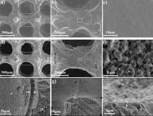
SEM images of the inner surface morphologies of (a-c) bare Ti scaffold and (d-f) BG200-coated Ti scaffold, (g) cross-sectional images of BG200 coatings at different magnifications.
“Although the annealed BG coating exhibited nonuniform thickness varying from 2 µm to 6 µm, in-vitro biological evaluation confirmed an improved osteoblast activity, probably owing to the bioactivity of the BG phase,” the researchers concluded. “Summarizing, flow-casting of BG coatings, possibly combined with other bioactive components or functional molecules, suggested a simple and effective approach for the biofunctionalization of PT or other porous devices, which will be advantageous for designing optimal scaffolds for biomedical applications.”

Co-authors of the paper are Haiou Yang, Qijie Zhu, Hongfei Qi, Xianhu Liu, Meixia Ma, and Qiang Chen.
Discuss this research and other 3D printing topics at 3DPrintBoard.com or share your thoughts in the Facebook comments below.