More Efficient Drug Screening with 3D Bioprinting

Taking a drug to market is a competitive, costly and challenging process involving preclinical laboratory and animal testing before the even more time-consuming and expensive four phases of human clinical trials, which can take as many as 7 to 15 years at price tags as high as $5.5 billion. Even if 10 viable drug compounds are identified for human trials, only 1 out of 9 will actually make it to market. Given this high attrition rate, can bioprinting save valuable time and resources by better identifying viable compounds in order to move only the most promising drugs to clinical trials?

The limitations of animal testing

During the initial stages of drug discovery, often referred to as preclinical trials, new chemical entities (NCEs) are monitored to determine the life cycle of the compounds inside and outside of the targeted system (pharmacokinetics) and their chemical reactions (metabolism). Because of the ethical issues surrounding human trials and their high costs, a significant number of these early tests are performed on animals.

While the transition from preclinical animal testing to clinical human trials has improved thanks to better research tools and the rise of artificial intelligence in target identification, there is still a real need for improved preclinical screening because animal testing often fails to recapitulate the complexity of the human metabolism, leading to false positives and negatives that do not accurately reflect the toxicity of drugs to human systems.

3D cell cultures are more relevant

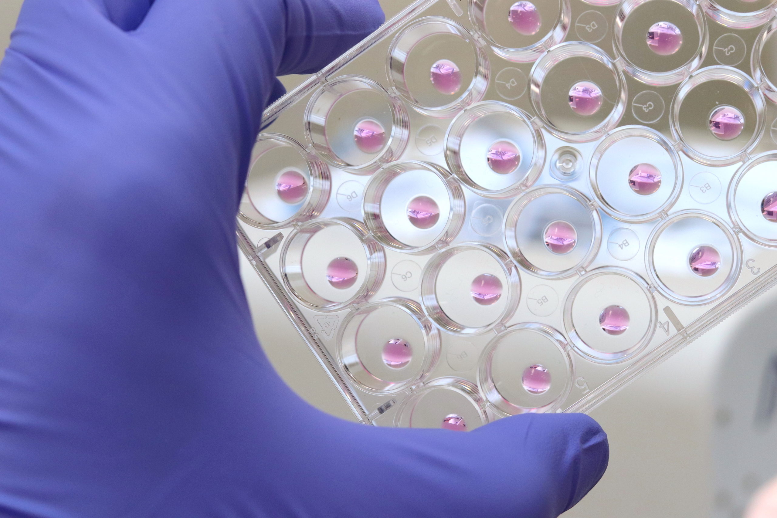
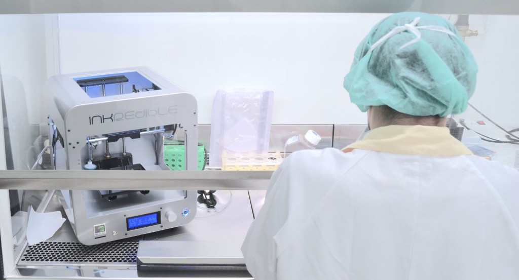
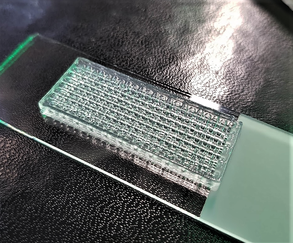
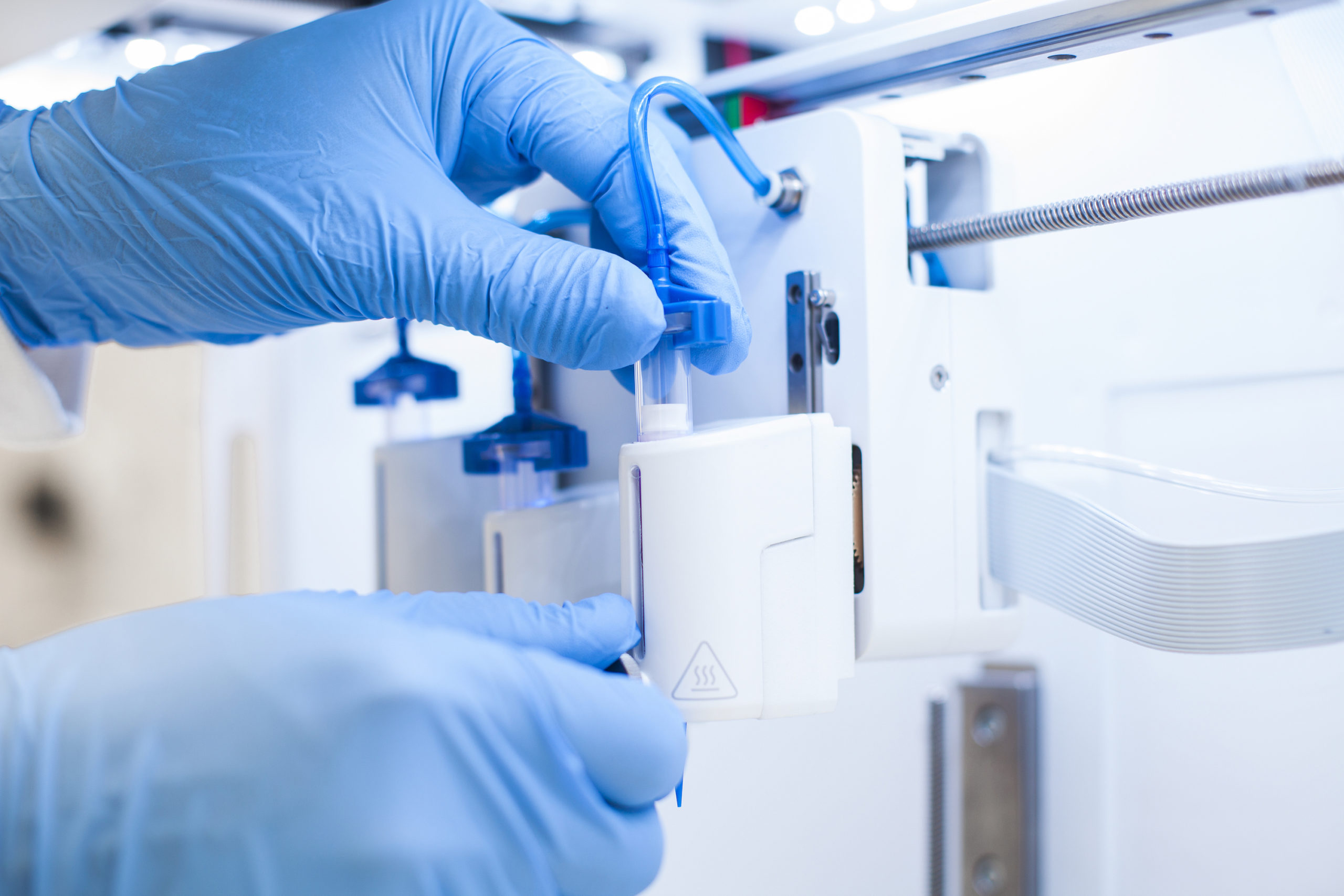
Given the limitations of animal models, it is no wonder that scientists have turned to human organ models. But although human cells have long been cultured in 2D, in recent years, a paradigm shift has led more and more scientists to recognize the importance of working with human cells in the 3D environments afforded by bioprinting in order to produce more physiologically relevant models. Combining the automation of cell culturing in 3D bioprinting with carefully tailored biomaterials, known as bioinks, has made it possible to grow, feed and maintain human organ models in larger quantities and in a lot less time, reducing time and labor spent on these tasks. Laboratory robotics can also now pick and place cell culture reagents or other NCEs and liquid samples in high numbers, enabling higher throughput screening and running a variety of other laboratory tasks more efficiently.

Bioinks better mimic ECM

Bioinks are another powerful tool that help researchers advance their drug discovery research. Tissue-specific bioinks improve cell adhesion and differentiation, helping with the formation of human organoids. Proteins and other biological factors can also be added to more accurately recreate extracellular matrices (ECM), once again better simulating the in vivo microenvironments. Furthermore, with multiple methods of crosslinking (chemical, light, thermal), the stiffness of constructs can be modulated to better serve specific cell types, like cartilage or bone tissue.

Learn more

Bioprinting’s more relevant human organ models can save the drug industry time and money by more efficiently identifying viable compounds in the initial stages of drug development in order to move only the most promising compounds to costly human clinical trials. The technology’s growing influence means that scientists continue to validate more and more applications. Dive deeper into how the bioprinting industry is changing drug screening and development. Watch our webinar on 3D bioprinting for COVID-19 studies or read our application note, which discusses the effectiveness of testing drug efficacy in 2D and 3D.

The post More Efficient Drug Screening with 3D Bioprinting appeared first on 3DPrint.com | The Voice of 3D Printing / Additive Manufacturing.

CELLINK acquires precision dispensing 3D printing company Scienion in €80m agreement

Swedish 3D bioprinter and materials developer CELLINK has agreed to acquire precision dispensing 3D printing firm Scienion in a deal worth €80 million.  The takeover, which is expected to take place by the end of August 2020, will see CELLINK pay €40 million in newly-issued shares, plus €40 million in cash for full control of […]

3D Printed Medicine Uses Fish Gelatin to Deliver Cancer Treatment

Japanese researchers Jin Liu, Tatsuaki Tagami, and Tetsuya Ozeki have completed a recent study in nanomedicine, releasing their findings in “Fabrication of 3D Printed Fish-Gelatin-Based Polymer Hydrogel Patches for Local Delivery of PEGylated Liposomal Doxorubicin.” Experimenting with a new drug delivery system, the authors report on new potential for patient-specific cancer treatment.

The study of materials science continues to expand in a wide range of applications; however, bioprinting is one of the most exciting techniques as tissue engineering is expected to lead to the fabrication of human organs in the next decade or so. Such research has also proven that bioprinting may yield much more powerful drug delivery whether in using hybrid systems, multi-drug delivery systems, or improved scaffolds.

Here, the materials chosen for drug delivery are more unique as the researchers combined printer ink with semi-synthesized fish gelatin methacryloyl (F-GelMA)—a cold fish gelatin derivative.

In providing aggressive cancer treatment to patients, the use of doxorubicin (DOX) is common as an anti-carcinogen for the treatment of the following diseases:

  • Breast cancer
  • Bladder cancer
  • Kaposi’s sarcoma
  • Lymphoma
  • Acute lymphocytic leukemia

DOX may also cause serious cardiotoxicity, however, despite its use as a broad-spectrum drug. As a solution, PEGylated liposomal DOX, Doxil has been in use for treatment of cancer with much lower cardiotoxity. The nanomedicine has also been approved by the FDA, and is used for targeting local tumors; for instance, this type of drug delivery system could be suitable for treating a brain tumor.

“PEGylating liposomes can prolong their circulation time in blood, resulting in their passive accumulation in cancer tissue, called the enhanced permeability and retention effect,” state the authors.

Using a 3D bioprinter, the authors developed liposomal patches to be directly implanted into cancerous cells.

(a) Synthesis of fish gelatin methacryloyl (F-GelMA). (b) Hybrid gel of cross-linked F-GelMA and carboxymethyl cellulose sodium (CMC) containing PEGylated liposome. The reaction scheme was prepared in previous studies

“We used a hydrogel containing semi-synthetic fish-gelatin polymer (fish gelatin methacryloyl, F-GelMA) to entrap DOX-loaded PEGylated liposomes. Fish gelatin is inexpensive and faces few personal or religious restrictions,” stated the authors.

Fish gelatin has not been used widely in bioprinting, however, due to low viscosity and rapid polymerization. To solve that problem, the authors created a bioink composite with elevated viscosity.

Viscous properties of drug formulations used as printer inks. (a) The appearance of F-GelMA hydrogels containing different concentrations of CMC. (b) The viscosity profiles of F-GelMA hydrogels containing different concentrations of CMC. The data represent the mean ± SD (n = 3).

And while hydrogels are generally attractive for use due to their ability to swell, for this study, the researchers fabricated a variety of different materials—with the combination of 10% F-GelMA and 7% carboxymethyl cellulose sodium (a thickening agent) showing the highest swelling ratio.

Swelling properties of hydrogels after photopolymerization. (a) Swelling ratio of different concentrations of F-GelMA. (b) Swelling ratio of mixed hydrogel (10% F-GelMA with different concentrations of CMC). The data represent the mean ± SD (n = 3).

Design of the different 3D geometries: (a) cylinder, (b) torus, and (c) gridlines.

Patches were printed in three different sample shapes, using a CELLINK bioprinter syringe as the authors tested drug release potential in vivo. Realizing that surface area, crosslinks density, temperature, and shaker speed would play a role, the team relied on a larger surface volume for more rapid release of drugs.

Printing conditions of patches.

While experimenting with the torus, gridline, and cylindrical sample patches, the researchers observed gridline-style patches as offering the greatest potential for sustained release.

Drug release profiles of liposomal doxorubicin (DOX). (a) Influence of shape on drug release. The UV exposure time was set to 1 min. (b) Influence of UV exposure time on drug release. The gridline object was used for this experiment. The data represent the mean ± SD (n = 3).

“These results indicate that CMC is useful for adjusting the properties of printer ink and is a useful and safe pharmaceutical excipient in drug formulations. We also showed that drug release from 3D-printed patches was dependent on the patch shapes and UV exposure time, and that drug release can be controlled. Taken together, the present results provide useful information for the preparation of 3D printed objects containing liposomes and other nanoparticle-based nanomedicines,” concluded the authors.

[Source / Images: ‘Fabrication of 3D Printed Fish-Gelatin-Based Polymer Hydrogel Patches for Local Delivery of PEGylated Liposomal Doxorubicin’]

The post 3D Printed Medicine Uses Fish Gelatin to Deliver Cancer Treatment appeared first on 3DPrint.com | The Voice of 3D Printing / Additive Manufacturing.

Rice university researchers develop sweet new way of 3D printing vascular networks 

Researchers from Rice University have developed a new method of using 3D printing to create artificial vascular networks from powdered sugar.  Replacing traditional production methods with Selective Laser Sintering (SLS) 3D printing, the team created sacrificial templates made from laser-sintered carbohydrate powders. These sugar-based constructs enable cell-laden hydrogels to be patterned with dendritic vessel networks, […]

Cellink Teams with Lonza to Advance Bioprinting Cell Cultures

For over four years biotech company Cellink has been helping researchers pursue creative ways to promote organ regeneration, test novel drugs, develop vascularized bioconstructs, and more. Through collaborative partnerships and pioneering research, the firm has quickly become a market leader for cell-based applications. Along that road, they have partnered with several startups and multinationals to open up a broader range of possibilities in the field. Their latest partnership with Lonza—a leading suppliers to the pharmaceutical, biotech, and specialty ingredients markets—will provide researchers and scientists with even more options to enhance bioprinting of complex 3D human tissue constructs.

The companies have joined forces to offer a comprehensive 3D bioprinting solution designed to optimize and increase access to complete 3D cell culture workflows. The solution integrates Cellink’s 3D bioprinting instruments and pioneering commercial bioinks with Lonza’s broad selection of human-derived primary cells and supporting culture media.

Credit: Lonza

“Everything we do at CELLINK, from live cell imaging to our innovative bioprinting systems and bioinks, is meant to support our customers with the products and services needed for them to more effectively and efficiently research solutions to some of the most important challenges of our time. Challenges such as cancer therapeutics, regenerative medicine and the testing and development of drugs, to name a few,” said Ginger Lohman, Biodispensing Product Manager at Cellink’s Gothenburg headquarters in Sweden. “When it came time to expand our portfolio into complete 3D cell culture workflows, we knew it was critical that we brought the right partner onboard. We’re confident that Lonza is that partner.”

According to Cellink, cell biologists will now be able to rely on a high-performing product portfolio to successfully execute some of the most demanding work on 3D bioprinting and boost their scientific research. The proposed solution under the partnership combines Cellink’s 3D bioprinting instruments and bioinks with Lonza’s primary cells and culture media, to meet the needs of cell biologists for enhanced bioprinting of complex 3D human tissue constructs.

Since the origins of cell culture more than a century ago, cells have been cultured in two-dimensions; however, 3D cell culture has proven to be a better model for representing in vivo conditions, offering a more accurate and reliable means of predicting and analyzing cell behavior. In fact, it has proven to be a great enabler for scientists to handle cells in vitro while obtaining results that are closer to the in vivo environment without relying on animals, thereby avoiding many ethical issues.

With so many benefits, 3D cell cultures are being widely adopted in numerous laboratories—even more so as researchers look to create increasingly complex 3D constructs and find solutions to structural and material engineering challenges. For years, Swiss-based Lonza has been developing the building blocks needed to create 3D cell culture models, offering an extensive array of human-derived primary cells and culture media, ethically sourced and authenticated thorough quality control testing.

Robust, viable cells are an essential component of any successful cell culture application, and so is 3D bioprinting. A big part of the 3D cell culture workflow involves 3D bioprinting, which is thriving as a powerful technology for engineering complex 3D tissues for in vitro drug discovery research. Cellink is currently providing a wide range of 3D bioprinting systems, like INKREDIBLE, Bio X6 systems, and LumenX, which stems from a collaboration with Volumetric, a startup established by Rice University professor Jordan Miller. Furthermore, since 2016, the company has successfully commercialized the world’s first universal bioink designed to print complex 3D human tissue constructs with any 3D bioprinting system. The biomaterial is useful for scientists as it can be modified with peptides and growth factors to develop a series of customized bioink formulations to meet varying application needs and is used in hundreds of labs around the world.

Cellink’s INKREDIBLE system (Credit: Cellink)

“Cell biology laboratories are constantly seeking innovative new technologies to enhance their experimental workflows and help deliver on their promise to drive the next research breakthrough,” explained Katrin Hoeck, head of marketing for Cell Analysis and Testing Solutions at Lonza. “Our broad panel of human-derived primary cells is specifically engineered to enable researchers to develop biological in vitro model systems that more closely reflect disease biology. This new collaboration with Cellink will enable our customers to build physiologically relevant 3D models to accelerate target identification/validation, investigation of mechanisms of action and safety testing in drug discovery.”

Under the agreement, Cellink will provide this complete solution through its global sales channels, supported by Lonza’s well-established logistics processes.

This is not the first partnership with the pharma industry for Cellink. The company also recently announced a collaboration with AstraZeneca, the British-Swedish multinational pharmaceutical and biopharmaceutical company, to utilize Cellink’s 3D bioprinting technology for liver organoid culture for drug discovery purposes in cardiovascular, renal and metabolic diseases.

According to Lonza, “The pharma and biomedical research laboratories are constantly seeking innovative new technologies to enhance their experimental workflows and help deliver on their promise to drive the next research breakthrough.” This collaboration could strengthen the processes and end products of research, offering substantial benefits for more predictive cell models and ultimately increasing the usefulness of 3D culturing cells for many applications in major areas of life sciences.”

The post Cellink Teams with Lonza to Advance Bioprinting Cell Cultures appeared first on 3DPrint.com | The Voice of 3D Printing / Additive Manufacturing.

Aalto University Develops a Novel Bioink for Cardiac Tissue Applications

Finland is one of Europe’s most forested nations. Over 70 percent of the country’s boreal forest is covered with spruce, pine, downy birch, and silver birch. But beyond the splendor of the Finnish woodlands, all these trees have one thing in common, and that is nanocellulose. A light solid substance obtained from plant matter which comprises cellulose nanofibrils (CNF) and is considered a pseudo-plastic that possesses the property of specific kinds of gels that are generally thick in normal conditions. Overall, it is a very environmentally friendly and non-toxic substance that is compatible with the human body and has the potential to be used for a range of medical applications.

In 2018, the Department of Bioproducts and Biosystems at Aalto University, located just outside Helsinki, began searching for new ideas to revitalize one of the country’s traditional economic engines, forests (which are handled sustainably thanks to renewable forest resources). At the time, they noticed that one of the possible applications could be working with nanocellulose. Forward two years and the researchers have come up with a new bioink formulation praising nanocellulose at its basis.

Thanks to the structural similarity to extracellular matrices and excellent biocompatibility of supporting crucial cellular activities, nanocellulose-based bioprinting has clearly emerged for its potential in tissue engineering and regenerative medicine. The qualities of the generally thick and fluid light substance make it an excellent match to develop bioinks that are both suitable and scalable in their production, but also have consistent properties. However, there have been major challenges in processing nanocellulose.

As described by Aalto University researchers in a recently published paper in the science journal ACS Publication, the unresolved challenges of bioink formulations based on nanocelluloses are what stops the substance from becoming one of the preferred components for 3D bioprinting structures. This is why Finnish researchers focused on developing a single-component bioink that could be used to create scaffolds with potential applications in cardiac biomedical devices, while fundamentally dealing with some of the limitations of using nanocellulose-based bioinks.

A co-author of the paper and a doctoral candidate at Aalto’s Department of Bioproducts and Biosystems, Rubina Ajdary, told 3DPrint.com that “other than natural abundance and as a renewable resource, nanocellulose has demonstrated to have an outstanding performance in tissue engineering.” She also suggested that “recent efforts usually consider the use of nanocellulose in combination with other biopolymers, for example, in multicomponent ink formulations or to encapsulate nanoparticles. But we were interested in investigating the potential of monocomponent nanocellulose 3D printed scaffolds that did not require crosslinking to develop the strength or solidity.”

In fact, the Biobased Colloids and Materials (BiCMat) research group at Aalto University, led by Orlando Rojas, proposed heterogeneous acetylation of wood fibers to ease their deconstruction into acetylated nanocellulose (AceCNF). As a unique biomaterial opportunity in 3D scaffold applications, the team considered using nanocelluloses due to the natural, easy to sterilize, and high stability porosity of the substance, and chose to introduce AceCNF for the generation of 3D printed scaffolds for implantation in the human body. The team then went on to evaluate the interactions of the scaffolds with cardiac myoblast cells.
“Most modifications make the hydrogels susceptible to dimensional instability after 3D printing, for instance, upon drying or wetting. This is exacerbated if the inks are highly diluted, which is typical of nanocellulose suspensions, forming gels at low concentrations,” went on Ajdary. “This instability is one of the main reasons why nanocellulose is mainly combined with other compounds. Instead, in this research, we propose heterogeneous acetylation of wood fibers to ease their deconstruction into acetylated nanocellulose for direct ink writing. A higher surface charge of acetylated nanocellulose, compared to native nanocellulose, reduces aggregation and favors the retention of the structure after extrusion even in significantly less concentration.”
This is exactly why it was important for the researches to develop a single component bioink. Nanocellulose has shown promises when combined with other biopolymers and particles. However, Ajdary insists that benefits including similarity to the extracellular matrix, high porosity, high swelling capacity, ease of surface modification, and shear thinning behavior of cellulose, encouraged them to study the potential of monocomponent surface-modified nanocelluloses.

Acetylated nanocellulose (Credit: Aalto University School of Chemical Engineering)

The team at Aalto University used the sustainable and widely available nanocelluloses to make several formulations of bioinks and evaluate them, including unmodified nanocellulose CNF, Acetylated CNF (AceCNF), and TEMPO-oxidized CNF.
To 3D bioprint the hydrogels, researchers used Cellink bioprinters, something Ajdary attributed to the user-friendliness of the device and because it provided a lot of flexibility to test different types of hydrogels and emulsions produced in the research group.
In this new process, the single-component nanocellulose inks were first 3D printed into scaffolds using Cellink’s BIO X bioprinter, which is equipped with a pneumatic print head was used to extrude single filaments and form the 3D structures. Then freeze-dried to avoid extensive shrinkage, and sterilized under UV light. After sterilization the scaffold was ready and cells seeded on the samples.
“3D structures of acetylated nanocellulose are highly stable after extrusion in far less concentrations. The lower concentration in wet condition facilitates the scaffold with higher porosity after dehydration which can improve the cell penetration in the structure and assist in nutrient transport to the cells as well as in the transport of metabolic waste,” specified Ajdary.
The researchers claim that the method was successful as the 3D printed scaffolds were compatible with the cardiomyoblast cells, enabling their proliferation and attachment, and revealing that the constructs are not toxic. Although still in research stages, these bioinks and technique can be used for the inexpensive, consistent fabrication and storage of constructs that can be applied as base materials for cardiac regeneration.
What is novel in this study is the particular focus on single-component nanocellulose-based bioinks that open up a possibility for the reliable and scale-up fabrication of scaffolds appropriate for studies on cellular processes and for tissue engineering. Since this is an ongoing research, we can expect to read more published material from Aalto University researchers as they continue testing their unique technique even further.

Scaffolds corresponding to 3D printed AceCNF (Credit: Aalto University)

The post Aalto University Develops a Novel Bioink for Cardiac Tissue Applications appeared first on 3DPrint.com | The Voice of 3D Printing / Additive Manufacturing.

UNICAEN: New Bioprinted Tumor Models Help Researchers in France Study Its Biology

Studying cancer biology is among the top priorities for researchers around the world. From consortiums to universities, pharma companies, newcomers in the drug development industry, and research institutions, current research to understand how tumors develop is crucial to progress against the disease. At the University of Caen Normandie (UNICAEN), in France, two teams of more than 30 researchers, clinicians, and doctoral students are developing a new 3D bioprinted tumor model that will provide a novel alternative tool for studying tumor biology and response to anti-cancer treatments.

Many of them are part of CERVOxy, one of the scientific teams of the Imaging and Therapeutic Strategies of Cerebral and Tumoral Pathologies (ISTCT) unit, which was created in early 2012 by the French National Centre for Scientific Research (CNRS), Commission for Atomic Energy and Alternative Energies (CEA) and the UNICAEN, and hosted at the GIP CYCERON imaging platform in Caen, France. CERVOxy’s scientific team focuses on hypoxia and its role in glioblastoma (a fast-growing brain tumor) and brain metastases.

All of these topics are developed in different axes to study tumorigenic or tumor-forming processes, to develop new therapeutic strategies. For example, they are researching how to use hadrontherapy (protons and carbon ions) to treat brain tumors. Moreover, the effects of these therapies on healthy brain tissue are also being evaluated using in vitro and in vivo methods, which is why they have started to develop new models based on bioprinting technology.

3DPrint.com spoke to Nolwenn Pasquet, a post-doctoral fellow from the University of Caen and one of the researchers at CERVOxy focused on studying the effects of radiotherapy and hadrontherapy on the brain healthy tissue in the context of a glioblastoma. Along with her colleagues, Pasquet is using Cellink’s INKREDIBLE+ to perform a great deal of the work.

“Despite recent improvements, treatment of glioblastoma is still challenging and the physiopathology of these tumors is so complex that the use of 2D in vitro models fails to recapitulate the in vivo situation,” indicated Pasquet. “Moreover, there is a lack of relevant models to mimic interactions between the cells, for example, it is not possible for the 2D models to reflect the tumor microenvironment such as the hypoxic gradient and the presence of surrounding cerebral and inflammatory cells. In this context, new 3D brain models obtained by bioprinting are very attractive for glioblastoma studies.”

For this study, Pasquet and fellow researchers used a murine glioblastoma cell line to develop a novel 3D bioprinted glioblastoma model. These cells were then embedded into specific bioinks from Cellink to mimic the extracellular matrix, and followed by bioprinting of the models, which was performed by the INKREDIBLE+ bioprinter, provided to CERVOxy by the LARIA team, part of the François Jacob Institute of Biology, and a collaborator in the development of the model.

According to Pasquet, in further experiments, it will be possible to observe the crosstalk between glioblastoma cells and surrounding cells (astrocytes, inflammatory cells, and more) by combining these cells in the same 3D model and analyzing cell progression, invasiveness, and interactions between them.

“In terms of preliminary results, we observed after bioprinting that glioblastoma cells have a homogeneous distribution until six days and then start to form cell clusters at the periphery of the model at 14 days of cell culture,” explained Pasquet. “Interestingly, these models recapitulate one of the most important features of glioblastomas: hypoxia. Indeed, 14 days after biobrinting we observed a hypoxic gradient in our model with hypoxic cells in the core of the model not observed in the periphery or at six days.”

Pasquet indicated that they also performed x-ray irradiation on these models. X-ray radiotherapy as a complement to surgery and chemotherapy is part of the standard protocol for the treatment of brain tumors. As in medical radiography, it involves delivering photons in different doses, except in this case it is to destroy cancer cells. Through these 3D bioprinted models, the researchers wished to evaluate the response and sensitivity of the cells to irradiation and thanks to specific markers, they were able to evaluate the proliferation of the cells which gives them indications on the evolution of the tumor in its environment.

Researcher at the CERVOxy lab (Image: CERVOxy)

“For now, we are starting with this new methodology and it is necessary to further characterize the model well and to know its limitations in order to reach a conclusion on the results obtained. For example, it is difficult to rule out the fact that cells interact with each other in this model and real-time microscopy experiments would allow us to verify it. This is an important point and is part of the reason why we decided to develop this type of model in order to recreate the microenvironment that these cells have within the patient’s brain tissue. These results are positive and encourage us to continue our research in this direction.”

The project is led by the laboratory, which is a French National Center for Scientific Research (CNRS) unit –a public-funded institution that covers all scientific disciplines. It is financed by several sponsors, notably the ARCHADE center for hadrontherapy in Caen; HABIONOR European project, co-funded by the Normandy County Council, the French State in the framework of the interregional development Contract “Vallée de la Seine”, and Région Normandie for the Normandy Network for Therapeutic Innovation in Oncology (ONCOTHERA) project.

Pasquet suggested that without bioprinting technology, the information obtained would not have been the same. She explained that “this technology is in full development,” and that “we’ve only been using it for a short period of time, just over a year, and there’s an important characterization step depending on what you want to study before you can do tests.”

The expert concluded that she “believes that there is a relevant distinction to be made about the models used in bioprinting, and many are going to be used to reproduce a fully functional organ in the fields of medicine and tissue engineering. The interest in 3D bioprinting is to create complex cellular structures through a process of superimposition of successive layers, and it is this aspect that is of particular interest to us in order to have a new and more complex study model for our research.”

The CERVOxy research team (Image: CERVOxy)

The post UNICAEN: New Bioprinted Tumor Models Help Researchers in France Study Its Biology appeared first on 3DPrint.com | The Voice of 3D Printing / Additive Manufacturing.

CELLINK in France: Expanding Their Portfolio in 2020

Seeking to strengthen their presence in Europe, 3D bioprinter provider and pioneer bioink company CELLINK, opened their new offices in Lyon, France, last October. Begining new partnerships and collaborations with universities, hospitals, pharmaceutical companies and more, is a big part of the core mission of CELLINK, as they combine their technology with research innovation everywhere. The city of Lyon offers a booming scene for bioprinting, with companies heavily focusing on microtumors and taking advantage of an established network that has collected more than 2000 tumors from patients in collaboration with 11 major cancer hospitals in the country via the IMODI initiative–a French consortium to develop new experimental models of cancer–which preserves and archives all tissue and cell samples developed by the consortium partners during the project.

3DPrint.com spoke to CELLINK’s Sales Director for France and Southern Europe, Edouard Zorn, who envisions a greater expansion this year, new partnerships and research collaborations: “We hope to really expand our portfolio this year in France.”

Cellink is amazing at building bridges between a product and researchers from different backgrounds and cultures. The company has a team-oriented mindset, looking to work with people from different nationalities,” sad Zorn, a biotechnology engineer with vast experience in scientific sales management.

Zorn and his five-people work team at the Lyon offices have their agenda full, with 30 customers in France, bioprinter installations, and training sessions for new CELLINK users, Zorn can’t help but highlight how fast the field of bioprinting is moving in Europe. “There are many researchers focusing on skin and cancer, that’s really big here, but lately I have also been in contact with companies working on biopharma, vaccines and some even trying to replace animal testing assays,” he stated. 

Since January 2018, CELLINK has been working with another major player in Lyon, CTI Biotech, using bioprinting to develop microtumors. CTI Biotech uses CELLINK technology exclusively for their work and now have three bioprinters at their lab, which is also located in Lyon. CELLINK and CTI Biotech had even signed a deal to 3D print customized cancer cells, with CELLINK assisting CTI in the production of patient-specific cancer tumor replicas, which will be 3D printed by combining CTI’s bioink with a sample of patients’ own cancer cells, promising to deliver personalized treatments for cancer on a custom, patient-by-patient basis.

“The aim of our collaboration is to give researchers an advantage in treating specific cancer types, and in the long term, take a serious step forward in the fight to cure cancer. CTI is moving really fast to develop models and commercialize them, and they choose our machines for their versatility, intuitiveness, and easily modifiable parameters. CTI Biotech is one of the customers we most grow with, and I believe it was a very good decision for both companies to work together,” explained Zorn. 

So far, they have already commercialized CELLINK skin for drug and cosmetic testing, which they have also been working to improve, and Zorn thinks that soon they will be working on introducing some human cells to the skin, as well as perhaps vascularizing the tissue.

Edouard Zorn (far right) and CELLINK bioprinters at the CTI Biotech labs (Credit: CELLINK)

The CELLINK office in Lyon is selling their machines to central Europe, working along the french-speaking part of Switzerland and Belgium, as well as in Spanish and Portuguese markets. 

Edouard Zorn at CELLINK France (Credit: CELLINK)

“We work with a lot of universities in France. For example, at the Medicine University of Montpellier, Xavier Garric, uses the INKREDIBLE+ bioprinter to teach master students how to design and print implantable medical devices and scaffolds for tissue engineering; and Alexandra Fuchs from the Hôpital St Louis employs a BIO X for tissue engineering.”

At the University of Grenoble, Vincent Haguet is generating skin, cornea and pancreas organoids for the modeling of organogenesis (organ formation) and pathogenesis (disease development), with a BIO X. Among these applications, organoids are used to screen and test new drugs. Also wielding the power of the BIO X is Anthony Treizèbre, from the University of Lille, for the bioprinting of Tumor-On-Chip and Blood-Vessels-on-Chip for the development of multicellular microfluidic biomimicry-based devices for the study of metastasis. Their idea is to reproduce blood vessels using human umbilical vein endothelial cells (HUVEC) and modulating the surrounding extracellular matrix.

The University of Nantes‘ Pierre Weiss also works with BIO X to print calcium phosphate-based personalized medical devices for maxillo-facial bone regeneration, as well as enzyme-based hydrogel formulation for the complex systems in bone regeneration.

Zorn believes that “there is a big demand from patients that expect the medical and bioengineering field to adapt treatments to patients. There is a lot of expectation for personalized medicine, especially with regard to microtumors for drug testing. Moreover, lately, we have seen researchers focusing heavily on immunotherapy, so I see a great future in that regard and consider that CTI Biotech is trying to position itself in that field.”

Fortunately, he suggests that there is collaboration in Europe. The European Union (EU) is financing joint collaboration projects with the objective to develop medical devices and applications with therapeutic solutions, and CELLINK wants to be a part of that.  Zorn emphasized the importance of the Silk Fusion Project, which unites scientists in the development of a technology that uses silk, a natural biocompatible and sustainable material, to produce a bioink and 3D print platelet production instrumentation, attempting to solve the limited supply of human platelets. Other projects which have CELLINK as a collaborator seek to solve problems for joint articulation, bone, and even bioprinting parts of the tendon and cartilage.

“We need people who understand cell biology, chemistry, hardware, electronics, and software, as well as a good comprehension and understanding the needs inherent to each country’s culture, that is the way in which we expand,” concluded Zorn.

Edouard Zorn at the new CELLINK offices in France (Credit: CELLINK)

The French branch of the company joins the other six worldwide offices of CELLINK, in Boston, Gothenburg, Freiburg, Blacksburg, Kyoto, and Stuttgart. Zorn hopes that the sales force along with the experienced network of professionals around the world working for CELLINK will result in a stronger presence of the company in central Europe as well as more joint efforts that could bring the future of bioprinting technology closer to our present.

The post CELLINK in France: Expanding Their Portfolio in 2020 appeared first on 3DPrint.com | The Voice of 3D Printing / Additive Manufacturing.

Brazilian scientists 3D bioprint functional mini-livers

Scientists from the Human Genome and Stem Cell Research Center (HUG-CELL) University of São Paulo (USP), have utilized 3D bioprinting to develop functional hepatic organoids, otherwise known as mini-livers.  Made from human blood cells, the mini-livers replicate normal functions such as producing vital proteins, storing vitamins, and secreting bile. Though still leagues away from a […]

CELLINK partners with Made In Space for microgravity 3D bioprinting

Swedish 3D bioprinter manufacturer CELLINK has announced a strategic collaboration with microgravity manufacturing specialist Made In Space. The aim of the collaboration is to identify 3D bioprinting development opportunities for the International Space Station (ISS) and future off-world platforms. The outcome of such projects is expected to have a real-world impact on drug screening and cancer research on Earth. […]