![]() One of the key players in the bioprinting field in Spain will be incorporating seven new biomaterials. In the coming months, REGEMAT 3D will launch a catalog of biomaterials that customers can buy and use along with their bioprinting systems. According to company officials, in recent years, advances in 3D bioprinting systems have become very important, as well as new biomaterials for regenerative medicine. The performance of the research groups with which they collaborate has produced results that were likely unheard of years ago. Still, they consider that these innovations in bioprinting systems must be accompanied by a progressive definition and characterization of the biomaterials being used. This year, one of REGEMAT 3D’s objective is to advance biomaterials for further research in the different applications derived from the 3D bioprinting sector, which is growing every year.
One of the key players in the bioprinting field in Spain will be incorporating seven new biomaterials. In the coming months, REGEMAT 3D will launch a catalog of biomaterials that customers can buy and use along with their bioprinting systems. According to company officials, in recent years, advances in 3D bioprinting systems have become very important, as well as new biomaterials for regenerative medicine. The performance of the research groups with which they collaborate has produced results that were likely unheard of years ago. Still, they consider that these innovations in bioprinting systems must be accompanied by a progressive definition and characterization of the biomaterials being used. This year, one of REGEMAT 3D’s objective is to advance biomaterials for further research in the different applications derived from the 3D bioprinting sector, which is growing every year.
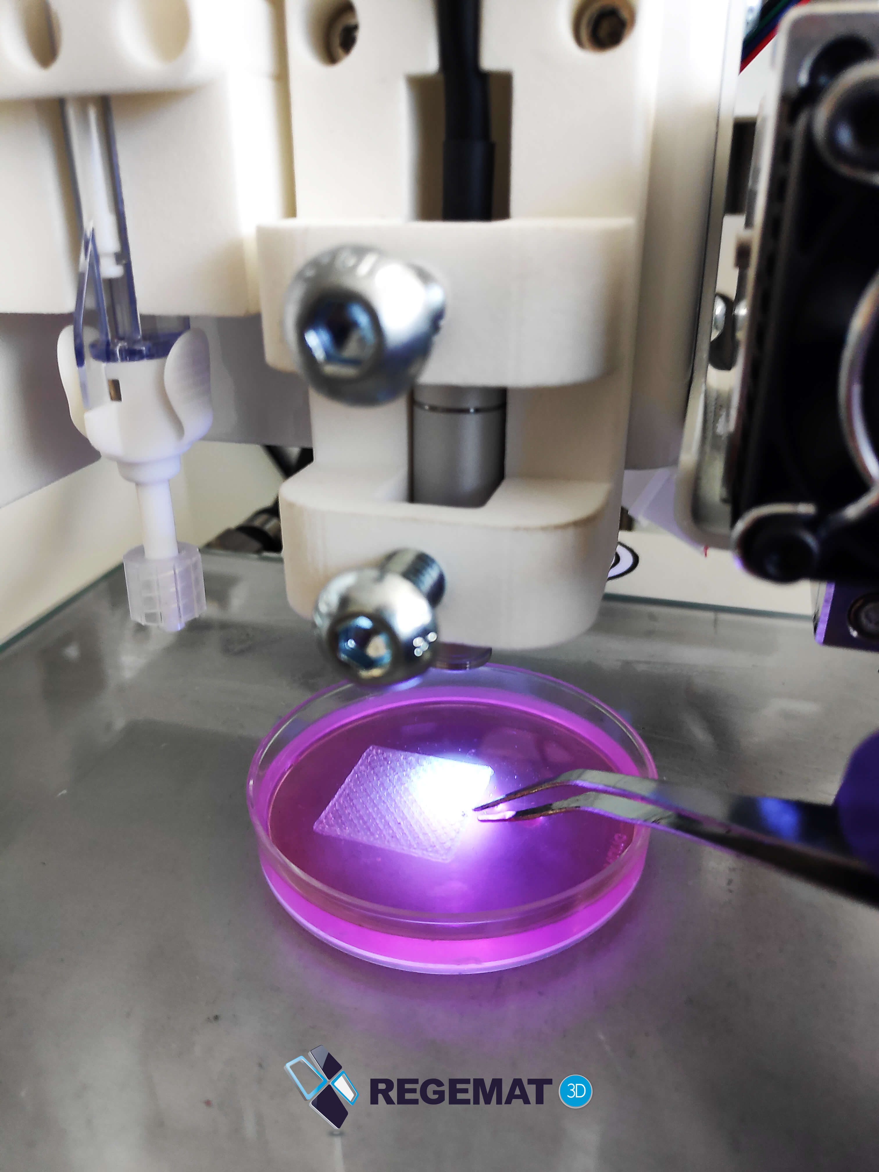
REGEMAT 3D bioprinting with new biomaterials
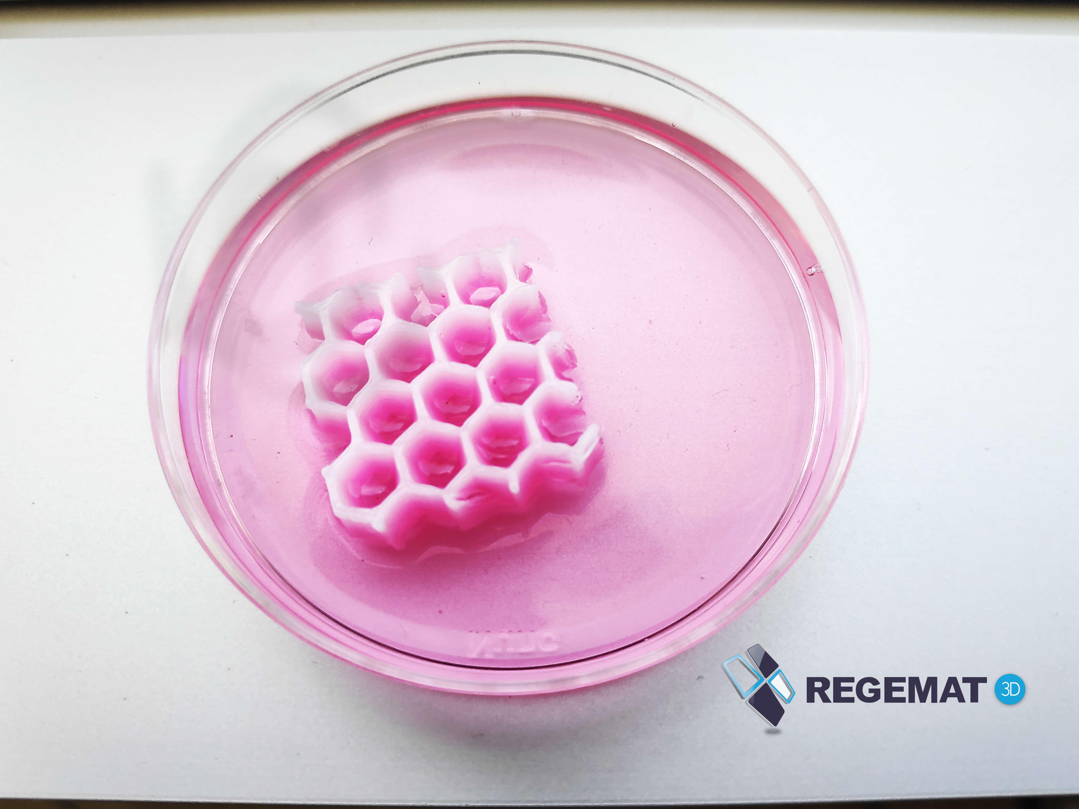
Each specific application requires different solutions and appropriate biomaterials to optimize processes. For instance, it is easy to understand that to regenerate skin components, hydrogels, cells and growth factors are different from those needed to regenerate muscle tissue, bone or cornea. So, it is essential to offer researchers and scientists different biomaterials to aid their work. REGEMAT is focusing on seven: thermoplastics, collagens, alginates, agaroses, gelatin methacryloyl (GelMA), nanocellulose, and different types of cell media compatible with the cells used. All of the biomaterials should be easy to print, handle and will allow researchers to tackle some of the challenges they face while working.
The new biomaterials for 3D bioprinting will be available on the company’s web page (which they will relaunch shortly), as well as through their offices. REGEMAT 3D has agreements with several national and international partners for the manufacture of these products. The first ones to be sold commercially will be nanocellulose, collagen, and alginate.
REGEMAT 3D new biomaterials
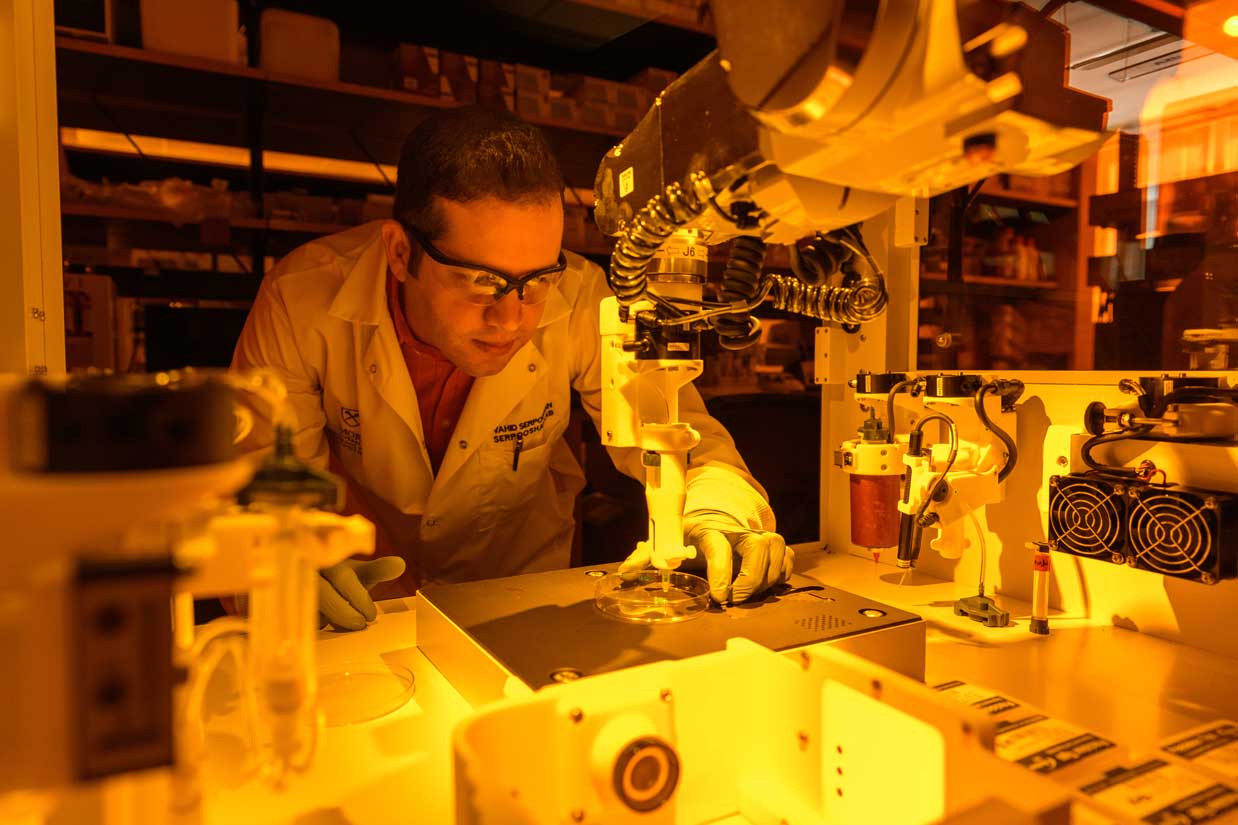
The Granada, Spain-based biotech company has sold its machines to users in more than 25 countries. For years, the company has been working with research groups at the University of Granada in advanced therapies, participated in a joint project for the development of new therapies for cartilage regeneration, and has collaborated with the University Hospital of La Paz, where REGEMAT 3D’s founder coordinates the 3D Tissue Engineering and Printing Platform (PITI3D), which provides ingredients and processes to generate functional tissues. Since its origin, the startup has been focusing on regenerative medicine, developing custom hardware and software required and demanded by some of the major hospitals and research universities in the region. They develop their own bioprinting systems – the BIO V1 machines – and customize them for their users’ applications according to the requirements of each investigation.
Last January, a group of researchers led by the University of Granada and REGEMAT 3D, published an academic article on the development of a volume-by-volume 3D biofabrication process that divides the printed part into different volumes and injects the cells after each volume has been printed, once the temperature of the printed thermoplastic fibers has decreased. This helps overcome problems that arise when working in 3D bioprinting with thermoplastics at high temperatures: one of the biomaterials they will soon begin commercializing, with which the company is very familiar and has worked with for a long time.
To continue developing new biomaterials and launching new products, the Spanish company, led by founder and CEO José Manuel Baena, has managed to raise 320,000 Euros in the midst of the latest financing round. REGEMAT 3D, along with its sister company Breca, are not only launching the new series of biomaterials, but they are also expanding their presence to Brazil, where the company has already started to market its products, and China, where they closed an agreement with Chinese distributor ApgBio, based in Shanghai, that’s responsible for introducing bioprinting equipment in the country for the regeneration of organs or tissues. Companies like REGEMAT 3D are gearing up to mass produce biomaterials, providing researchers with more options when it comes to bioprinting for regenerative medicine and advanced therapies, usually keeping in mind how patients bodies will react to the new materials, and whether they will be compatible. Spain, like many other European countries, is quickly catching up to the world of bioprinting, which today is led by US-based companies but shows promise in many developed countries.
[Images: REGEMAT 3D]
The post REGEMAT 3D Will Start Selling Biomaterials appeared first on 3DPrint.com | The Voice of 3D Printing / Additive Manufacturing.