EnvisionTEC-exocad Partnership Signals Industry Integration

For Lab Day 2020, a number of 3D printing companies are rolling out their dental 3D printing product lines. This year, there’s the sense that there is a change taking place within the dental additive manufacturing (AM) space as systems manufacturers move beyond simply presenting their latest wares and, instead, present integrated product suites that tackle entire dental workflows. This reflects a larger trend within the AM space in which the industry is working to create integrated solutions for each vertical.

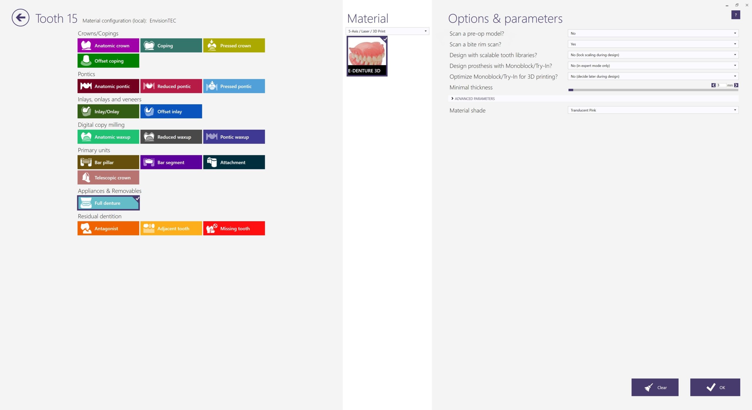
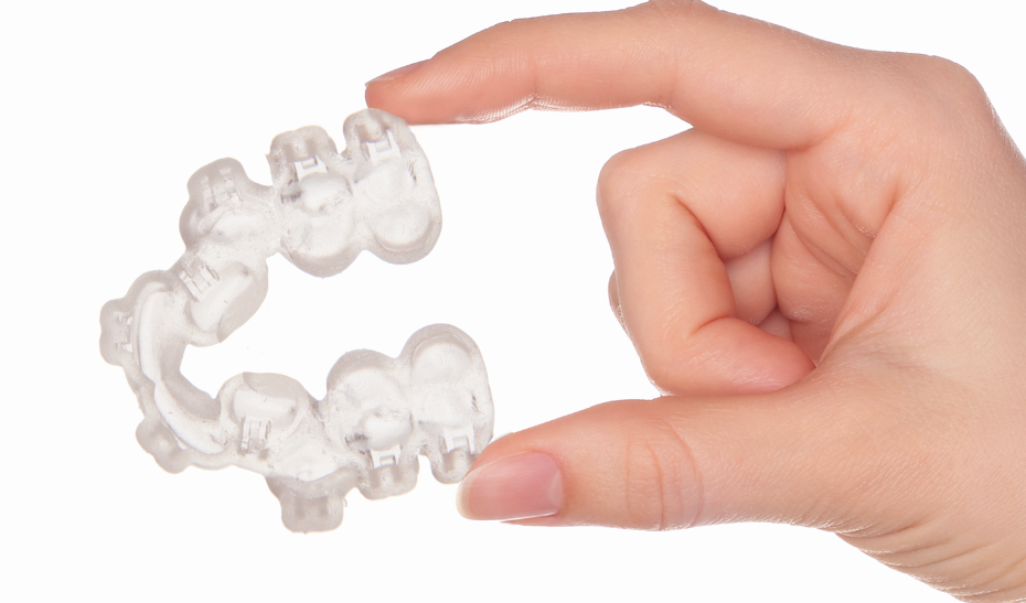
Dental materials from EnvisionTEC are pre-loaded in exocad’s exoprint module. Image courtesy of EnvisionTEC.

The latest Lab Day announcement comes from EnvisionTEC, who has partnered with dental CAD/CAM software developer exocad to integrate their products. For the past year, EnvisionTEC and exocad collaborated to develop a “seamless workflow” between exocad’s DentalCAD software and EnvisionTEC’s Envision One RP software using an open XML-based interface. The exoprint tool is meant to streamline the digital workflow from an intraoral scan through design and 3D printing.

EnvisionTEC is positioning its Envision One cDLM Dental 3D printer as the best system for this workflow. Using a continuous digital light processing (DLP) technique, the Envision One prints at rapid speeds and produces objects without visible layers and with isotropic properties.

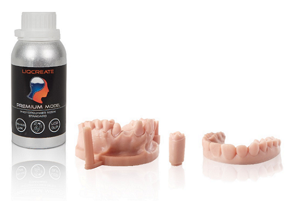
The new exoprint tool from EnvisionTEC represents continued industry integration, which is also evidenced by 3D Systems’ release of a new software workflow for 3D printing up to 30 orthodontic models in a single print. The workflow relies on the NexDent 5100 3D printer, NextDent Model 2.0 Software, and 3D Sprint software, the last of which includes a new auto-stacking feature that automatically prepares and places dental models on the build plate using smart nesting and proprietary support structures.

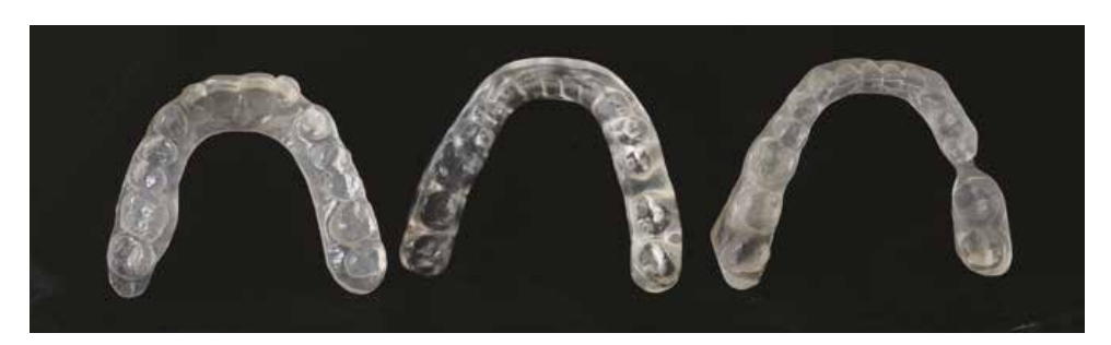
Neither of these tools is quite as extensive as the latest solution from Prodways, which announced the introduction of its Clear Aligners Manufacturing Ecosystem at the event. The ecosystem is made up of a complete workflow for the fabrication of patient-specific dental aligners, from scanning to thermoforming the aligners on 3D-printed models. Throughout the process there are automated and semi-automated steps, including an automatic platform loader and unloader, a semi-automatic thermoforming module, and an automatic laser marking and trimming module.

Now that 3D printers themselves are becoming industrialized and capable of large batch production, the next step seems to include integrating them into target industries. The medical and dental industries, as SmarTech predicted, has been a prime sector for AM growth, often acting as a prototype for how the technology will be integrated in other verticals. Former 3D Systems CEO Vyomesh Joshi, for instance, was extrapolating the business practices of his company’s Healthcare division onto other verticals.

We are also seeing vertical integration in the footwear segment, specifically with orthotics, in which foot scanner manufacturers, orthotic software developers, 3D printer hardware manufacturers and traditional orthotics companies are partnering to create complete ecosystems. Prosthetics makers are following a similar suit, as are general industry manufacturers with regard to custom grippers.

There is a more complex integration of AM into aerospace manufacturing, though the process is more involved and rely on specific business-to-business interactions. Honeywell, for instance, is qualifying the AM processes of a number of companies, while also qualifying quality control software and hardware from Sigma Labs.

At the same time, metal AM hardware manufacturers are attempting to create elaborate ecosystems for more general production environments. These ecosystems include the AM Factory of Tomorrow concept from GE Additive, the DMP 8500 Factory Solution from 3D Systems, and the Scale4Series from Additive Industries. All of these feature automated workflows for powder supply and part removal, as well as stations for heat treatment and finishing of metal parts.

As much as one might be skeptical about companies actually achieving these complex, automated ecosystems, it’s always surprising to see the milestones reached on the way to the complete picture. Prodways’ aligner ecosystem itself is an impressive one that represents just one accomplishment on the road to complete automation. Whether complete automation will ever be reached, given our current ecological state, or even desirable, given the issues faced by labor in our existing economic system, are different questions altogether.

The post EnvisionTEC-exocad Partnership Signals Industry Integration appeared first on 3DPrint.com | The Voice of 3D Printing / Additive Manufacturing.

New Partnership: BEGO’s Dental Materials Allow Formlabs Customers to 3D Print Crowns & Bridges

BEGO, headquartered in Germany, has been a leader in the dental field for 130 years—and as pioneers in 3D printing for the last two decades, they now specialize in all types of dental fabrication techniques for prosthodontics and implant dentistry. Today, they have announced a partnership with Formlabs, the US-based designer and manufacturer of 3D printing systems—along with a wide-ranging product line and other strong partnerships around the world.

This alliance, just announced, means that Formlabs dental customers will be able to make on-demand dental products for their patients–using BEGO’s dental materials to 3D print both temporary and permanent crowns and bridges.

Formlabs customers who are already using the Form 3B and Form 2 3D printers will have access to fabricating permanent single crowns, inlays, onlays and veneers, and temporary crowns and bridges. Appreciating many of the benefits of 3D printing, they will be able to save on the bottom line, while making customized products, faster—and better.

“Directly printing temporary crowns and bridges are one of the most sought-after applications from Formlabs customers. By partnering with BEGO and leveraging their 130 years of dental experience, we will be able to not only address this need but take it a step further by offering materials for permanent crowns. We are excited to see how this partnership can continue to advance the dental industry and overcome the major challenges labs and dentists face as digital dentistry becomes a standard for patient care,” said Dávid Lakatos, Chief Product Officer at Formlabs.

Patients will enjoy:

  • Excellent aesthetics
  • Less tendency for dental work to age and discolor
  • Low-plaque accumulation
  • More comfort, thanks to less cold and heat sensitivity

“The ability to use completely digital dental workflows with 3D printed temporary restorations will enable a rapid, low cost, iterative process that improves patient care and case acceptance rates,” states the BEGO team in a press release sent to 3DPrint.com.

Dávid Lakatos, Chief Product Officer at Formlabs with Axel Klarmeyer, Chief Executive Officer of BEGO Dental

The two companies will be in attendance at LMT Lab Day in Chicago from February 21-22, presenting materials for attendees to see and feel, as well as showcasing their new partnership, which is also part of the ‘natural evolution’ of the also recently announced Dental Business Unit and Form 3B, meant to ensure that dentists are able to offer their patients the best in care.

“We could not be happier to partner with Formlabs, especially at this time, where digital dentistry is reaching a breakthrough. It took some time and a lot of effort and commitment of all involved people to be able to offer to the market a fully validated workflow for final restorations. This partnership underlines BEGO’s leading position in the dental 3D printing materials market,” said Axel Klarmeyer, Chief Executive Officer of BEGO Dental.

Find out more about these products here, and please visit BEGO and Formlabs at LMT Lab Day at Formlabs booth (#P-1) and BEGO booth (#H-13).

Both BEGO and Formlabs stay in the news with continue product development and successes from BEGO’s recent launch of the Varseo XL to new resins and other recent acquisitions by Formlabs.

What do you think of this news? Let us know your thoughts! Join the discussion of this and other 3D printing topics at 3DPrintBoard.com.

[Source / Images: BEGO]


The post New Partnership: BEGO’s Dental Materials Allow Formlabs Customers to 3D Print Crowns & Bridges appeared first on 3DPrint.com | The Voice of 3D Printing / Additive Manufacturing.

Prodways Announces Clear Aligners Manufacturing Ecosystem

At Lab Day 2020 in Chicago, Prodways has announced the development of its Clear Aligners Manufacturing Ecosystem, meant to cover all aspects of the design and production of clear aligners. Developed in partnership with prevalent companies in the digital dentistry space—3Shape, Full Contour, Imes-Icore and Dreve—the product ecosystem can deliver up to 1200 aligners daily, according to Prodways.

As SmarTech Analysis has reported, the market for additive manufacturing in dentistry was on track to surpass $2.7 billion in 2019, with an anticipated 500 million dental devices and restorations projected to be produced annually by 2022. If you’ve seen an TV ads lately, you’ve likely caught one 3D-printed dental item that is already in high demand.

Clear dental aligners were among the first consumer products to be mass manufactured with the use of 3D printing. These devices are typically made by the following workflow: an intraoral scan of a patient’s mouth captures the 3D data used to design a model of their teeth; this model is 3D printed before the aligner thermoformed on top of the mold.

Given the success of clear aligners, not only are there now numerous competing aligner brands, but also 3D printing companies vying for their business. Prodways aims to stake out its own territory in the segment with what it considers the first end-to-end digital manufacturing solution. This includes 3D scanning, data preparation, treatment design, 3D printing, thermoforming, laser marking and trimming, with hardware and software integration and automation along each step. Part of this automation comes in the form of the automatic platform loader and unloader (seen in the video below), a semi-automatic thermoforming module and an automatic laser marking and trimming module.

With Prodways linking together 3Shape, Full Contour, Imes-Icore and Dreve, each companies’ hardware and software are interoperable. This results in the ability to manufacture up to 1200 aligners per day at what Prodways suggests is a low cost-per-part and a short time to market.

This ecosystem is a significant one for Prodways, which has been building its spot in the dental market for some time. The company’s MOVINGLight DLP technology is known for its high throughput, making it ideal for batch production of products like dental molds for aligners. The release of the LD3 system marked the company’s entry into smaller systems that can be used in dentistry, as well as jewelry and other fields. The acquisition of Solidscape also added to its footprint in the dental market.

As Prodways works to fully automate the process of making clear dental aligners, they may have some stiff competition, particularly from the likes of HP, which is already claiming to make 50,000 molds for aligners per day via its customer, the Smile Direct Club.

The post Prodways Announces Clear Aligners Manufacturing Ecosystem appeared first on 3DPrint.com | The Voice of 3D Printing / Additive Manufacturing.

Switzerland: in vivo Analysis of Intraoral Scanners for the Dental Arch

In the recently published ‘Trueness and precision of intraoral scanners in the maxillary dental arch: an in vivo analysis,’ authors Jonas Winkler and Nikolaos Gkantidis from the Department of Orthodontics and Dentofacial Orthopedics at the University of Bern in Switzerland are exploring ways to improve dental devices via 3D technology.

Intraoral 3D imaging is beginning to offer significant impacts within the dental field, especially because it allows for patients to receive oral cavity imprints that are without risk—as well as offering the following:

  • Diagnosis
  • Growth assessment
  • Outcome evaluation
  • Potential for use in 3D printing applications

“Digital dental models can overcome certain drawbacks associated with plaster models, such as patient discomfort and vulnerability. Being also advantageous in terms of cost, time, and space required, digital models will probably soon become the new standard in clinical practice,” explain the authors.

Intraoral scans are quickly becoming more useful in clinical settings; however, the researchers note that there is still a concerning amount of inaccuracy associated with such processes. Evaluating accuracy and precision is the goal of this study, and specifically in the fabrication of dental arches.

Box plots showing the trueness of the repeated intraoral scans with two different scanners in millimeters (n = 12, p = 0.176, Friedman test). The upper limit of the black line represents the maximum value, the lower limit the minimum value, the box the interquartile range, and the horizontal line the median value. Outliers are shown as black dots or stars, in more extreme cases. CS1, CS2, CS3: CS3600 repeated scans. TR1, TR2: TRIOS3 repeated scans. Artec: Artec Space Spider scan.

Eight male patients and four female patients participated, ranging in age from 27 to 52 years old. Three different scanners were used:

  • CS 3600 (Carestream, Atlanta USA, Software CS Imaging Version 7.0.23.0.d2)
  • TRIOS 3 (3Shape, Copenhagen, Denmark, Software Version 1.4.7.5)
  • Artec Space Spider (Artec3D, Luxembourg, Software ArtecStudio 12 Professional Version 12.1.6.16)

“All scans were obtained by the first author who had more than two years of experience with regular clinical use of intraoral scanners,” explained the authors. “The same investigator performed all the steps of data generation following relevant training and under close supervision by the senior author.”

Irrelevant data like soft tissue were cropped out using Artec Studio 12 Professional software, and then the files were transferred for 3D printing. Final models were then superimposed in Viewbox 4, and the researchers used these settings:

  • 100% estimated overlap of meshes
  • Matching point to plane
  • Exact nearest neighbor search
  • 100%-point sampling
  • Fifty iterations

Colour maps showing the trueness assessment measured through the MAD of the CS3600 and TRIOS3 intraoral scanners from the gold standard scans, in the upper buccal front teeth area. Three scans obtained through CS3600 (CS1, CS2, CS3) and two scans through TRIOS3 (TR1, TR2) were assessed (n = 12 for each set of scans).

The research team noted ‘significant difference’ between the TRIOS 3 and the CS 3600, with the first demonstrating superior precision in two out of three cases.

“No specific imprecision patterns were observed in the respective color maps, since the errors were distributed equally between the different areas of the arch. However, in certain cases the imprecision was higher than in others. There was also significant variation within the arches, with local imprecisions reaching in several cases relatively high values,” explained the researchers.

Colour maps showing the precision of the intraoral scanners. Three scans form CS3600 (CS1, CS2, CS3) and two scans from TRIOS3 (TR1, TR2) were assessed. The region for superimposition was the whole maxillary dental arch.

The Artec Spider was noted as ‘excellent’ in terms of scanning; in fact, it was chosen as a gold standard by the researchers.

(a) Box plots showing the precision (millimeters) assessment of the gold standard scanner versus the intraoral scanners (n = 3), measured through the MAD of the whole dental arch area between repeated scans, when only the upper buccal front teeth area was used as superimposition reference. The upper limit of the black line represents the maximum value, the lower limit the minimum value, the box the interquartile range, and the horizontal line the median value. CS1, CS2, CS3: CS3600 repeated scans. TR1, TR2: TRIOS3 repeated scans. Artec1, Artec2: Artec Space Spider repeated scans. (b) Colour maps showing the absolute distances between corresponding points of surface models obtained through repeated scans with the Artec Spice Spider scanner.

“Both intraoral scanners showed comparable trueness in the upper buccal front region. Precision of the whole upper dental arch was also comparable when the repeated models were superimposed in the upper buccal front region. TRIOS 3 showed slightly higher precision (approximately 10 μm), when the whole model was used as superimposition reference, but it also showed the highest imprecision in a specific case,” concluded the researchers.

“However, in individual cases and in various, not spatially defined areas of the arch, higher imprecision was evident. In a small spatial scale, there is considerable variation in scanner performance between and within cases. Thus, the usage of intraoral scanners in highly demanding clinical applications, considering the whole arch, remains questionable.”

3D printing technology within the dental industry just continues to grow as some manufactures target mass production of products, improve fabrication of implants, continue to expand materials, and more.

What do you think of this news? Let us know your thoughts! Join the discussion of this and other 3D printing topics at 3DPrintBoard.com.

[Source / Images: ‘Trueness and precision of intraoral scanners in the maxillary dental arch: an in vivo analysis’]

The post Switzerland: in vivo Analysis of Intraoral Scanners for the Dental Arch appeared first on 3DPrint.com | The Voice of 3D Printing / Additive Manufacturing.

Structo & pro3dure Partnering: Dental Customers Benefit from Expansion of Materials for Velox Ecosystem

Innovative materials once again merge with 3D printing hardware in an international collaboration as Structo announces a partnership with pro3dure to jointly offer products for the dental industry.

Structo is a leading dental 3D printing solutions provider, headquartered in Singapore, that we caught up with late last year after they completed a funding round that would not only allow them to continue with additive manufacturing innovations but also to expand their presence worldwide. Over the past couple of years they have also offered innovation in creating orthodontic aligners, automated post-processing, and more.

Germany’s pro3dure, founded in 2013, offers a focus on generative and subtractive manufacturing, while integrating them into digital fabrication. Their team specializes in matching customized materials to the workflow of each customer, emphasizing 3D printing. Pro3dure also offers the greatest selection of 3D printing resins for dental applications.

“We strive to make our range of dental materials as widely available as possible,” said Dr. Martin Klare, CEO of pro3dure. “The Structo Velox is a revolutionary ecosystem that will change how 3D printing is adopted in the dental office and we are proud to introduce our range of 3D printing resins with the world’s largest spectrum of dental applications available on that platform.”

Now, dental labs using the Velox ecosystem will be able to use pro3dure materials too as many new applications open to customers, allowing 3D printing of the following also:

  • Splints
  • Surgical guides
  • Indirect bonding trays
  • Temporaries

Indirect bonding trays

Huub van Esbroeck emphasizes how critical partnerships are, especially with closely related companies like pro3dure, allowing them to ‘push the envelope of chairside 3D printing’ within the dental industry.

“We are committed to continue developing the Velox ecosystem of hardware, software and materials to empower the delivery of same day dentistry” said Huub van Esbroeck, Founder at Structo. “Today’s announcement will see Velox users gain access to pro3dure’s range of industry-leading dental 3D printing materials which gives them a range of options for their in-office 3D printing needs.

“This is another step for us to provide more options to all Velox users worldwide and we will continue working with industry leaders to add more value and streamline the chairside 3D printing workflow for our industry,” continued van Esbroeck. “We started off with just the hardware that does the print, wash and cure cycle of a 3D printing workflow. With more partnerships like these, we are confident that the Velox ecosystem will be able to empower dentists to deliver better patient care with revolutionary new solutions,” Huub added.

Many innovations continue within 3D printing, and specifically for the dental industry with progress in creating dental implants, targeting mass production of digital fabrication in dentistry, and more. What do you think of this news? Let us know your thoughts! Join the discussion of this and other 3D printing topics at 3DPrintBoard.com.

[Source – Images: Structo/pro3dure]

The post Structo & pro3dure Partnering: Dental Customers Benefit from Expansion of Materials for Velox Ecosystem appeared first on 3DPrint.com | The Voice of 3D Printing / Additive Manufacturing.

Early Bird Discount for AMS 2020, “The Business of 3D Printing,” Ends Tomorrow, February 5th

3DPrint.com and SmarTech Analysis will be holding the third annual Additive Manufacturing Strategies event next week, February 11th-12th,  at the Seaport World Trade Center in Boston, Massachusetts. This year, AMS 2020 is titled “The Business of 3D Printing: Medicine, Dentistry and Metals,” and will be centered around 3D printing business practices and applications in both the medical and dental fields, as well as industries, such as automotive, which use metal often.

AMS 2019

Tomorrow, February 5th, is the last day for attendees to take advantage of the final Early Bird registration discount for this event. Full access to the entire Additive Manufacturing Strategies summit, which will include an exhibit hall, multiple keynote presentations, the annual Startup Competition, a networking reception, fireside chats, SmarTech’s forecasts on 3D printing for the medical and dental industries, and more, requires paid registration. You can attend the competition and stop in to visit the exhibit hall for free, but you’ll still need to register.

Members of the 3DPrint.Com and SmarTech Analysis teams will be present during the event to meet with other attendees, and more than 60 expert speakers will be on hand to share their expertise with us at AMS 2020 in panels, presentations, and fireside chats. About 30 exhibitors and sponsors, including Platinum Sponsor Global Advanced Metals and Gold Sponsor EOS, will be there as well.

While AMS traditionally covers 3D printing in the medical and dental fields, the metals track is new this year. In fact, SmarTech just released a new report on metal additive manufacturing service bureaus, which will be one of many topics addressed at AMS 2020 next week.

“This is SmarTech’s latest business assessment and market forecast of metal additive manufacturing services,” SmarTech writes about its report. “It updates our analysis and market projections and also extends the coverage to include a broader range of services and profiles of leading AM service firms.  It also assesses the impact on the business of service bureaus on the changes that have occurred in end user industries and on the dramatic shift of AM to become a fully-fledged manufacturing process.”

Image: SmarTech Analysis

To gain full access to our AMS 2020 event, and receive the final Early Bird discount, you need to register by tomorrow, February 5th.

Discuss this news and other 3D printing topics at 3DPrintBoard.com or share your thoughts in the Facebook comments below.

The post Early Bird Discount for AMS 2020, “The Business of 3D Printing,” Ends Tomorrow, February 5th appeared first on 3DPrint.com | The Voice of 3D Printing / Additive Manufacturing.

Formlabs Tells Us How to Make Good Looking 3D Printed Dentures

More than 36 million Americans do not have any teeth, and 120 million people in the US are missing at least one tooth. With these numbers expected to grow in the next two decades, the market for 3D printed dentures is expected to grow significantly.

Sam Wainwright, Dental Product Manager at Formlabs, suggested during the company’s latest webinar that he wouldn’t “be surprised to see 40% of dentures in America made with 3D printing,” claiming that it makes sense “at the technology level because there is no loss of material.” The expert delved into some of the techniques that have proven to work for aesthetically better 3D printed dentures. The webinar, titled Can 3D printed dentures look good?, offered dentists, technicians, and anyone interested in using 3D printing to improve dentures, tips on how to cut material costs by up to 80% (compared to traditional denture cards and acrylic); perform fewer steps to attain high-quality results, and overall prevent teeth from looking unnatural. 

“This is an ever expanding market with many options. 3D printed dentures are a very new thing, especially for removable prosthetics (something that has never been digitalized) so it is going to take some time for labs, dentists and patients to become used to it. The material is indicated for long term use but the most rapid adoption of this technology will be immediate conversion and provisional dentures, which have lower risk allowing dental professionals to walk not run into this new technology. We also expect the resins to get better, stronger and more aesthetic in time,” said Wainwright.

In fact, in the last year, Formlabs has already managed to upgrade the resins it sells for medical professionals to make oral prostheses, called Digital Dentures. These new FDA-approved resins not only resemble traditional dentures but they are also cheaper than other options. At $299 for the denture base resin and $399 for the teeth resin, the company estimates that the total resin cost for a maxillary denture is $7.20. Moreover, Formlabs also recently released the new Form 3 printer, which uses light touch supports: meaning post-processing just became much easier. Support removal is going to be quicker on the Form 3 than the Form 2, which translates to fewer materials costs and time.

“We are trying to prevent teeth from looking unnatural, and sometimes with these 3D printed dentures, the aesthetics are really suffering from it. We like to think that dentures should have life-like gingiva, natural cervical margins, individual looking-teeth, and be easy to assemble,” Wainright said.

The general basic workflow proposed by Wainright is to follow the traditional workflow until the final models are poured and articulated with wax rim, that set-up needs to be made digital with a desktop dental 3D scanner allowing for the digital design in any open CAD dental system, followed by 3D printing the base and teeth, and finally post-processing, assembling and finishing the piece. 

“After making so many parts, printing a ton of denture teeth and bases, and assembling them, we’ve come up with three techniques for an aesthetic 3D printed denture. What we want is to avoid some of the outcomes of today’s digital dentures, like products with an opaque base or gingiva, which is a bit of a mess in my opinion. Or you come about a semi transluscent base which leaves the roots exposed, and lastly when you use the splinted tooth workflow you can end up with a bulky interproximal connection. And since the papillae are a really thin printed parts, it’s really easy to see the teeth connecting, looking unnatural.”

The three aesthetic denture techniques suggested by Wainwright include:

  1. Natural gingival connection and cervical margin are based on the CAD output for optimal result
  2. Splinted arch ease of assembly without a bulky interproximal
  3. Life-like gingiva, inspired by “Brazilian Dentures”

Wainright suggests that for his first aesthetic dental technique, users can control the depth of penetration of the tooth as well as the angle it comes in or goes out, by using a new function in the 3Shape Dental System CAD software (version 2018+). The option is called coupling mechanism, and gives the user much more control than before, something which comes in very handy considering that “the more subgingival length the tooth has, the stronger the bond is with the base.” 

“The reason why 3D printed dentures are different than traditionally made dentures is that resins for the base and the teeth are like cousins. When the parts come out of the printer and you wash them, they are almost soft and even sticky, because they are only partially cured, between 25 and 35 percent. But during the final UV curing process, the tooth and the base become one solid part.”

In fact, the dentures specialist indicates that users should cure the combined base and teeth with a handheld UV cure light, moving towards the interior, just to really hold the parts together. Once the user has checked that all the cavities have been filled up and removes any residual base resin, the denture is complete and ready to be submerged for 30 minutes in glycerine at 80 degrees celsius, for a total hour of cure time. At that point, the piece can be finished up with a UV glaze or wheel for a high shine polish.

The second recommended aesthetic denture technique involves a splinted arch ease of assembly without a bulky interproximal.

Wainright explained that he sets up “these cases up in CAD so they are 100% splinted together because it is so much easier to have consistent placement of teeth, instead of doing it one by one which can be labor-intensive. I first export the arch splinted, but the question here is how to make the connection between the teeth interproximally look natural, especially when you have a very thin papilla. So before assembly, during our support removal part of the process, we’ll take a cutting disk and reduce the interproximal connection down from the cervical margin up towards the incisal. This really helps the aesthetics of the denture without worrying about any spaces.”

He also recommends that during the assembly process, users can easily brush in gingiva resin in the spaces to make sure there is no air, gaps or voids, maintaining the strength.

“Keep your eye out for bubbles,” repeated Wainright many times, explaining that “if you do minimal interaction to get the resin in the spaces, it really reduces the bubbles.”

He also added that the key is to “flow in more resin at first, instead of just wetting it, and when it’s squeezed together it will flow into that area. Finally, the overflow can be wiped away with a gloved finger.” 

“It seems quite simple but this are the things we learn over time. I repeated many of these processes a handful of times and got better, today it may take me up to 10 minutes at the most to finish up one denture. Moreover, if you think about the soft touch supports in the Form 3, post processing will be even easier, as anyone will be able to rip them off and add very little finishing to the product.”

For the last aesthetic denture technique, Wainwright suggested following up the “Brazilian dentures” example, which offers an inspiring way to create life-like gingiva. He says he noticed Brazilians have become experts in creating dentures, adding translucent resins in the base that allow for the patient’s own gingiva color to show through. He proposed the LP resin Formlabs resin is also quite translucent, but when tested on a model or patient’s mouth, “it adds a nice depth to the gingiva itself giving a reflection of light useful in aesthetics.”

“When the denture is seated intraorally, the patient’s natural gingiva shows through making the prosthetic come to life.”

Formlabs is known for creating reliable, accessible 3D printing systems for professionals. According to the company, in the last decade, the dental market has become a huge part of the company’s business and that Formlabs is trusted by dental industry leaders across the globe, “offering over 75 support and service staff and more than 150 engineers.” 

It has shipped over 50,000 printers around the world, with tens of thousands of dental professionals using Form 2 to improve the lives of hundreds of thousands of patients. Additionally, using their materials and printers in more than 175,000 surgeries, 35,000 splints and 1,750,000 3D printed dental parts. One of the aims at Formlabs is to expand the access to digital fabrication, so anyone can make anything, this is one of the reasons why the company is making webinars, to help everyone get there.

Wainright also revealed that Formlabs will be releasing two new denture bases, RP (reddish pink) and DP (dark pink), as well as two new denture teeth shapes, A3 and B2, that will complement the already existing A1, A2, A3.5, and B1. 

If you are a big fan of webinars, make sure to check out more at 3DPrint.com’s webinars under the Training section.

Discuss this article and more on 3DPrintBoard.com or comment below to tell us what you think.

[Images: Formlabs]

The post Formlabs Tells Us How to Make Good Looking 3D Printed Dentures appeared first on 3DPrint.com | The Voice of 3D Printing / Additive Manufacturing.

3D Printed Occlusal Splint Provides Relief in Just a Few Weeks

In the recently published ‘Digital manufacturing of occlusal splint: from intraoral scanning to 3D printing,’ researchers explored the use of a new digital workflow for creating intraoral occlusal splints. These devices are used by dentists to treat temporomandibular disorders as they force muscles to relax, protect the teeth, and more.

As 3D technology continues to progress, so do manufacturing processes within dentistry, featuring 3D scanning, CAD technology, and 3D printers for fabrication of dental prostheses and a range of other devices.

For this case, the researchers chose to study a 44-year-old man presenting with issues in his jaw muscles. As doctors examined his masticatory system, they were unable to find any health conditions, except for pain emanating from his anterior temporalis muscles and external pterygoid muscles. It was suspected that he may have been in the habit of clenching his jaw, thus leading to the pain. The doctors decided to fit him with an intraoral occlusal splint to stop the pain and begin ‘repositioning’ the mandible.

“With the accuracy and efficiency of the digital workflow, the splint would be fabricated with digital intraoral scanners and a 3D printer,” stated the researchers.

The team used a scanner to photograph all the teeth, saving an .stl file read for 3D printing. The first scans, however, related both the lower and upper virtual models. The initial setting allowed parameters to include:

  • 40 °TMJ eminence angle
  • 10° Bennet angle
  • 40° incisal guide angle

“The bite plane was designed to be retained by the mandibular teeth, according to patient’s preference, and the maxillary teeth had a single contact with the appliance,” explained the researchers.

Splint designed on the digital casts.

A 3D ProJet MJP 3600 Dental by 3D Systems was used to 3D print the intraoral splint, with VisiJet® M3 Stoneplast acrylic resin, made for dental practices. Once inserted and evaluated, the doctors found that the intraoral occlusal splint was in need of little adjustment. The patient was to wear the splint while sleeping or while at home during the day. Pain subsided after three weeks; however, the patient was instructed to keep using the splint for another six months.

“The presented workflow allowed for a noticeable reduction of the complexity and of the total time of the laboratory procedures. Although in this case the authors sent the impression scan to the dental laboratory, the possibility of producing the same appliance in the dental clinic should be considered as 3D printers are becoming more popular within the dental office. However, chairside production could be time consuming for the dentist and the authors prefer at this time to delegate the design and the production to the dental technician more familiar with CAD software and 3D printers,” concluded the researchers.

“After centric relation was recorded with bimanual manipulation, the technique included intraoral scans of the maxillary and mandibular arches, digital registration of vertical relationship, computer-aided design of the intraoral occlusal splint and then manufactured with a multi-jet 3D printer. The presented technique allows for time efficient laboratory manufacturing, which could also be performed chair-side in the dental office. The delivered splint is accurate and precise and could be reproduced anytime if needed.”

3D printing has made huge impacts in the dental world, and has improved the quality of life for many patients from dental implants to orthodontics to new manufacturing systems. What do you think of this news? Let us know your thoughts; join the discussion of this and other 3D printing topics at 3DPrintBoard.com.

Splint produced with a multi-jet 3D printer and verified on the prototyped casts.

Intraoral pictures of the splint immediately after delivery

[Source / Images: ‘Digital manufacturing of occlusal splint: from intraoral scanning to 3D printing’]

The post 3D Printed Occlusal Splint Provides Relief in Just a Few Weeks appeared first on 3DPrint.com | The Voice of 3D Printing / Additive Manufacturing.

3D Printing News Briefs: September 2, 2019

In this edition of 3D Printing News Briefs, we’ve got stories to share about a new material, a case study, and an upcoming symposium. Liqcreate has released a new 3D printing material for dental professionals. FELIXprinters published a case study about its automotive 3D printing work with S-CAN. Finally, ASTM International will soon be hosting an AM symposium in Washington DC.

Liqcreate Releasing New Dental 3D Printing Resin

Manufacturer of professional-grade 3D printing materials Liqcreate has been hard at work on a new 3D printing resin to help dental professionals optimize their digital workflow and scale up their in-house manufacturing. The hard work has paid off, as the company is announcing the release of its newest material, Liqcreate Premium Model – an accurate, low shrinkage resin for fabricating dental and aligner models.

The opaque photopolymer is matte, and the color of skin. Parts 3D printed with Liqcreate Premium Model have low shrinkage and excellent dimensional stability, and its low odor makes it great for office use. Other benefits include high detail and accuracy, and temperature resistant for aligner production. The resin is compatible with the Anycubic Photon, Wanhao D7, and Kudo3D Bean 3D printers, in addition to all open source 385 – 420nm LCD and DLP systems. You can purchase Liqcreate Premium Model through the company’s distributor network starting September 2nd.

FELIXprinters Publishes Case Study

Dutch 3D printer manufacturer FELIXprinters published a case study about its work with reverse engineering and 3D scanning company S-CAN 3D Ltd, a UK customer which uses FELIX’s AM platforms to manufacture jigs, create casting molds and masters, and prototypes. Founded in 2012, S-CAN also uses FELIX technology to manufacture automotive parts, like the pictured engine block. FELIXprinters offers a range of systems for industrial prototyping and production applications, inlcuding its Pro 3 & Tec 4 series of AM platforms and its new, larger Pro L and XL models.

“We have found FELIXprinters AM platforms to be very easy to use. You can be up and running within a few minutes of getting them out of the box. We run all of our printers through Simplify3D software so you load the profile, pick a material and you are ready to go. In-house we now have the first machine we bought from FELIX back in 2015 (the Pro 1), and a Tec 4.1, a Pro 3 and the new Pro XL. Our first Pro printer has paid for itself 10 times over,” stated James Senior, MD of S-CAN 3D.

“Internally, S-CAN 3D use FELIX 3D printers for prototyping designs. We might do five or more different concept designs of a particular part or component, as it’s much easier to visualise a part when it’s in your hand. We are putting a lot of work through the newly purchased XL printer and it’s opening up things which we wouldn’t have been able to do before (at least to the same quality and size), so things are very encouraging. We have found FELIX machines to be very repeatable which is our most fundamental requirement for any application, and we also haven’t noticed any accuracy degradation over time.”

At the upcoming TCT Show in Birmingham, September 24-26, the two partnering companies will exhibit together at Stand E50 in Hall 3. Visitors will be able to view FELIXprinters’ Pro series of 3D printers, as well as its new advanced, customizable 3D bioprinting platform.

ASTM International’s AM Symposium

Speaking of industry events, ASTM International, which recently announced that it will be hosting its second Additive Manufacturing Center of Excellence Workshop in France, will also host a symposium in the Washington DC area. The Fourth ASTM Symposium on Structural Integrity of Additive Manufactured Materials and Parts, held by the ASTM International Additive Manufacturing Center of Excellence (AMCOE) from October 7-10 at the Gaylord National Resort and Convention Center, National Harbor, Maryland, is designed to give AM professionals a forum to exchange ideas about the structural integrity of 3D printed components and materials, focusing on quality and certification criteria and the lack of design principles and industry standards.

Paper topics for the symposium include the effect of anomalies, process optimization to improve performance, feedstock and its related effects on mechanical behavior and microstructure, and the applicability of existing test methods. Sessions will be organized by sector-specific applications, such as aviation, consumer, maritime, and spaceflight. Registration for the event will be open until October 2nd, 2019.

Discuss these stories and other 3D printing topics at 3DPrintBoard.com or share your thoughts in the Facebook comments below.

The post 3D Printing News Briefs: September 2, 2019 appeared first on 3DPrint.com | The Voice of 3D Printing / Additive Manufacturing.

Germany: Research Shows Good Response from Students Using 3D Printed Dental Traumatology Training

Authors M. Reymus , C. Fotiadou, R. Hickel, and C. Diegritz explore the uses of 3D printed models in dental traumatology training, with their findings outlined in the recently published ‘3D printed model for hands-on training in dental traumatology.’ For their study, they used an SLA printer to create a 3D printed model of a dental patient’s maxilla, mimicking several different traumatic dental injuries.

Being able to create accurate models exhibiting significant trauma offers a host of benefits to dental students who can take their time in a deliberate learning mode rather than waiting to rush in to see what could be a relatively small number of injured patients on-site. This also accentuates the enormous amount of learning gained from lectures. The hope is that more knowledge can be gained about dental traumatology, as the researchers point out that dental accidents are often treated by general dentists who may not have an adequate education or experience to deal with such cases overall.

The researchers wanted to make a model that was not only realistic but would allow for students to practice both diagnosis and treatment too. They also wanted to design a product that would translate from educational settings to dental clinics. With these hands-on tools available, the authors also created another level to their study regarding the use of dentaltraumaguide.org, offering the resource to only half of the students participating in the study—and comparing their knowledge.

The model was designed and 3D printed as follows to show dental trauma for a 16-year-old boy:

“The data generated were exported as single DICOM files and imported to Invesalius for Mac (Centre for Information Technology Renato Archer, Amarais, Brazil) to convert it into one .stl file. This file was subsequently imported to Meshmixer for Mac 11.0 (Autodesk, San Rafael, CA, USA) and trimmed to a region extending from the right first premolar to the left premolar. The right lateral incisor, the left first incisor as well as the left second incisor were cut out of the STL-mesh and exported as single STL-files.

Using the function ‘Boolean difference’, these teeth were cut out, leaving imitation tooth sockets in their original position. Additionally, the right lateral incisor was positioned at a 30° angle towards the palatal from its original position, and again, the function ‘Boolean difference’ was used to imitate a lateral luxation of the tooth perforating the buccal bone. The left lateral incisor was separated into two parts at its apical third imitating a horizontal root fracture. The extracted left incisor was not changed, imitating an avulsion. The mesial edge of the right incisor was removed, exposing the pulp chamber to imitate a complex crown fracture.”

Computer-aided designed model with empty tooth sockets, buccal perforation and complicated crown fracture.

The 32 undergraduate students were tasked to work on the case, even simulating a conversation with the mother of the injured boy as they practiced asking the correct questions about the accident, as well as advising on post-traumatic behavior. Upon examining the 3D printed model, they were given information about every tooth, and asked to offer the following:

  • Diagnosis
  • Treatment plan
  • Recall regime
  • Prognosis of each injured tooth

The assessment was considered in these areas:

  • Pre-treatment
  • Therapy
  • Post-treatment
  • Recall
  • Complications

“The presented workflow allowed the manufacturing of a radiopaque model that imitated a luxation injury, a complicated crown fracture, an avulsion, and a horizontal root fracture in a realistic way,” stated the authors.

Radiograph of the right lateral incisor with a luxation injury (left) and Radiograph of the empty tooth socket of the left incisor and the left lateral incisor with a horizontal root fracture (right).

And while their goal was for such a workflow to be easily transferred to another dental school, they would need to own a CBCT and a stereolithographic printer, along with software that could be offered free. The 32 students were asked to evaluate the model, with 57 percent reporting it to be ‘very realistic,’ and 43 percent choosing ‘rather realistic.’

“The diagnosis of the lateral luxation was evaluated to be the most difficult of all injuries, whereas the avulsion was the easiest injury to diagnose. Concerning treatment planning, the horizontal root fracture was rated as being the most difficult injury. When listing possible complications, the students had serious problems with the horizontal root fracture.

Students’ evaluations of difficulty in diagnosis, treatment planning, therapy and knowledge about complications for each injury.

“All participants reported to have gained new knowledge on dental traumatology, and 97 percent felt better prepared for treating traumatic dental injuries in the future.

“Students seem to focus especially on the diagnosis and treatment of traumatic injuries to teeth when dealing with dental traumatology. This is logical because these steps are of outmost importance for immediate care when confronted with a trauma case. Fortunately, both groups of students in the present study achieved their best results in these fields. The group without access to dentaltaumaguide.org, however, had only poor results when faced with developing a recall regime and knowing about possible complications,” concluded the researchers.

Many dentists and orthodontists rely on 3D printing today for digital dentistry, dentures, and even grafts for issues like alveolar augmentation. What do you think of this news? Let us know your thoughts! Join the discussion of this and other 3D printing topics at 3DPrintBoard.com.

Printed model with gingival mask.

[Source / Images: ‘3D-printed model for hands-on training in dental traumatology

The post Germany: Research Shows Good Response from Students Using 3D Printed Dental Traumatology Training appeared first on 3DPrint.com | The Voice of 3D Printing / Additive Manufacturing.