![]() The 3D-Bioplotter from EnvisionTEC has been at the heart of over 333 scientific papers, from 3D printing human tissue to 3D printing an ovary for mouse implantation. It is one of the most commonly used (and earliest) 3D bioprinters in the industry for tissue engineering and biofabrication research.
The 3D-Bioplotter from EnvisionTEC has been at the heart of over 333 scientific papers, from 3D printing human tissue to 3D printing an ovary for mouse implantation. It is one of the most commonly used (and earliest) 3D bioprinters in the industry for tissue engineering and biofabrication research.
Now, the global provider of professional-grade 3D printing solutions is expanding the bioprinting capabilities of their star product just in time for the European Society for Biomaterials (ESB) Annual Conference in Dresden, Germany. Launched in 2000, the 3D-Bioplotter is probably one of the most seasoned bioprinters in the market, and now it’s getting two new print head options that will help advance biomaterial research.
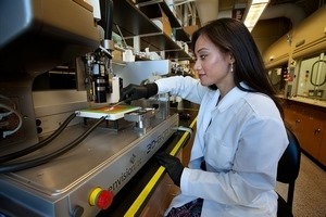
Researcher using EnvisionTEC 3D-Bioplotter
The first is an upgrade of the Photo-Curing head, now allowing up to five wavelengths or combinations thereof during one project. The second is an Ink-Jet Low-Temperature head designed to dispense materials through a non-contact process.
One of the several solidification processes available to 3D-Bioplotter customers is photo-curing. According to EnvisionTEC, while the wavelength of 365 nanometers (nm) remains the most commonly required by photoinitiators used by academic and industrial users, this wavelength has a negative impact on cell survivability during prolonged or repeated use, especially in the research field of bioprinting.
Therefore, the company sought out a way to solve this need to shift towards photoinitiators that react to the visible light range, to which cells can be exposed to with minimal biological effect.
To avoid the constant manual exchange of light sources when using different materials, as well as to allow combinations of photoinitiators, EnvisionTEC came up with an upgrade for their machine, a multi-wavelength pen upgrade which now provides five wavelengths into one single source pen. Through the 3D-Bioplotter software, individual wavelengths, or combinations thereof, are user-selectable and can be assigned to individual parts.
Current customers with existing photo-curing heads can have their existing heads upgraded to allow the use of the new source pens. The wavelengths included are 365, 385, 395, 405 and 455 nm.
The firm’s second highlight, the Ink-Jet Low-Temperature head, is aimed at dispensing low viscosity hydrogels as coatings while 3D printing parts or for hybrid scaffold fabrication. The key is that the built-in microdispensing valve can be controlled through the software to dispense individual, unconnected dots of material, or to connect them into lines of dispensed droplets.
With a 100 micron aperture, this head is restricted to low viscosity materials, such as hydrogels. Additionally, all components in contact with the dispensing material can be autoclaved, allowing for cell-suspensions to be dispensed as well. And depending on the choice of materials, the Ink-Jet head can be used to create fast dot printing projects, to dispense materials in specific positions on the platform (organ-on-a-chip projects), to fill pores in hybrid scaffolds, or to dispense coatings onto simultaneously printed 3D scaffolds.
EnvisionTEC states that the whole cartridge mount is heatable, from a room temperature range of up to 70 degrees centigrades, in order to keep the materials in the cartridge at their proper processing temperatures. Also, this head was designed to use 10 ml cartridges but also fits 3 ml disposable cartridges, with a dispensing duration of between 0.4 ms to 100 ms, and a frequency range of 1-100 Hz.
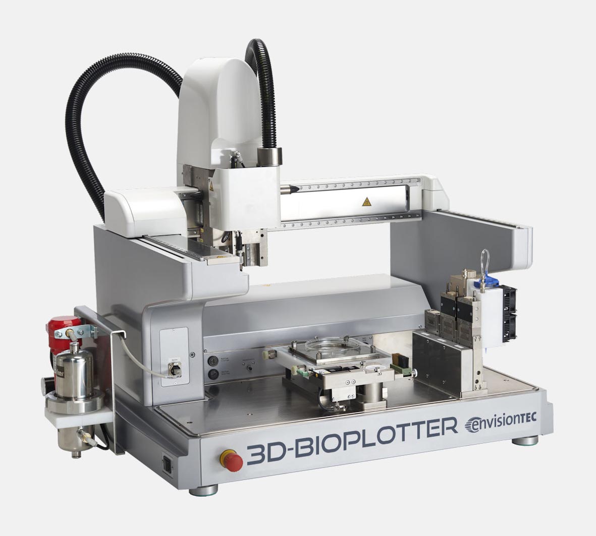
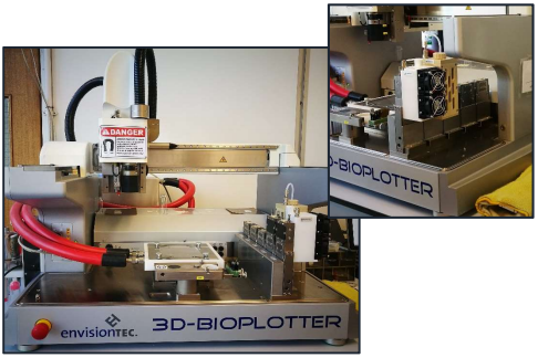
The 3D-Bioplotter
The 3D-Bioplotter family of printers consists of three models: the Starter series, the Developer series and the Manufacturer series, each with increasing capabilities. The original 3D-Bioplotter is now in its fourth generation, and more than 15 years of hardware and software development have gone into it.
One of the frequent users of the 3D-Bioplotter, Teja Guda, Assistant Professor of Biomedical Engineering at the University of Texas at San Antonio, said recently that “what’s so unique about the printer is that it is capable of printing living cells within the material as you print it.”
The modular 3D printer is easy to use while being capable of advanced research at the same time. The Bioplotter series prints with open-source biomaterials, using air or mechanical pressure to extrude them through a variety of syringes. Both new heads are currently on display in Dresden, at the EnvisionTEC booth, where attendees can see them in action and learn more about the research these new additions will make possible.
[Images: EnvisionTEC]
The post Envisiontec 3D-Bioplotter: New Bioprinting Capabilities appeared first on 3DPrint.com | The Voice of 3D Printing / Additive Manufacturing.