Rice Researchers 3D Print with Lasers and Sugar to Build Complex Vascular Networks

A team of researchers from Rice University has uncovered a promising strategy to generate vascular networks, one of the most daunting structures in the human body. Using powdered sugar and selective laser sintering, the researchers were able to build large structures from complex, branching, and intricate sugar networks that dissolve to create pathways for blood in lab-grown tissue.

This is the team’s latest effort to build complex vascular networks for engineered tissues to show that they could keep densely packed cells alive for two weeks. The findings of their study—published in the Nature Biomedical Engineering journal—prove that developing new technologies and materials to mimic and recapitulate the complex hierarchical networks of vessels gets them closer to providing oxygen and nutrients to a sufficient number of cells to get a meaningful long-term therapeutic function.

“One of the biggest hurdles to engineering clinically relevant tissues is packing a large tissue structure with hundreds of millions of living cells,” said study lead author Ian Kinstlinger, a bioengineering graduate student at Rice’s Brown School of Engineering. “Delivering enough oxygen and nutrients to all the cells across that large volume of tissue becomes a monumental challenge. Nature solved this problem through the evolution of complex vascular networks, which weave through our tissues and organs in patterns reminiscent of tree limbs. The vessels simultaneously become smaller in thickness but greater in number as they branch away from a central trunk, allowing oxygen and nutrients to be efficiently delivered to cells throughout the body.”

Overcoming the complications of 3D printing vascularization has remained a critical challenge in tissue engineering for decades, as only a handful of 3D printing processes have come close to mimic the in vivo conditions needed to generate blood vessels. Without them, the future of bioprinted organs and tissues for transplantation will remain elusive. Many organs have uniquely intricate vessels, like the kidney, which is highly vascularized and normally receives a fifth of the cardiac output, or the liver, in charge of receiving over 30% of the blood flow from the heart. By far, kidney transplantation is the most common type of organ transplantation worldwide, followed by transplants of the liver, making it crucial for regenerative medicine experts to tackle vascularization.

Ian Kinstlinger with a blood vessel template he 3D printed from powdered sugar (Credit: Jeff Fitlow/Rice University)

In the last few years, extrusion-based 3D printing techniques have been developed for vascular tissue engineering, however, the authors of this study considered that the method presented certain challenges, which led them to use a customized open-source, modified laser cutter to 3D print the sugar templates in the lab of study co-author Jordan Miller, an assistant professor of bioengineering at Rice.

Miller began work on the laser-sintering approach shortly after joining Rice in 2013. The 3D printing process fuses minute grains of powder into solid 3D objects, making possible some complex and detailed structures. In contrast to more common extrusion 3D printing, where melted strands of material are deposited through a nozzle, laser sintering works by gently melting and fusing small regions in a packed bed of dry powder. According to Miller, “both extrusion and laser sintering build 3D shapes one 2D layer at a time, but the laser method enables the generation of structures that would otherwise be prone to collapse if extruded.”

“There are certain architectures—such as overhanging structures, branched networks and multivascular networks—which you really can’t do well with extrusion printing,” said Miller, who demonstrated the concept of sugar templating with a 3D extrusion printer during his postdoctoral studies at the University of Pennsylvania. “Selective laser sintering gives us far more control in all three dimensions, allowing us to easily access complex topologies while still preserving the utility of the sugar material.”

Assistant professor of bioengineering at Rice University, Jordan Miller (Credit: Jeff Fitlow/Rice University)

Generating new 3D printing processes and biomaterials for vascularization is among the top priorities for the researchers at Miller’s Bioengineering Lab at Rice. The lab has a rich history of using sugar to construct vascular network templates. Miller has described in the past how sugar is biocompatible with the human body, structurally strong, and overall, a great material that could be 3D printed in the shape of blood vessel networks. His original inspiration for the project was an intricate dessert, even going as far as suggesting that “the 3D printing process we developed here is like making a very precise creme brulee.”

To make tissues, Kinstlinger chose a special blend of sugars to print the templates and then filled the volume around the printed sugar network with a mixture of cells in a liquid gel. Within minutes, the gel became semisolid and the sugar dissolved and flushed away to leave an open passageway for nutrients and oxygen. Clearly, sugar was a great choice for the team, providing an opportunity to create blood vessel templates because it is durable when dry, and it rapidly dissolves in water without damaging nearby cells.

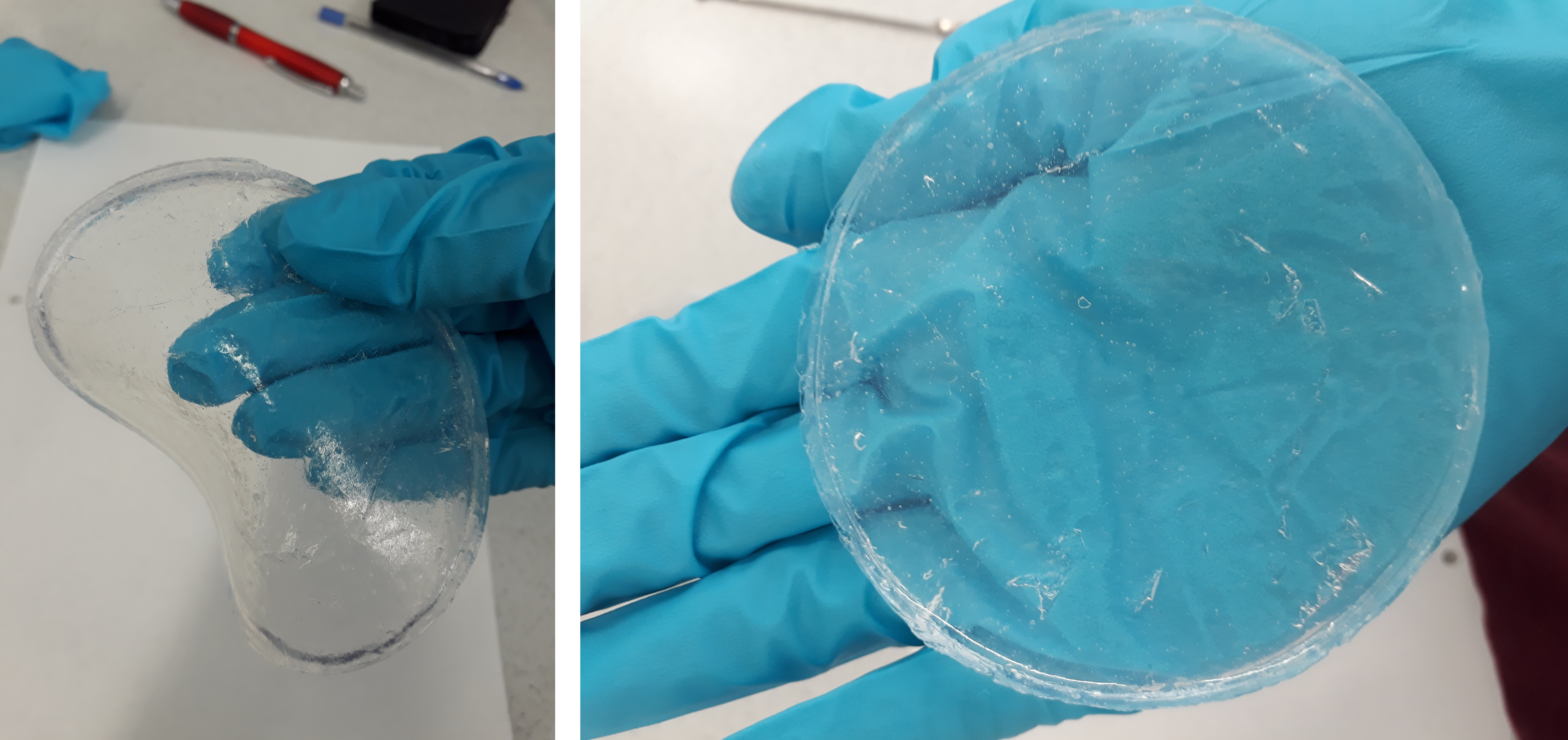
A sample of blood vessel templates that Rice University bioengineers 3D printed using a special blend of powdered sugars. (Credit: B. Martin/Rice University)

In order to create the treelike vascular architectures in the study, the researchers developed a computational algorithm in collaboration with Nervous System, a design studio that uses computer simulation to make unique art, jewelry, and housewares that are inspired by patterns found in nature. After creating tissues patterned with these computationally generated vascular architectures, the team demonstrated the seeding of endothelial cells inside the channels and focused on studying the survival and function of cells grown in the surrounding tissue, which included rodent liver cells called hepatocytes.

The hepatocyte experiments were conducted in collaboration with the University of Washington (UW)’s bioengineer and study co-author Kelly Stevens, whose research group specializes in studying these delicate cells, which are notoriously difficult to maintain outside the body.

“This method could be used with a much wider range of material cocktails than many other bioprinting technologies. This makes it incredibly versatile,” explained Stevens, an assistant professor of bioengineering in the UW College of Engineering, assistant professor of pathology in the UW School of Medicine and an investigator at the UW Medicine Institute for Stem Cell and Regenerative Medicine.

The results from the study allowed the team to continue their work towards creating translationally relevant engineered tissue. Using sugar as a special ingredient and selective laser sintering techniques could help advance the field towards mimicking the function of vascular networks in the body, to finally deliver enough oxygen and nutrients to all the cells across a large volume of tissue.

Miller considered that along with the team they were able to prove that “perfusion through 3D vascular networks allows us to sustain these large liverlike tissues. While there are still long-standing challenges associated with maintaining hepatocyte function, the ability to both generate large volumes of tissue and sustain the cells in those volumes for sufficient time to assess their function is an exciting step forward.”

The post Rice Researchers 3D Print with Lasers and Sugar to Build Complex Vascular Networks appeared first on 3DPrint.com | The Voice of 3D Printing / Additive Manufacturing.

FRESH News: SLAM Used to Fabricate Complex Hydrogel Structures With Gradients

There has been plenty of research on creating 3D printed hydrogels and using them to fabricate functional tissues. Biopolymer hydrogels, with properties that can be tailored and controlled, can be crosslinked to replicate tissue structures, and extrusion-based 3D printing is often used. But, the use of biopolymer hydrogels as 3D printing bioinks is tough, due to issues like low viscosity and trouble controlling microstructure variations. Some researchers have turned to embedded 3D printing methods, but this comes with its own laundry list of problems, such as having difficulty extracting the final product.

UK researchers Jessica J. Senior, Megan E. Cooke, Liam M. Grover, and Alan M. Smith from the University of Huddersfield and University of Birmingham created a method called suspended layer additive manufacturing, or SLAM, that can extrude low viscosity biopolymers into a self‐healing fluid‐gel matrix. The team recently published a paper on their work, titled “Fabrication of Complex Hydrogel Structures Using Suspended Layer Additive Manufacturing (SLAM).” It is worthwhile to note here that FRESH (Freeform Reversible Embedding of Suspended Hydrogels) is a super remarkably similar if not identical technology. So far we’re not getting involved with calling out which term should win here but we are leaning towards using FRESH because it will make it simpler for everyone going forward.

A schematic showing the production of a 3D bioprinted scaffold by use of SLAM.

The abstract states, “There have been a number of recently reported approaches for the manufacture of complex 3D printed cell‐containing hydrogels. Given the fragility of the parts during manufacturing, the most successful approaches use a supportive particulate gel bed and have enabled the production of complex gel structures previously unattainable using other 3D printing methods. The supporting gel bed provides protection to the fragile printed part during the printing process, preventing the structure from collapsing under its own weight prior to crosslinking. Despite the apparent similarity of the particulate beds, the way the particles are manufactured strongly influences how they interact with one another and the part during fabrication, with implications to the quality of the final product. Recently, the process of suspended layer additive manufacture (SLAM) is demonstrated to create a structure that recapitulated the osteochondral region by printing into an agarose particulate gel. The manufacturing process for this gel (the application of shear during gelation) produced a self‐healing gel with rapid recovery of its elastic properties following disruption.”

SLAM works like this: shear cooling a hot agarose solution throughout the sol–gel transition creates the fluid-gel print bed, as fluid gels behave like liquids once stress is applied. Then, the solution is put into a container in order to support the scaffold. Hydrogel and cells are mixed to produce a bioink, which is added to a bioprinter cartridge and extruded into the self-healing, fluid-gel matrix. The bioink is then suspended in its liquid state, and solidification is induced through crosslinking and cell media, which also provides the cell scaffold with metabolites. The construct is released from the supporting gel through low shear washing with deionized water.

The method prevents the initiation of gelation during 3D printing, which allows for great layer integration and “the production of constructs from two or more different materials that have dissimilar physicochemical and mechanical properties,” which creates a part with anisotropic behavior.

“To demonstrate clinical application, we recently created a structure that recapitulated the osteochondral region (the microstructure of which changes across a hard/soft tissue interface) as directed by microcomputed tomography (micro‐CT) imaging to provide accurate dimensions and was tailored to support specific cell phenotypes by controlling the microenvironment,” the researchers explained. “These complex scaffolds feature mechanical gradients that were similar to those found within the ECM and play a crucial role in preventing mechanical failure between interconnecting tissues as well as maintaining cell phenotype.”

The researchers needed to consider the mechanical properties of the fluid-gel print bed during SLAM 3D printing, as “they can impact on construct resolution and complexity.”

“Another embedded printing technique, freeform reversible embedding of suspended hydrogels (FRESH), developed by Hinton et al., uses a gelatin slurry support bath, however the rheological behavior of such material as a suspending agent for 3D bioprinting has not been investigated in depth,” the researchers wrote.

Additionally, they prepared fluid gels at different concentrations of agarose in order to find the best formulation for 3D printing uniform particle sizes, and investigated using needles with differing inner diameters, and a low viscosity dye solution, to find the optimal print resolution.

Optimizing print parameters within agarose fluid gel. A–G) Resolution of bioink printed within fluid bed support using multiple needle diameters and H–J) diffusion of dye through the gel at given time points.

“As the needle inner diameter was increased, the potential resolution decreased. Further, with larger needles, the printable filament thickness was more variable. This is likely due to both more material being extruded and also greater deformation of the fluid‐gel print bed. In very low viscosity solutions of low Mw (molecular weight), diffusion is also a limiting factor for resolution,” the researchers explained.

A) Intricate lattice prior to (left) and following extraction (right) from the bed. B) T7 intervertebral disc as CAD file (left) and lateral (middle) and apical (right) views. C) Intricate bulk structure in the form of a gellan spider. D) Carotid artery as CAD file (left) and during printing (right). D) Tubular structure (left) demonstrating material durability (middle) and perfusibility.

Alginate, collagen, gellan gum, and i‐carrageenan bioinks were used to demonstrate how many complex structures could be made. An intricate lattice structure showed off the scale and complexity that SLAM can achieve, while a T7 intervertebral disc was manufactured to show how the system can print large bulk structures and a spider was an example of 3D printing smaller, more intricate parts.

Several hollow and bifurcating structures, like a carotid artery model and a thick-walled tubular structure, were printed to show how the system can create geometries which are impossible without a 2D collector.

“These structures highlight the capability of this technique for freeform fabrication as large overhanging structures can be printed without the need for additional support structures,” the researchers explained.

The SLAM method can also deposit multiple layers laterally, horizontally, and within “a previously deposited extrude,” which allows constructs to be fabricated with the same biochemical and mechanical gradients that can be found in native tissues. The technique also uses microextrusion to continuously dispense bioinks, which caused better dispensing precision than the use of inkjet printing, and allows for more freedom with cell densities inside the bioink.

“Previous studies have indicated that the use of inkjet printers enables a reduction in print‐induced shear stresses applied to suspended cell populations compared with microextrusion methods, however, there are key features of the supporting bed utilized for SLAM that enable shear minimization utilizing microextrusion,” the researchers wrote.

Due to SLAM’s supporting fluid‐gel bed, low viscosity bioinks can be used – material viscosity can cause shear induction in bioinks, so it’s optimal for fabricating hydrogels.

“Using our system, it has therefore been demonstrated that the issues associated with cell shearing during microextrusion can be easily reduced, achieving admirably low shear stresses on cells that rival those seen during other forms of biofabrication including drop‐on‐demand techniques such as inkjet printing,” the team wrote.

A,B) Bilayer scaffolds using combinations of collagen‐alginate and collagen‐gellan. C) Large collagen‐core gellan‐shell scaffold and D) small collagen‐core alginate‐shell scaffold. E) Schematic of diagram showing control of cell behavior with attachment motif bearing complexes in the upper collagen gel and no attachment motifs for cell suspension within an alginate gel. F) Micro‐CT showing gradient porosity within a lyophilized collagen‐alginate scaffold. G) Confocal micrographs of Hoechst/actin cell staining of HDFs attached in the collagen layer and suspended in the alginate regions of a dual layer scaffold. H) Stress versus showing variations in gel strength and elasticity across a collagen‐alginate scaffold.

The SLAM method can also incorporate multiple biopolymer hydrogels into a single structure, which is important to “satisfy the mechanical, chemical, and biological variations that occur throughout native tissue.” The researchers demonstrated this capability by 3D printing an osteochondral construct, with ex vivo chondrocytes deposited into a gellan gum layer and osteoblasts into one of gellan‐hydroxyapatite. But they went even further, and used SLAM to 3D print integrated structures with different chemistries and gelatin mechanisms.

“Ionotropically gelled (alginate, gellan, and ι‐carrageenan) and thermally gelled (collagen) biopolymers were successfully integrated to form interfacing, dual‐phase scaffolds,” the researchers wrote.

The materials blended enough that mechanical failure did not occur – an environment that closely mimics the native tissue environment.

“Furthermore, this technique of printing integrated layered structures is not only compliant to printing different materials layer upon layer, but also deposition of a second material into the center of another. For example, in addition to producing layered constructs, it was possible to create 3D printed core–shell structures comprising a cylindrical core of collagen encapsulated within a gellan or alginate cylinder with various dimensions,” they continued.

“Another advantage of being able to deposit scaffold material precisely is that cell behavior can be spatially manipulated. Polymers such as collagens that are saturated with integrin binding domains allow cell attachment to the scaffold, whereas alginate and gellan do not naturally possess cell attachment motifs and instead, encapsulate cells with minimum attachment to the surrounding material.”

To learn more about the team’s use of SLAM to 3D print multilayer gradient scaffolds, I suggest you read the paper – they can explain it far better.

“In summary, we have demonstrated that the SLAM technique can be used to overcome the problems associated with using low viscosity bioinks in extrusion‐based bioprinting,” the researchers concluded. “The method enabled the successful fabrication of bulk, intricate, dual phase, and phase‐encapsulated hydrogels from a variety of biopolymer materials that are currently widely investigated in regenerative medicine. Furthermore, it was shown that controlled spatial gradients in mechanical and chemical properties can be produced throughout a single part with interface integrity between different materials. This allows for physicochemical properties of the structure to be designed accordingly with the ability to control porosity, mechanical gradients, cell distribution, and morphology.”

Discuss this and other 3D printing topics at 3DPrintBoard.com or share your thoughts below.

The post FRESH News: SLAM Used to Fabricate Complex Hydrogel Structures With Gradients appeared first on 3DPrint.com | The Voice of 3D Printing / Additive Manufacturing.

Nervous System Works with Rice University Researchers 3D Printing Vascular Networks

Nervous System has been heavily engaged in experimenting with 3D and 4D printing of textiles in the past years, and all their research is paying off now as they find themselves engaged in the realm of tissue engineering. The Somerville, MA company is known for their generative design process, combining both programming and art within most of their serious projects, drawing bioengineers from Rice University to turn to them for added expertise.

Assistant professor Jordan Miller invited the Nervous System team to join his researchers on an incredible journey to fabricate examples of possible vascular networks via bioprinting—harnessing their knowledge of software and materials to find a way to create soft hydrogels. Kind of not a phantom but more a path towards ideas that can lead to concepts that may let us build true vascularized structures at one point. As Miller explains, in their research they were able to create large tissue blocks easily, but as so many scientists engaged in bioprinting today have discovered before them, it is extremely challenging to keep cells alive. Viability becomes the goal, and as that becomes more comprehensively mastered overall in bioprinting, it may finally unlock the door to true fabrication of organs that can be transplanted into the human body.

Open-source technology, mainly centered around 3D printing has offered huge opportunity for the bioengineers from Rice University to make progress in their work—and that was what drew them to Nervous System in the first place. Jordan became ‘captivated’ with the structures they were creating, specifically in their Growing Objects series, which was featured as an exhibit at the Simons Center for Geometry and Physics in Stonybrook, NY in August and September of 2014. In speaking with Nervous System, his proposal involved what they describe as an ‘epic task,’ to create simulated synthetic tissue and human organs.

Rendering showing lung-mimicking structures generated within different volumes

“The idea of taking our generative systems which are inspired by nature and using them to actually make living things was a dream come true,” states the Nervous System team in their case study.

Elsewhere the research did,

“…show that natural and synthetic food dyes can be used as photoabsorbers that enable stereolithographic production of hydrogels containing intricate and functional vascular architectures. Using this approach, they demonstrate functional vascular topologies for studies of fluid mixers, valves, intervascular transport, nutrient delivery, and host engraftment.”

As Miller and his expanding team continued to work on developing the necessary tools for bioengineering, part of their research resulted in a new 3D printing workflow called SLATE (stereolithography apparatus for tissue engineering). Their proprietary hardware can bioprint cells encased in soft gels that act just like vascular networks. Nervous System accompanied them (going back as far as 2016) in this bioprinting evolution by designing the materials for the networks—but with their background in programming, the contribution went far beyond designed materials and included customized software for creating ‘entangled vessel networks.’ These networks can be connected to both inlets and outlets for oxygen and blood flow, as they use specific algorithms to ‘grow’ the branching airways.

“Air is pumped into the network and it pools at the bulbous air sacs which crown each tip of the network,” states Nervous System in their case study. “These sacs are rhythmically inflated and deflated by breathing action, so called tidal ventilation because the air flow in human lungs is reminiscent of the flows of the ocean tides.

“Next we grow dual networks of blood vessels that entwine around the airway. One to bring deoxygenated blood in, the other to carry oxygen-loaded blood away. The two networks join at the tips of the airway in a fine mesh of blood vessels which ensheathes the bulbous air sacs. These vessels are only 300 microns wide!”

This project, bringing together scientists and art designers, was featured in the American Association for the Advancement of Science (AAAS) in ‘Multivascular networks and functional intravascular topologies within biocompatible hydrogels,’ authored by Bagrat Grigoryan, Samantha J. Paulsen, Daniel C. Corbett, Daniel W. Sazer, Chelsea L. Fortin, and Alexander J. Zaita.

The recently published article goes into great detail about SLATE 3D printing, indicating that this hardware is capable of rapid bioprinting, and offering possible sustainability to human cells—along with maintaining functionality of stem cells and necessary differentiation.

The project was created by Jordan Miller at Rice University and Kelly Stevens at the University of Washington, and included 13 additional collaborators from Rice, University of Washington, Duke University, and Rowan University.

Nervous System is undeniably one of the most fascinating companies producing 3D printed innovations today. Their versatility has led them to create everything from 4D textiles and 3D printed stretched fabrics to their famed Kinematics Petal Dress. With their latest project delving into 3D printed tissue, the stakes become higher—and their impact on the world much greater. Find out more here.

What do you think of this news? Let us know your thoughts! Join the discussion of this and other 3D printing topics at 3DPrintBoard.com.

The Miller Lab fabricated and tested the architectures we generated showing that they can withstand more than 10,000 ventilation cycles while being perfused with human red blood cells. Study of the printed gels shows that the architecture we designed promotes red blood cells mixing and bidirectional flow which is hypothesized to occur in the human lung.

[Source / Images: Nervous System]

Tel Aviv University: Researchers 3D Print Cardiac Patches & Cellularized Hearts

Researchers at Tel Aviv University continue to try to meet the ongoing challenges in cardiac tissue engineering. In ‘3D Printing of Personalized Thick and Perfusable Cardiac Patches and Hearts,’ authors Nadav Noor, Assaf Shapira, Reuven Edri, Idan Gal, Lior Wertheim, and Tal Dvir outline the steps they took to match technology with tissue.

Cardiovascular disease is the leading killer of patients in the US, and organ donor and transplantation processes can still mean a long wait for those suffering from heart failure. Here, the authors demonstrate the need for alternative ways to treat the infarcted (usually referring to clogging of one of more arteries) heart. And while tissue engineering has pointed the way to freeing many patients from terrible physical suffering and organ donor waiting lists, creating the necessary scaffolds with true biocompatibility has presented obstacles.

The authors have created an engineered cardiac patch meant to be transplanted directly onto the patient’s heart, integrating into the ‘host,’ with excess biomaterials degrading over time. This leaves the cardiac patch, full of live, healthy tissue, regenerating a previously defective heart. Because there is always the threat of rejection when implanting anything into the body though, the authors emphasize the need for appropriate materials:

“Most ideally, the biomaterial should possess biochemical, mechanical, and topographical properties similar to those of native tissues,” state the researchers. “Decellularized tissue‐based scaffolds from different sources meet most of these requirements. However, to ensure minimal response of the immune system, completely autologous materials are preferred.”

The researchers were able to create patient-specific cardiac patches in their recent study, extracting fatty tissue from cardiac patients—and then separating cellular and a-cellular materials.

“While the cells were reprogrammed to become pluripotent stem cells, the extra‐cellular matrix (ECM) was processed into a personalized hydrogel,” stated the researchers.  “Following mixture of the cells and the hydrogel, the cells were efficiently differentiated to cardiac cells to create patient‐specific, immunocompatible cardiac patches.”

In using the patient-specific hydrogel as bioink, the researchers were able to create patches, but ultimately, they were also able to 3D print comprehensive tissue structures that include whole hearts.

An omentum tissue is extracted from the patient and while the cells are separated from the matrix, the latter is processed into a personalized thermoresponsive hydrogel. The cells are reprogrammed to become pluripotent and are then differentiated to cardiomyocytes and endothelial cells, followed by encapsulation within the hydrogel to generate the bioinks used for printing. The bioinks are then printed to engineer vascularized patches and complex cellularized structures. The resulting autologous engineered tissue can be transplanted back into the patient, to repair or replace injured/diseased organs with low risk of rejection.

The authors used two different models in their study, with one serving as proof-of-concept, with pluripotent stem cells (iPSCs)‐derived cardiomyocytes (CMs) and endothelial cells (ECs). The other model relied on:

  • Rat neonatal CMs
  • Human umbilical vein endothelial cells (HUVECs)
  • Lumen‐supporting fibroblasts

One bioink, laden with cardiac cells, printed parenchymal tissue, while the other extruded cells for forming blood vessels. The researchers were successful in 3D printing the patient-specific cardiac patches but found when a higher degree of complexity was necessary for fabrication of organs or other tissues, the hydrogels were not strong enough. They created a new process for organs and more complex tissues where they could print in a free-form manner and cure structures at varying temperatures; they were able to overcome previous challenges and 3D print accurate, personalized structures.

Bioinks characterization. A human omentum a) before and b) after decellularization. c) A personalized hydrogel at room temperature (left) and after gelation at 37 °C (right). d) A SEM image of the personalized hydrogel ultrastructural morphology, and e) a histogram of the fibers diameter. f) Rheology measurements of 1% w/v and 2.5% w/v omentum hydrogels, showing the gelation process upon incubation at 37 °C. g) Stromal cells originated from human omental tissues were reprogrammed to become pluripotent stem cells (red: OCT4, green: Ki67 and blue: nuclei). h) Differentiation to ECs as determined by CD31 (green) and vimentin staining (red). Differentiation to cardiac lineage: i) staining for sarcomeric actinin (red), j) staining for NKX2‐5 (red), and TNNT2 (green). Scale bars: (e) = 10 µm, (g,i,j) = 50 µm, (h) = 25 µm.

This study carries substantial weight, considering the researchers were able to create cellularized hearts with ‘natural architectures.’ This furthers the potential for cardiac transplants after heart failure, along with encouraging the process for drug screening. The authors point out that more long-terms studies and research with animal models are necessary.

“Although 3D printing is considered a promising approach for engineering whole organs, several challenges still remain,” conclude the researchers. “These include efficient expansion of iPSCs to obtain the high cell number required for engineering a large, functioning organ. Additionally, new bioengineering approaches are needed to provide long‐term cultivation of the organs and efficient mass transfer, while supplying biochemical and physical cues for maturation.”

“The printed blood vessel network demonstrated in this study is still limited. To address this challenge, strategies to image the entire blood vessels of the heart and to incorporate them in the blueprint of the organ are required. Finally, advanced technologies to precisely print these small‐diameter blood vessels within thick structures should be developed.”

Imaging of the heart and patch modeling. CT image of a) a human heart and b) left ventricle coronary arteries. c) A model of oxygen concentration profile in an engineered patch. d) Replanning of the model showed better oxygen diffusion, sufficient to support cell viability.

Without good heart health, it is very difficult to survive. Responsible for transporting nutrients, oxygen, and more to cells populating the human body, the heart also removes waste like carbon dioxide and more. 3D printing is assisting scientists and doctors in researching and treating a variety of different diseases and conditions, whether they are using 3D printed metamaterials for fabricating heart valves, creating better cardiac catheters, or experimenting with new types of phantoms.

What do you think of this news? Let us know your thoughts! Join the discussion of this and other 3D printing topics at 3DPrintBoard.com.

Printing thick vascularized tissues. a) A top view of a lumen entrance (CD31; green) in a thick cardiac tissue (actinin; pink). b) A model of a tripod blood vessel within a thick engineered cardiac tissue (coordinates in mm), and c) the corresponding lumens in each indicated section of the printed structure. d) Tissue perfusion visualized from dual viewpoints. e–k) A printed small‐scaled, cellularized, human heart. e) The human heart CAD model. f,g) A printed heart within a support bath. h) After extraction, the left and right ventricles were injected with red and blue dyes, respectively, in order to demonstrate hollow chambers and the septum in‐between them. i) 3D confocal image of the printed heart (CMs in pink, ECs in orange). j,k) Cross‐sections of the heart immunostained against sarcomeric actinin (green). Scale bars: (a,c,h, i,j) = 1 mm, (g) = 0.5 cm, (k) = 50 µm.

[Source / Images: 3D Printing of Personalized Thick and Perfusable Cardiac Patches and Hearts]

Bioprinting 101: Part 4 – Alginate


Alginate is derived from brown algae

Alginate is a polysaccharide distributed widely in cell walls of brown algae. When it bonds with water it typically forms a viscous gum. Alginate absorbs water quickly, which makes it useful as an additive. Alginate can be used as a hydrogel as well. It has very interesting applications. It is a hydrogel consisting of microparticles or in bulk gels combined with nerve growth factor in bioengineering research. It has been used as scaffolding for tissue engineering, as a delivery vehicle for drugs, and as model extracellular matrices for basic biological studies. These applications require tight control of a number of material properties including mechanical stiffness, swelling, degradation, cell attachment, and binding or release of bioactive molecules.

The chemical structure of alginate is composed of two types of uronic acid: Mannuronic acid unit (M) and Guluronic acid unit (G). Differences in M/G ratio and block configuration account for the differences in alginate properties and functionality, especially in gelling capability and gel strength. The M/G ratio is dependent upon such factors as is the species of seaweed (imagine the difference of a species found near water around remote East Asian Islands vs. seaweed found in a nice tropical location near the Bahamas), the part of the seaweed used, the harvest location, and the harvesting season. The carboxyl groups within the M and G units are easily ion-exchanged, and can react with several kinds of cations. Cations are essential to consider when it comes to material strength.

Alginate Bioink

Alginates have been used as scaffolds for tissue engineering extensively. We have discussed hydrogels previously within this series, but what makes an alginate different? Alginate based products possess excellent shear-thinning properties, and can be easily extruded through a nozzle. There they also have good initial shape fidelity. Alginate is widely used for most bioprinting extrusion processes due in part to this. There is also a focus on alginate due to the ability to mix materials readily with these extrusion methods. Alginate is biodegradable, has controllable porosity, and may be linked to other biologically active molecules. Encapsulation of certain cell types into alginate beads may actually enhance cell survival and growth. Outside of biodegradability, alginates are also non-toxic. This is vital for biomedical purposes.

Sodium Alginate Chemical Structure

Alginate is a great biomaterial. As with the other biomaterials we have analyzed, it is typically used in combination with other materials. This is related to what we previously mentioned in this article in terms of bioreactivity. It allows a material scientist or engineer to experiment and make substances with various tensile strengths and viscosity levels. With the benefits of alginate, there come some major cons. The most important thing to consider is how much water a hydrogel derived from alginate can contain. Depending on the hydrophilicity, a hydrogel from alginate may not be able to maintain absorbance levels for larger scale builds. This is why we cannot make a bioink or hydrogel from solely alginate. Stay tuned for info on more materials used within bioprinting. This field has such variety within it, so we will do our best to shed a light on important materials and processes.

This article is part of a series that ultimately wants to update 3DPrint.com readers in the most vital and relevant information in bioprinting. We hope that this will prompt people to bioprint at home which in turn may accelerate the bioprinting revolution. Essentially, this article has as its goal to kickstart a DIY bioprinting revolution. Please spread the word. The first article Bioprinting 101 is here, while Part Two Hydrogels is here and part three Industrial printers is here.

Bioprinting 101: Part 2 Hydrogels


hydrogels

Simple Hydrogel

A hydrogel is a macromolecular polymer gel constructed of a network of crosslinked polymer chains. Hydrogels are synthesized from hydrophilic monomers by either chain or step growth, along with a functional crosslinker to promote network formation. A net-like structure along with void imperfections enhance the hydrogel’s ability to absorb large amounts of water via hydrogen bonding. As a result, hydrogels are self-healing in nature. They also develop firm yet have great elastic mechanical properties. Self-healing refers to the spontaneous formation of new bonds when old bonds are broken within a material. The ability to self-heal is very attractive to bio-printing and systems related to the body. Human biology is a system based on reproductive and self-healing components, thus the need for self-healing agents and materials is paramount.

The chemical structure of a hydrogel allows for a variety of chemical reactions to occur. Depending on their composition, they are responsive to various stimuli including heating, pH, light, and other chemical agents. The important property to understand within a hydrogel is the ability to capture water while simultaneously maintaining its chemical bonding structure and therefore its three-dimensional structure.

Engineered Hydrogel

The stability of a three-dimensional structure is important for 3D printing. The main usage of hydrogels within bioprinting are as scaffolds within tissue engineering. Tissue engineering is defined as a combination of materials, engineering, and cells to improve or replace biological organs. This needs finding proper types of cells and culturing them in a suitable scaffold under appropriate conditions. Hydrogels are an appealing scaffold material because their structures are similar to the extracellular matrix of many tissues, they can often be processed under relatively mild conditions, and they may be delivered in a minimally invasive manner.


Synthetic Hydrogel Scaffold

In terms of bio-engineering, scaffold creation with hydrogels can be done through two methods: natural derivation or synthetic derivation. For a 3D bio-printing enthusiast, it is beneficial to have a synthetic method. These methods are easy to control in terms of chemistry and structure. This allows for manipulable properties. Synthetic bio-engineering is the most probable route for anything 3D bio-printing related within a home setting. It is easier to experiment within this context. It is also easier to create specific skin properties for different areas of the body with easily manipulable synthetic hydrogels.

In summary, the benefits of hydrogels and 3D bio-printing are tremendous in terms of easily engineer-able materials within synthetic methods. This allows for a variety of uses for hydrogel materials. Within the context of materials it is optimal and gives us a lot of flexibility on its own. We will discuss in our next article different 3D bio-printing setups. This will also allow us to understand hydrogels in the context of a machine and why they are beneficial, as well as some limitations that need to be understood.

This article is part of a series Bioprinting 101 which hopes to teach people how to bioprint in the home, the first article is here.

3D Printing Cell Cultures Using PulMA Hydrogels

Biomimetic hydrogels offer several opportunities in medical applications, according to a group of researchers in a paper entitlted “Polysaccharide hydrogels for multiscale 3D printing of pullulal scaffolds.” But the generation of synthetic microenvironments that simulate the effects of natural tissue niches on cell growth and differentiation requires new methods to control hydrogel feature resolution, biofunctionalization and mechanical properties. In the paper, the researchers show how this can be achieved by using a 3D printable pullulan-based hydrogel with tunable mechanical properties.

Pullulan is a non-ionic linear polysaccharide that is naturally produced from starch. It has many advantages: it is biodegradable, edible, bio- and blood-compatible, non-toxic, non-immunogenic, non-mutagenic and non-carcinogenic, as well as being easily soluble in water, which allows a clear and viscous hydrogel to be prepared. It has been proposed as an anti-adhesive barrier layer for the prevention of postoperative problems, and is widely used in cosmetics. It has anti-free-radical properties and has been used in nanofibers or coatings for food preservation, as well as in applications in tissue engineering and regenerative medicine.

“We reasoned that pullulan could be custom modified on its hydroxyl groups with desired chemical moieties (for example chemical polymerizable groups) in order to generate 3D crosslinked structures of tunable mechanical properties,” the researchers state. “Indeed, in spite of these attractive chemical features, the potential of pullulan for the generation of scaffolds or culture systems with specific 2D and 3D shapes and morphologies has so far remained unexplored.”

The researchers synthesized methacrylated pullulan (PulMA) hydrogels that had been printed by multiscale light assisted 3D printing techniques. Using stereolithography and two-photon lithography, they produced 3D patterns from millimeters down to a few microns as well as suspended structures.

“Using further engineering the material into a photosensitive formulation, we built complex 3D shapes through spatially controlled irradiation (SL), overcoming current limitations in structuring along the third dimension,” they continue. “Mechanical properties, in particular rigidity, were controlled by the addition of a bifunctional crosslinker that enabled to tune elastic modulus and water absorption of PulMA hydrogels.”

Cell lines and mesenchymal stem cells (MSCs) were seeded both on micro- and macro-metric 3D structures to test biological response. Testing showed that the cells were viable and metabolically active, adhering only when in the presence of fibronectin or fibrin functionalization.

“As the integration of multi-scale patterns in the same structure can be an important step forward in tissue engineering and regenerative medicine fields and hydrogel fabrication with these two different scale sizes and technologies have not commonly reported, this study confirms PulMA hydrogels as excellent substrate supporting cell culture after proper functionalization, with endowed mechanical properties and 3D printing feature sizes,” the researchers conclude. “On the other hand, the non-adhesive property of PulMA could allow to fine tuning of adhesive areas and patterning of individual cells and of monolayers in both 2D and 3D, giving vast opportunities for cell engineering through microfabrication. In future, fabrication by SL and TPL in presence of pre-embedded cells is potentially feasible with PulMA at no or low toxicity given the use of visible and infrared radiation, respectively.”

Authors of the paper include G. Della Giustina, A. Gandin, L. Brigo, T. Panciera, S. Giulitti, P. Sgarbossa, Delfo D’Alessandro, Luisa Trombi, Serena Danti and G. Brusatin.

Discuss this and other 3D printing topics at 3DPrintBoard.com or share your thoughts below.


Creating Vascular Structures Using Low Cost Desktop 3D Printers

In a thesis entitled “Engineering of vascular networks within biocompatible hydrogels using 3D printing technology,” a PhD student named Juan Liu discusses the need for new technologies in wound healing. While skin flaps and grafts are the “gold standard” in clinical treatment of large skin and subcutaneous tissue defects, there are many complications that can arise from these treatments, and there is also the issue of not having enough skin to be able to harvest to cover particularly large wounds.

3D printing, however, using stem cells, allows for an unlimited amount of tissue to be created to heal large wounds. Using cells from the patient reduces the risk of rejection, as well. In order to create and maintain live tissue, however, vascularization is required, meaning that blood vessels need to be established, which is the tricky part of tissue engineering. Liu hypothesizes that open source desktop 3D printing technology can be used to design and fabricate “customized bio-artificial multicellular tissues with embedded vessel-like supply channels and corresponding bio-reactors for long-term 3D tissue culture.”

Liu outlines the following objectives:

  • Use open source software to design 3D tissues with vascular patterns and fabricate them with desktop 3D printers
  • Analyze cell survival and function over time in 3D tissues in respect to vascular patterns
  • Generate 3D printable bioreactors that allow online monitoring of the process
  • Prevent shrinking of 3D tissues
  • Generate multilayer 3D tissues with different cell types

Liu goes on to demonstrate how 3D printing technology can be used to precisely form vascular structures within cell-laden hydrogels. He also creates “customized PLA and PDMS bioreactors for continuous perfusion and real-time operation.” The vascular structures formed within the cell-laded hydrogels, which were created by 3D printing, were able to increase the viability of the cells surrounding the vascular structures.

“In addition, the final system based on PDMS has been proven to sustain long-term 3D cell culture, which is the basic for 3D cell proliferation and tissue formation in vitro,” says Liu. “Among others this approach exhibits unique advantages to fabricate a hydrogel-based multilayer vascular device in a cost-effective and fast manner, which has a huge potential for viable 3D cell culture, complex tissue engineering, disease modeling as well as drug screening.”

This model, he continues, could also be used to mimic natural tissue architecture and create bigger multilayer hydrogel constructs with corresponding layers of functional cells to study cell morphology, differentiation, and potential function of engineered tissue constructs.

“Further optimization of the hydrogel concentration in each layer, cell density and perfusion parameters may enable the preparation of 3D vascular tissues under the conditions mimicking the natural environment with better functions and matrix compositions,” he adds. “In order to overcome the tendency of shrinking in soft hydrogels, combination with eletrospun fibers are promising. The system presented in this thesis allows fabrication of vascular networks in both soft and stiff cell laden hydrogels. It may serve as a novel platform for vascularized tissue engineering, that facilitates the generation of more functional, engineered vascular tissue. It may be useful for studies of wound coverage and tissue regeneration and eventually aid the treatment of wounds.”

Discuss this and other 3D printing topics at 3DPrintBoard.com or share your thoughts below.


Marine-Based Hyrdogels Used to Develop New Bioink for 3D Bioprinting

One of the challenges in the field of bioprinting is developing bioinks that are safe and effective. In a paper entitled “Marine Biomaterial-Based Bioinks for Generating 3D Printed Tissue Constructs,” a group of researchers discusses the development of a bioink using alginate and fish gelatin (f-gelatin). They created a marine-based interpenetrating polymer network (IPN) consisting of alginate and f-gelatin methacryloyl (f-GelMA) networks via physical and chemical crosslinking methods, respectively.

“In this study, four different concentrations of alginate (1%, 2%, 3%, and 4%) and three low concentrations of f-GelMA (4%, 5%, and 6%) were investigated and found to form double networked alginate/f-GelMA hydrogels,” the researchers explain. “In the mechanical properties test, the pure alginate hydrogel showed a typical increase in mechanical strength with the increase of concentration and low mechanical strength when its compressive modulus was around 40 kPa, even at 4% alginate, compared with alginate/f-GelMA IPN hydrogel where the modulus of alginate/f-GelMA was approximately 40 kPa at 1% alginate.”

This showed that the mechanical strength of hydrogels was significantly increased by employing an alginate and f-GelMA double network. According to the researchers, the tunable mechanical strength range in alginate/f-GelMA hydrogel would be sufficient to meet the diverse requirements of different tissues.

The researchers also performed swelling tests with pure alginate hydrogel as a control group, and found that the mass swelling ratio decreased with the increase in concentration of alginate.

“For the alginate/f-GelMA hydrogel, the mass swelling ratio for all tested groups was lower than for the pure alginate group,” the researchers continue. “This was due to the increased crosslinking density from the addition of f-GelMA which generated additional polymeric networks via covalent bonding…The swelling properties of hydrogel mainly depend on the hydrogel pore size, polymer concentration, density of cross-linking, and the interaction with solvents.”

The researchers also investigated the degradation characteristics of the hyrdogels. The degradation rate of the alginate/f-GelMA in a saline solution was similar for 2% and 4% alginate, though the 2% degraded faster. The morphology of the hydrogels was tested as well, and the alginate/f-GelMA exhibited a highly porous structure, which can provide enough space for the transport of nutrients and gas exchange for cell survival.

“To assess the cell behavior and examine the feasibility (cell viability, adhesion, and cell proliferation) of alginate/f-GelMA hydrogel, cell adhesion and 3D cell encapsulation assays were performed to examine the ability to bind to alginate/f-GelMA scaffold which is crucial for cell survival,” the researchers state. “…Encapsulated NIH-3T3 cells were cultured for seven days and cell viability was determined using LIVE/DEAD assay kits. As shown in Figure 3C, cells maintained high viability during the culture period (one, three, five, and seven days) and demonstrated that cells can maintain long-term survival rates in alginate/f-GelMA hydrogel.”

The results of the testing showed that alginate/f-GelMA hydrogel has a lot of promise for tissue engineering applications, including 3D bioprinting. To further confirm the morphology and cell viability in the process of 3D printing, a two-layer scaffold was printed and Live/Dead assay was carried out to investigate the cell survival ratio. The scaffold displayed a satisfactory 3D arrangement under microscopy with high cell viability.

This study was the first incidence of using alginate and f-GelMA for 3D bioprinting, and the successful results mean that marine biopolymers could feasibly replace biopolymers from mammalian resources, which can carry diseases or be subject to religious restrictions.

Authors of the paper include Xiaowei Zhang, Gyeong Jin Kim, Min Gyeong Kang, Jung Ki Lee, Jeong Wook Seo, Jeong Tae Do, Kwonho Hong, Jae Min Cha, Su Ryon Chin and Hojae Bae.

Discuss this and other 3D printing topics at 3DPrintBoard.com or share your thoughts below. 


Researchers Build Inexpensive Open Source Bioprinter for 3D Printing Branching, Hydrogel-Based Vascular Constructs

While 3D bioprinting is not yet able to fabricate full human organs just yet, it can be used to manufacture several different kinds of human tissue, such as heart and bile duct. One of the main barriers of forming viable tissues for clinical and scientific use is the development of vasculature for engineered tissue constructs, mainly due to generating branching channels in hydrogel constructs that can later produce vessel-like structures after being seeded with endothelial cells.

But thanks to 3D bioprinting, it’s now possible to 3D print complex structures on multiple length scales within a single construct. This enables the generation of branching, interconnected vessel systems of small, vein-like microvessels and larger macrovessels, which couldn’t be done with former tissue engineering methods. However, the best sacrificial material for fabricating branching vascular conduits in constructs based in hydrogel has yet to be determined.

A team of researchers from the University of Toronto recently published a paper, titled “Generating vascular channels within hydrogel constructs using an economical open-source 3D bioprinter and thermoreversible gels,” in the Bioprinting journal. Co-authors of the paper include Ross EB FitzsimmonsMark S. Aquilino, Jasmine Quigley, Oleg ChebotarevFarhang Tarlan, and Craig A. Simmons.

The abstract reads, “The advent of 3D bioprinting offers new opportunities to create complex vascular structures within engineered tissues. However, the most suitable sacrificial material for producing branching vascular conduits within hydrogel-based constructs has not yet been resolved. Here, we assess two leading contenders, gelatin and Pluronic F-127, for a number of characteristics relevant to their use as sacrificial materials (printed filament diameter and its variability, toxicity, rheological properties, and compressive moduli). To aid in our assessment and help accelerate the adoption of 3D bioprinting by the biomedical field, we custom-built an inexpensive (< $3000 CAD) 3D bioprinter. This open-source 3D printer was designed to be fabricated in a modular manner with 3D printed/laser-cut components and off-the-shelf electronics to allow for easy assembly, iterative improvements, and customization by future adopters of the design. We found Pluronic F-127 to produce filaments with higher spatial resolution, greater uniformity, and greater elastic modulus than gelatin filaments, and with low toxicity despite being a surfactant, making it particularly suitable for engineering smaller vascular conduits. Notably, the addition of hyaluronan to gelatin increased its viscosity to achieve filament resolutions and print uniformity approaching that with Pluronic F-127. Gelatin-hyaluronan was also more resistant to plastic deformation than Pluronic F-127, and therefore may be advantageous in situations in which the sacrificial material provides structural support. We expect that this work to establish an economical 3D bioprinter and assess sacrificial materials will assist the ongoing development of vascularized tissues and will help accelerate the widespread adoption 3D bioprinting to create engineered tissues.”

3D Bioprinter Hardware.

Existing 3D bioprinters have different technical advantages and deposition methods, which influence their prices and available applications. Extrusion-based 3D printers are good for tissue engineering, but the cost is usually too high for the field to experience significant growth.

For this experiment, the researchers chose to create their own open source 3D bioprinter, which costs roughly $3,000 and can be used for lower resolution applications, such as 3D printing perfusable microvessels in tissue constructs.

Printer operational overview.

Both the chosen method and material have to meet a certain number of requirements to successfully 3D print complex branching vessel systems within hydrogel constructs. First, sacrificial materials, which need to be non-toxic and maintain a uniform filament diameter during printing, have to be deposited in the desired vascular design during printing, then flushed away once the construct is done.

In addition, the 3D printer needs to have enough resolution to print all the channels – even those that will act as the small artery vessels of ~0.5–1 mm. It also needs to be able to deposit at least two materials, though more is better when it comes to creating heterogeneous tissues with different regions of varying cell and hydrogel composition.

The team investigated formulations of gelatin and PF127 due to their potential advantages as sacrificial materials in hydrogel-based tissue constructs. Gelatin, which has been used in several biomedical applications, is a thermoreversible (the property of certain substances to be reversed when exposed to heat) biopolymer of several hydrolyzed collagen segments, and can be 3D printed at ~37 °C, which is a temperature compatible with cells.

PF127 is a surfactant, meaning that it could have potential cytotoxic effects on embedded cells. But, it has inverse thermal gelation, which means it can be 3D printed at an ambient temperature, and then removed at ~4 °C to create void vascular channels.

According to the paper, “By using our custom-built printer in order to assess the printability of these materials and assessing mechanical properties, we aimed to establish which may be the best option for creating branching vascular channels within engineered tissues.”

The team’s modular 3D bioprinter includes extruding systems, 3D printed out of ABS on a MakerBot 3D printer, which were designed specifically to hold commercially-available, sterile 10 mL syringes, instead of custom-made reservoirs that would need to be specially made and repeatedly sterilized. An open-source Duet v0.6 controller board controls the system, and the print heads are isolated from the XYZ movements executed by the lower part of the chassis.

Fabricating perfusable channels.

For testing purposes, water droplets were 3D printed in a defined pattern with each extruder system, and the average distance between the droplets’ centers in the X and Y directions were measured; then, the mean distances were compared to the pre-defined CAD model distances.

“In conclusion, we found that PF127 is generally superior to gelatin as a sacrificial material for creating vascularized tissues by merit of its filament uniformity during printing and its greater compressive modulus,” the paper concluded.

Discuss this and other 3D printing topics at 3DPrintBoard.com or share your thoughts in the Facebook comments below.