regenHU CEO: Bioprinting Will Strengthen OrganTrans Project to 3D Print Liver Organoid

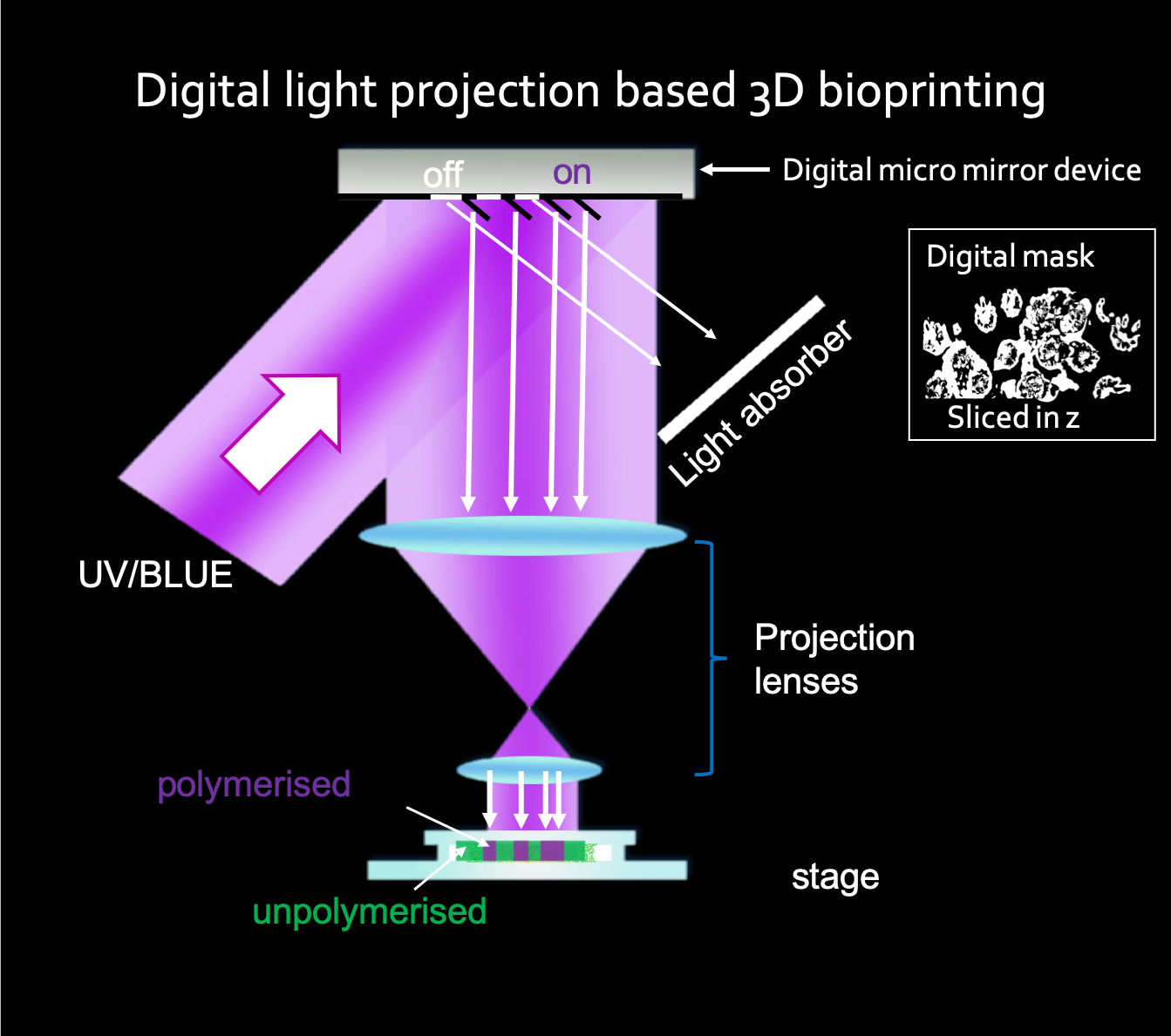
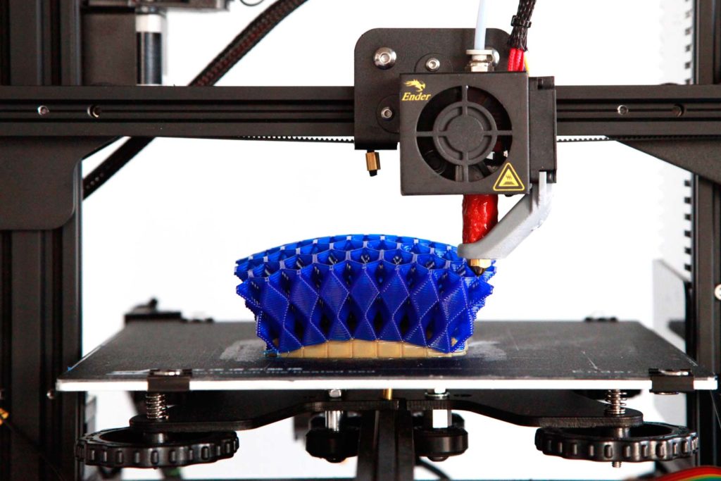
The European consortium OrganTrans is preparing to develop a tissue engineering platform capable of generating liver tissue. The proposed automated and standardized disruptive alternative solution to organ donation for patients with liver disease will stand on 3D bioprinting know-how from Swiss biomedical firm regenHU. Coordinated by Swiss research and development center CSEM, the eight partners and two transplantation centers engaged in the consortium will be using regenHU’s 3D bioprinters to produce organoid-based liver constructs with organoid laden bioinks.

In April 2020, we reported that OrganTrans would tackle the important healthcare challenge of end-stage liver disease (ESLD) by capitalizing on advancements in the regenerative medicine field, like using biofabricated liver tissue, to develop an entire value chain from the cell source to tissue engineering, biofabrication, post-processing and testing, and liver transplantation under the “compassionate use exemption” regulation (which provides an important pathway for patients with life-threatening conditions to gain access to unproven human cells and tissue products). To understand the key role of biofabrication in this innovative project, 3DPrint.com asked regenHU’s new CEO, Simon MacKenzie, to tell us more about the challenges that lie ahead for the European consortium and his company.

regenHU CEO Simon MacKenzie (Image courtesy of regenHU)

The project officially began in January 2020, what can we expect when it ends in December 2022?

The current goal of this project is to create a functional biofabricated liver construct that can be implanted into a mouse model. I consider that the OrganTrans team will accelerate new solutions for patients with liver failure. It is challenging, but we do envision successful in vivo trials. Of course, this major achievement will not be the end of the story; significant work and research will still be required to transfer these results to human clinical trials. The major remaining challenges will probably be the process scale-up to produce larger tissue and regulatory aspects.

Will this research be groundbreaking to treat liver disease in the future?

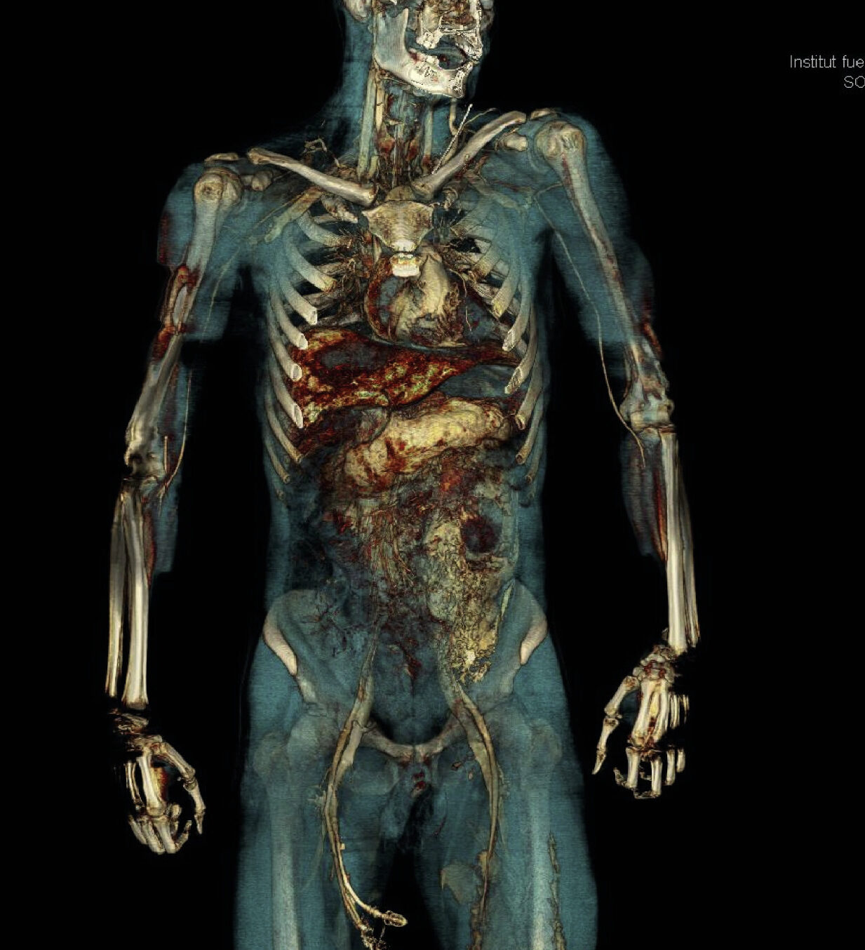
Demonstrating the feasibility of the approach in a mouse model will be groundbreaking for the disease because it will demonstrate its potential as an alternative to transplantation. Diseases like NASH [nonalcoholic steatohepatitis, an aggressive form of fatty liver disease] are increasing dramatically, and likely to be a leading cause of death within the next few years. Moreover, the difficulty of detecting the disease until it is potentially too late leads to significant challenges for therapeutic intervention, meaning transplantation will remain the main option for severely affected patients. This well-recognized need, along with the lack of donor organs will ensure bioprinted livers will continue to be well funded. But the value of the project goes beyond liver disease, as the new technologies developed in the frame of OrganTrans will not be limited to liver applications. They relate to the challenges of biofabrication of any organoid-based tissue, which can potentially be beneficial for a large variety of indications.

Can you tell me more about the role of regenHU within the OrganTrans consortium?

Such a complex and ambitious endeavor needs very different and complementary knowledge and competences. Teamwork will be a central element, first to enable, then to accelerate, these new solutions. With this in mind, we have been reorganizing regenHU to bring better project collaborative capabilities to this project, and others like it that we are engaged in. regenHU is a pioneer and global leader in tissue and organ printing technologies converging digital manufacturing, biomaterials, and biotechnology to lead transformational innovations in healthcare. We focus on delivering advancements in the instruments and software required for tissue engineering, and our technology evolving along with the biological research of our partners. We, therefore, consider these partnerships with the scientific community critical for our development.

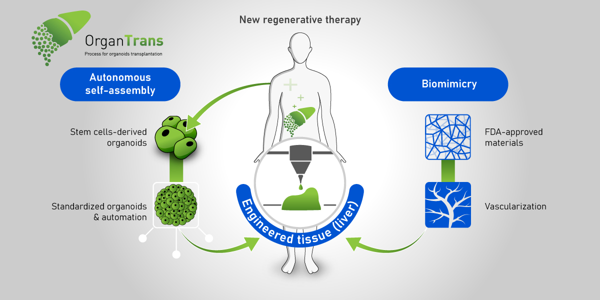
An outline of the OrganTrans project (Image courtesy of OrganTrans)

regenHU is one of the largest contributors to this project, is this part of the company’s commitment to regenerative medicine?

We can see the need for biotechnology solutions for a wide range of disease states. Our strengths are in engineering the instruments and software necessary to allow the producers of biomaterials and the suppliers of cells to combine their products to achieve functional tissues and organs. Our commitment is to provide disruptive technologies that will enable the community to make regenerative medicine a reality, with precision and reproducibility in mind, for today’s researchers and tomorrow’s industrial biofabrication needs. One of the key challenges is the current limitation in the scale and volume of bioprinting which is linked to the reproducibility of the print. To progress into the manufacture of medical products, bioprinters will need to operate at a scale beyond current capabilities. We design our instruments with these goals in mind and have assembled a team to solve the many challenges to achieve this.

How advanced is the bioprinting community in Europe?

The 3D bioprinting field is several years behind mainstream 3D printing, with the industrialization of the instruments, biomaterials, and cells required before bioprinting can progress to commercial-scale biofabrication. However, as with continued development seen in 3D printing, the technology convergence required for tissue and organ printing that changes medical treatments will become a reality through the efforts of engineering companies like regenHU, biomaterial developers, and human cell expansion technologies, being combined in projects such as OrganTrans.

As the newly appointed CEO of the company, how do you feel taking on this project?

Successfully entering the OrganTrans consortium is just one part of the company. regenHU investors see my arrival as the catalyst to bring regenHU to the next stage in its evolution. Our goal remains the production of industrial biofabrication instruments capable of delivering the medical potential of bioprinting, novel bioinks, and stem cells. To achieve this, we are enhancing the team and structure of the company, bringing forward the development of new technologies and increasing our global footprint to better support our collaborative partners. I have spent many years in regenerative medicine and pharma and can see the potential of bioprinting to revolutionize many areas of medical science, so joining regenHU was an easy choice. As CEO, my main role is to provide the right support structure to enable our entrepreneurial engineering teams to thrive and be brave enough to push boundaries. Additionally, as we cannot achieve our end goal on our own, I am here to nurture the important connections with our user community. Only by listening to their valuable insights and solving problems with them, we will push the technology onward.

The post regenHU CEO: Bioprinting Will Strengthen OrganTrans Project to 3D Print Liver Organoid appeared first on 3DPrint.com | The Voice of 3D Printing / Additive Manufacturing.

Rice Researchers 3D Print with Lasers and Sugar to Build Complex Vascular Networks

A team of researchers from Rice University has uncovered a promising strategy to generate vascular networks, one of the most daunting structures in the human body. Using powdered sugar and selective laser sintering, the researchers were able to build large structures from complex, branching, and intricate sugar networks that dissolve to create pathways for blood in lab-grown tissue.

This is the team’s latest effort to build complex vascular networks for engineered tissues to show that they could keep densely packed cells alive for two weeks. The findings of their study—published in the Nature Biomedical Engineering journal—prove that developing new technologies and materials to mimic and recapitulate the complex hierarchical networks of vessels gets them closer to providing oxygen and nutrients to a sufficient number of cells to get a meaningful long-term therapeutic function.

“One of the biggest hurdles to engineering clinically relevant tissues is packing a large tissue structure with hundreds of millions of living cells,” said study lead author Ian Kinstlinger, a bioengineering graduate student at Rice’s Brown School of Engineering. “Delivering enough oxygen and nutrients to all the cells across that large volume of tissue becomes a monumental challenge. Nature solved this problem through the evolution of complex vascular networks, which weave through our tissues and organs in patterns reminiscent of tree limbs. The vessels simultaneously become smaller in thickness but greater in number as they branch away from a central trunk, allowing oxygen and nutrients to be efficiently delivered to cells throughout the body.”

Overcoming the complications of 3D printing vascularization has remained a critical challenge in tissue engineering for decades, as only a handful of 3D printing processes have come close to mimic the in vivo conditions needed to generate blood vessels. Without them, the future of bioprinted organs and tissues for transplantation will remain elusive. Many organs have uniquely intricate vessels, like the kidney, which is highly vascularized and normally receives a fifth of the cardiac output, or the liver, in charge of receiving over 30% of the blood flow from the heart. By far, kidney transplantation is the most common type of organ transplantation worldwide, followed by transplants of the liver, making it crucial for regenerative medicine experts to tackle vascularization.

Ian Kinstlinger with a blood vessel template he 3D printed from powdered sugar (Credit: Jeff Fitlow/Rice University)

In the last few years, extrusion-based 3D printing techniques have been developed for vascular tissue engineering, however, the authors of this study considered that the method presented certain challenges, which led them to use a customized open-source, modified laser cutter to 3D print the sugar templates in the lab of study co-author Jordan Miller, an assistant professor of bioengineering at Rice.

Miller began work on the laser-sintering approach shortly after joining Rice in 2013. The 3D printing process fuses minute grains of powder into solid 3D objects, making possible some complex and detailed structures. In contrast to more common extrusion 3D printing, where melted strands of material are deposited through a nozzle, laser sintering works by gently melting and fusing small regions in a packed bed of dry powder. According to Miller, “both extrusion and laser sintering build 3D shapes one 2D layer at a time, but the laser method enables the generation of structures that would otherwise be prone to collapse if extruded.”

“There are certain architectures—such as overhanging structures, branched networks and multivascular networks—which you really can’t do well with extrusion printing,” said Miller, who demonstrated the concept of sugar templating with a 3D extrusion printer during his postdoctoral studies at the University of Pennsylvania. “Selective laser sintering gives us far more control in all three dimensions, allowing us to easily access complex topologies while still preserving the utility of the sugar material.”

Assistant professor of bioengineering at Rice University, Jordan Miller (Credit: Jeff Fitlow/Rice University)

Generating new 3D printing processes and biomaterials for vascularization is among the top priorities for the researchers at Miller’s Bioengineering Lab at Rice. The lab has a rich history of using sugar to construct vascular network templates. Miller has described in the past how sugar is biocompatible with the human body, structurally strong, and overall, a great material that could be 3D printed in the shape of blood vessel networks. His original inspiration for the project was an intricate dessert, even going as far as suggesting that “the 3D printing process we developed here is like making a very precise creme brulee.”

To make tissues, Kinstlinger chose a special blend of sugars to print the templates and then filled the volume around the printed sugar network with a mixture of cells in a liquid gel. Within minutes, the gel became semisolid and the sugar dissolved and flushed away to leave an open passageway for nutrients and oxygen. Clearly, sugar was a great choice for the team, providing an opportunity to create blood vessel templates because it is durable when dry, and it rapidly dissolves in water without damaging nearby cells.

A sample of blood vessel templates that Rice University bioengineers 3D printed using a special blend of powdered sugars. (Credit: B. Martin/Rice University)

In order to create the treelike vascular architectures in the study, the researchers developed a computational algorithm in collaboration with Nervous System, a design studio that uses computer simulation to make unique art, jewelry, and housewares that are inspired by patterns found in nature. After creating tissues patterned with these computationally generated vascular architectures, the team demonstrated the seeding of endothelial cells inside the channels and focused on studying the survival and function of cells grown in the surrounding tissue, which included rodent liver cells called hepatocytes.

The hepatocyte experiments were conducted in collaboration with the University of Washington (UW)’s bioengineer and study co-author Kelly Stevens, whose research group specializes in studying these delicate cells, which are notoriously difficult to maintain outside the body.

“This method could be used with a much wider range of material cocktails than many other bioprinting technologies. This makes it incredibly versatile,” explained Stevens, an assistant professor of bioengineering in the UW College of Engineering, assistant professor of pathology in the UW School of Medicine and an investigator at the UW Medicine Institute for Stem Cell and Regenerative Medicine.

The results from the study allowed the team to continue their work towards creating translationally relevant engineered tissue. Using sugar as a special ingredient and selective laser sintering techniques could help advance the field towards mimicking the function of vascular networks in the body, to finally deliver enough oxygen and nutrients to all the cells across a large volume of tissue.

Miller considered that along with the team they were able to prove that “perfusion through 3D vascular networks allows us to sustain these large liverlike tissues. While there are still long-standing challenges associated with maintaining hepatocyte function, the ability to both generate large volumes of tissue and sustain the cells in those volumes for sufficient time to assess their function is an exciting step forward.”

The post Rice Researchers 3D Print with Lasers and Sugar to Build Complex Vascular Networks appeared first on 3DPrint.com | The Voice of 3D Printing / Additive Manufacturing.

NASA Wants Aerojet Rocketdyne to Make More Rocket Engines with 3D-Printed Parts

On its first launch, NASA‘s uncrewed Space Launch System (SLS) mega-rocket will go on a trip around the Moon as part of the initial test flight for the Artemis 1 mission. It will mark the beginning of one of the most talked-about space programs this year, Artemis, an ongoing government-funded crewed spaceflight initiative with the goal of landing the first woman and the next man on the Moon by 2024, particularly, on the lunar south pole region. The most powerful rockets ever built, the SLS is in turn powered by four super engines that are designed to handle some of the most extreme temperatures as they move massive amounts of propellants to generate enough energy for the rocket to escape Earth’s gravity.

As part of a years-long working relationship with NASA, Aerojet Rocketdyne of Sacramento, California, will be building a total of 24 RS-25 rocket engines to support as many as six SLS flights for a total contract value of almost $3.5 billion. Originally slated to produce six new RS-25 engines, the company has recently been awarded a $1.79 billion contract modification to build 18 additional RS-25 rocket engines to support future deep space exploration missions.

“This contract allows NASA to work with Aerojet Rocketdyne to build the rocket engines needed for future missions,” said John Honeycutt, the SLS program manager at NASA’s Marshall Space Flight Center in Huntsville, Alabama. “The same reliable engines that launched more than 100 space shuttle missions have been modified to be even more powerful to launch the next astronauts who will set foot on the lunar surface during the Artemis missions.”

Although the Space Shuttle Endeavour is now at a museum exhibit at the California Science Center in Los Angeles, its engines—along with those that used to power space shuttles Discovery and Atlantis—have been maintained for SLS. However, unlike the shuttles, SLS will not reuse its engines. Once the core stage falls away at around eight minutes after launch, the engines will disintegrate during reentry. There are currently 16 RS-25 engines remaining from NASA’s Space Shuttle Program that Aerojet Rocketdyne has upgraded, tested, and that are ready to support the first four SLS missions. Yet, with more SLS missions expected to launch well into the end of the decade, Aerojet Rocketdyne has been asked to build more engines; actually, six new expendable RS-25 engines are already being assembled using advanced manufacturing techniques, including 3D printing, that reduces both the cost and time for manufacturing each engine.

The additional 18 engines will continue to leverage supply chain optimization and the incorporation of additive manufacturing (AM) techniques that were already introduced in the initial SLS engine production.

Initial SLS Configuration, powered by RS-25 rocket engines (Credits: NASA)

Employing AM technology to reduce costs and improve the efficiency of its engines is among the top priorities of the aerospace and defense company. Aerojet Rocketdyne’s senior engineer on the Additive Manufacturing team, Alan Fung, told 3DPrint.com that hundreds of people have been working on the design, development, and manufacture of the engines which relies mainly on laser powder bed fusion technology to additively manufacture at least 35 parts on each engine. 

“Our primary focus is to make reliable, robust printed parts, that will work 100 percent of the time. We started designing some of these pieces a couple of years ago to make sure they were tested and certified for NASA’s space program, which is crucial to the safety of the upcoming crewed missions,” said Fung. 

AM Team at Aerojet Rocketdyne, from left to right: Bryan Webb, Ivan Cazares, and Alan Fung (Credit: Aerojet Rocketdyne)

With the delivery of these new engines scheduled to begin in 2023, the team is not wasting any time. Fung said that “part of the big quest in the first round was to work with NASA closely on developing the certification processes.” Revealing that “we now have a process to make parts using AM that we know is safe and it is exactly what we need to make sure that our parts will work on the engines that will power future SLS missions.”

3D printing simplifies the production of several RS-25 parts and components, making the engine more affordable to produce while increasing reliability. With fewer part welds, the structural integrity of the engine increases. This is a very manual, complex manufacturing process. In fact, rocket engines are so complicated to build, that only a handful of countries have been able to manufacture them.

“That’s where AM really shined for us. We were able to get rid of many welding joints and just incorporate the processes automatically, getting down the part count and reducing the load across the engine,” said Fung.

One of the largest 3D-printed components of the engine was the critical “Pogo” accumulator assembly. Roughly the size of a beach ball, the complex piece of hardware acts as a shock absorber to reduce oscillations caused by propellants as they flow between the vehicle and the engine. Fung described the 3D-printed component as a critical part of the engine because it helps smooth the ride for astronauts and the vehicle ensuring a safe flight. Moreover, he explained that the Pogo used to demand more than 100 weld joints that had to be done manually and took almost four years to make, while the 3D-printed Pogo developed at Aerojet Rocketdyne’s factory in Los Angeles, brought the welds down to just three, and was finished in less than a year.

Some of these modified components have already been tested during engine tests that replicate the conditions of flight. For example, during a 400-second test at NASA’s Stennis Space Center, Aerojet Rocketdyne was able to successfully evaluate the performance of the 3D printed Pogo accumulator assembly.

“We expect that more and more engines will be additively manufactured in the future, leaving behind a lot of traditional rocket engine manufacturing processes that are very difficult, and allowing us to print more engines. Eventually, the time to build is going to go down even more, especially as the industry gears towards incorporating more lasers and bigger machines; which is good for us, because our engines keep getting a little bit bigger than the last ones. So, when those machines get to be bigger, use more lasers, and print parts faster, then that’s when we will see a really big shift in the way we make rocket engines,” went on Fung. 

Artemis I RS-25 Engines (Credits: Aerojet Rocketdyne)

Working with NASA, Aerojet has implemented a plan to reduce the cost of the engines by more than 30% on future production when compared to the versions that flew on the Space Shuttle, all thanks to more advanced manufacturing techniques, like AM, that help the engineers modify some of the rocket components.

During the flight, the four engines will provide the SLS with around two million pounds of thrust to send the heavy-lift rocket to space. The rocket engines are mounted at the base of a 212-foot-tall core stage, which holds more than 700,000 gallons of propellant and provides the flight computers that control the rocket’s flight.

The AM team at Aerojet is using GE Concept Laser and EOS machines for its selective laser melting requirements. Fung said they were using superalloys, mostly nickel-based for the engine parts being 3D printed, due to its outstanding corrosion resistance, high strength, and ability to resist hydrogen embrittlement due to the hydrogen fuels found in most of Aerojet Rocketdyne’s liquid propellant rocket engines.

“These new RS-25 engines are an upgrade from the Space Shuttle engines, which were already some of the most reliable engines made in history. Engineers spent 40 years making the shuttle engines as reliable, safe and high performance as possible; but with additive manufacturing we thought we could also try to get the cost down. This technology will revolutionize the way we build engines” 

With so many challenges ahead, having certified rocket engines to take the next lunar explorers to orbit feels like a stepping stone for the journey that lies ahead. After all, the SLS rocket is part of NASA’s backbone for deep space exploration and will prepare humans for long-duration space travel and the eventual journey to Mars.

Space Launch System (SLS) (Credits: NASA)

The post NASA Wants Aerojet Rocketdyne to Make More Rocket Engines with 3D-Printed Parts appeared first on 3DPrint.com | The Voice of 3D Printing / Additive Manufacturing.

3DPOD Episode 27: Terry Wohlers

Max and I really enjoyed our chat with Terry Wohlers. Terry has been writing the Wohlers Report for 25 years. This report is the definitive yearly 3D printing report, and gives us all an annual update on market developments, breakthroughs, and new applications worldwide. Additionally, Terry consults for many businesses globally, helping them to implement and understand 3D printing. His company has worked with over 275 clients in 27 countries including the likes of Airbus, GE, Lockheed, Apple, Procter & Gamble and NASA. I’ve known Terry for a long time and he always has insight and concise analysis of developments in the industry. Max and I talked with him about when to use additive, what is holding the technology back, the general state of the industry, growth today, some key highlights of the Wohlers Report, and his America Makes involvement.

The post 3DPOD Episode 27: Terry Wohlers appeared first on 3DPrint.com | The Voice of 3D Printing / Additive Manufacturing.

Marine Biologist Modifies Bioprinting for the Creation of Bionic Coral

Corals are dying globally. In the face of climate change and global warming, we can expect some severe consequences, which in turn directly affects marine life. In what is panning out to be a mass extinction event, coral reefs have been dangerously threatened by toxic substances and excess carbon dioxide for years, causing the certain death of may of these diverse marine invertebrates. Once the coral is dead, the reefs will also die and erode, destroying important marine life, that would otherwise feed and spawn on it.

Considering that scientists have predicted that nearly all coral reefs will disappear in 20 years, it is crucial that we protect corals and learn from them. For the expanding field of biotechnology, untapped resources like corals hold great potential, as bioactive compounds for cancer research or simply as an inspiration for the production of bioenergy and bioproducts.

In an interview with 3DPrint.com, interdisciplinary marine biologist Daniel Wangpraseurt, from the University of California San Diego (UCSD)’s Department of NanoEngineering, explained how bioprinting technology was a pivotal point in his work to develop bionic 3D printed corals as a new tool for coral-inspired biomaterials that can be used in algal biotechnology, coral reef conservation and in coral-algal symbiosis research. 

“For many years I have been studying how corals optimize light management and discovered that there are lots of interesting evolutionary tricks, such as different growth forms and material properties, so I became interested in copying these strategies and developing artificial materials that could host living microalgae, just like corals do in nature,” revealed Wangpraseurt.

Daniel Wangpraseurt

As one of the most productive ecosystems globally, coral reefs use photosynthesis to convert carbon dioxide into energy that they in turn use for food. Even though light provides the energy that fuels reef productivity, key nutrients such as nitrogen and phosphorus are also required, but are found in very low quantities in warm tropical oceans where coral reefs are generally found, making scientists wonder how these marine animals have managed to create a competitive habitat with such limited resources.

A laser beam is intensely scattered by elastic coral tissue and aragonite skeleton. (Credit: Daniel Wangpraseurt)

Wangpraseurt described that, while different corals have developed a plethora of geometries to achieve such capabilities, they are all characterized by an animal tissue-hosting microalgae, built upon a calcium carbonate skeleton that serves as mechanical support and as a scattering medium to optimize light delivery toward otherwise shaded algal-containing tissues.

“Taking what we learned about corals and biomaterials, we began working on a project to develop a synthetic, symbiotic system using a 3D bioprinting approach. We know corals have both animal cells and algal cells, and, so far, we have mimicked the animal part of the corals, that is, the physical and chemical microhabitat that partially controls the activity of the algal cells.”

At UCSD, Wangpraseurt expects to continue recreating coral-inspired photosynthetic biomaterial structures using a new bioprinting technique and a customized 3D bioprinter capable of mimicking functional and structural traits of the coral-algal symbiosis. Along with fellow researchers from UCSD, the University of Cambridge, the University of Copenhagen and the University of Technology Sydney, and thanks to a grant from the European Union’s Horizon 2020 research and innovation program, and the National Institutes of Health (NIH), the team reported the results of their work on bioinspired materials that was published in the journal Nature Communications earlier this year.

“We want to go further and not just develop similar physical microhabitat but also modulate cellular interactions, by mimicking biochemical pathways of symbiosis. We hope that this allows us to not only optimize photosynthesis and cell growth, but also to gain a deeper understanding of how the symbiosis works in nature. By doing so, we can improve our understanding of stress phenomena such as coral bleaching, which is largely responsible for global coral death.”

Living colonies of Symbiodinium are visible within the 3D bioprinted tissues (Credit: Daniel Wangpraseurt)

So, how did bioprinters become the go-to technology for this project? Wangpraseurt explains that, while working as a researcher at the University of Cambridge’s Department of Chemistry Bio-Inspired Photonics lab, he noticed that scientists were using cellulose as a biomaterial with interesting optical responses. He was wondering how he could use cellulose to develop a material with very defined architectural complexity. 

“In the beginning, the main aim was to develop a coral-inspired biomaterial, that has a similar optical response as natural coral, and then to grow algae on it or within it. Thereby, we started off with simple techniques, using conventional 3D printers; however, it wasn’t very easy to recreate the spatial resolution we needed for corals.”

Inspired by 3D bioprinting research in the medical sciences, Wangpraseurt reached out to scientists at the UCSD NanoEngineering lab that were developing artificial liver models, and who later became collaborators in the project.

A laser beam is intensely scattered by elastic coral tissue and aragonite skeleton (Credit: Daniel Wangpraseurt)

The team went on to develop a 3D printing platform that mimics morphological features of living coral tissue and the underlying skeleton with micron resolution, including their optical and mechanical properties. It uses a two-step continuous light projection-based approach for multilayer 3D bioprinting and the artificial coral tissue constructs are fabricated with a novel bioink solution, in which the symbiotic microalgae are mixed with a photopolymerizable gelatin-methacrylate (GelMA) hydrogel and cellulose-derived nanocrystals (CNC). Similarly, the artificial skeleton is 3D printed with a polyethylene glycol diacrylate-based polymer (PEGDA).

Close up of coral polyps and living photosynthetic biomaterials. Living colonies of Symbiodinium are visible within the 3D bioprinted tissues (Credit: Daniel Wangpraseurt)

Based in San Diego, Wangpraseurt has spent months trying to recreate the intricate structure of the corals with a distinguished symbiotic system that is known to grow as it creates one of the largest ecosystems on the planet. 

“We used a 3D bioprinter that had been developed for medical purposes, which we modulated and further developed a specific bioink for corals. A lot of the work was related to the optimization of the material properties to ensure cell viability. Having the right bioink for our algal strains was crucial as if we were to use mixtures commonly used for human cell cultures, the cells will not grow very well and can die rapidly.”

The implications of the newly developed 3D printed bionic corals capable of growing microalgae are many. Wangpraseurt said he plans to continue working on bionic corals and potentially scale up the process for his startup, called mantaz, as well as for commercial properties; or to develop coral-inspired materials at a larger scale to have a more immediate impact on efforts related to coral reef restoration, and also for biotechnology.

SEM images of the skeleton structure of the coral Stylophora pistillata and the coral-inspired, 3D-printed material (Credit: Daniel Wangpraseurt)

Wangpraseurt is looking to scale the bioprinting system to have a more immediate impact on algae biotechnology, bioenergy, and bioproducts. He claims that he and his colleagues can “customize the environment of the algae and fine-tune the production of a certain bioproduct to potentially tap into the algae bioproduct market and scale the system for bioenergy production.” 

“Another interest of mine is to further develop a 3D bioprinted synthetic coral-algal symbiosis system, which can provide important insight into the mechanisms that lead to coral death, but can also result in the development of future technology for coral reef restoration.”

The researcher talks about coral reefs with a reverent passion that today goes beyond his lab work. When he is not moving the research along at USCD, Wangpraseurt is working with his social enterprise in Panama, as he and his team try to restore coral reef ecosystems to help coastal communities in the tropics, including local fishermen, by harvesting algae biomass that can be sold for different purposes, such as natural fertilizer, which contributes to an organic and sustainable chain of production. Furthermore, the coral-inspired aspects of Wangpraseurt’s research and startup company are really coalescing to enable him and his team to understand how corals work and, in turn, how we can learn from them for the benefit of our planet.

Nutrient sampling at a polluted reef in Panama (Image: Daniel Wangpraseurt)

The post Marine Biologist Modifies Bioprinting for the Creation of Bionic Coral appeared first on 3DPrint.com | The Voice of 3D Printing / Additive Manufacturing.

Start Non-Planar 3D Printing Today on your Ender 3 with nonplanar.xyz

One of the most exciting developments in 3D printing is non-planar 3D printing for FDM. Fused Deposition Modeling (FFF, Material Extrusion) is the most popular, affordable and widespread 3D printing technology. FDM can print many different materials and has a large number of possible applications. There are some limits to FDM however. Z-axis strength is not optimal and the esthetics of parts are not really great. Regular FDM printers print using a cartesian system that moves upward layer by layer. This works well enough if you consider a printer a box that makes things. But, what if you wanted to make things that were more suited to the real world? Parts such as braces, bioprints, orthotics, insoles, helmets and the like all have to fit humans and humans are rather curvy with many organic round parts. Organic designs generally are a challenge to printers as is reducing stepped surfaces and increasing in layer and intralayer adhesion. What if we could make stronger FDM parts that simultaneously looked better? Non-planar FDM may just do all of that for us. Work on slicing nonplanar surfaces for FDM and testing parts is very promising already.

An Airfoil made by Daniel Ahrens and Co. shows us some of the benefits of non-planar 3D printing.

Additional work on non-planar 3D printing shows us that it may be useful for transparent displays and optics, can be done using existing robot arms, and thanks to Grasshopper and a Slic3r plugin there is even software for it. The combination of non-planar printing with microstructures, gradients, multiple materials and lattices could make FDM an even more versatile technology.

Now nonplanar.xyz hopes to make non-planar 3D printing much more widespread. The company was started by Gabriel Boutin who worked on the Kupol helmet we’ve written about before. Getting your current FDM printer to print non-planar is actually quite complex as you can see from this awesome video below. It also illustrates the results one can get.

At nonplanar.xyz they want to sell us the elongated nozzles we would need, teach us how to go non-planar and give us sample G-Code to get us started. I’m incredibly excited about non-planar FDM and hope that the firm spreads non-planar far and wide. You can buy a nozzle starting from $8.49. An introductory course into path design and path programming is $30. You can also buy Gcode for $10 or $2. The Gcode doesn’t really seem like an amazing deal to me but the course and nozzles seem like a steal given what non-planar can do to expand your 3D printing arsenal.
We asked Boutin why he and the Kupol team got into non-planar printing.  He responded that:
“I have been looking for the magic bullet among all the additive manufacturing technologies for the last few years. Like most of us, I was captivated by the brand new and expensive machines. They are capable of amazing results but are they value creation tools?Being part of the NFL helmet challenge to create the most protective helmet ever created, I have screened all the possibilities for manufacturing unique lattices I have created for Kupol Inc.”

nonplanar.xyz’s kit on an Ender 3

It took him a while to get the hang of it.
“I have always disregarded FDM, thinking it was not a real manufacturing process. I was completely wrong. The solution was to push the boundaries of FDM to use its full potential, meaning nonplanar paths. It was a scary thing at first because I did not have any knowledge of programming. Nevertheless, I have investigated Rhino Grasshopper to see if a guy like me could succeed in printing a nonplanar shape. And I just did it.”
Also, the way that Gabriel looks at non-planar is squarely from the view of a designer:
“Nonplanar printing is in fact a workflow that you design or adaptt to the product you want to create. There will never be a single solution for everything because it requires intent. If you want to create an insole for example, you need to define what you want the product to look like and how to achieve this.  Once you have invested time into your ”design”, you can use it to print similar shapes and therefore you open the door to custom products at scale.”
I love that he sees it as a workflow and indeed looking at it in this manner would make it seem as an option and “path” to certain products. I feel that this is a far more realistic and healthier approach than to look at everything as a “technology” to replace all others. I was very surprised that Gabriel opted for a low cost 3D printer to introduce the technology with.

I have selected the Ender3 for a simple reason, it was dirt cheap and easy to modify. There are other DIY printers on the market that could be very good choices too. I expect nonplanar.xyz to offer compatible nozzles for those other choices in the very near future.

It is now time to leave the surface!
You can order nozzles now and sign up for the tutorial at their site.

The post Start Non-Planar 3D Printing Today on your Ender 3 with nonplanar.xyz appeared first on 3DPrint.com | The Voice of 3D Printing / Additive Manufacturing.

CELLINK in France: Expanding Their Portfolio in 2020

Seeking to strengthen their presence in Europe, 3D bioprinter provider and pioneer bioink company CELLINK, opened their new offices in Lyon, France, last October. Begining new partnerships and collaborations with universities, hospitals, pharmaceutical companies and more, is a big part of the core mission of CELLINK, as they combine their technology with research innovation everywhere. The city of Lyon offers a booming scene for bioprinting, with companies heavily focusing on microtumors and taking advantage of an established network that has collected more than 2000 tumors from patients in collaboration with 11 major cancer hospitals in the country via the IMODI initiative–a French consortium to develop new experimental models of cancer–which preserves and archives all tissue and cell samples developed by the consortium partners during the project.

3DPrint.com spoke to CELLINK’s Sales Director for France and Southern Europe, Edouard Zorn, who envisions a greater expansion this year, new partnerships and research collaborations: “We hope to really expand our portfolio this year in France.”

Cellink is amazing at building bridges between a product and researchers from different backgrounds and cultures. The company has a team-oriented mindset, looking to work with people from different nationalities,” sad Zorn, a biotechnology engineer with vast experience in scientific sales management.

Zorn and his five-people work team at the Lyon offices have their agenda full, with 30 customers in France, bioprinter installations, and training sessions for new CELLINK users, Zorn can’t help but highlight how fast the field of bioprinting is moving in Europe. “There are many researchers focusing on skin and cancer, that’s really big here, but lately I have also been in contact with companies working on biopharma, vaccines and some even trying to replace animal testing assays,” he stated. 

Since January 2018, CELLINK has been working with another major player in Lyon, CTI Biotech, using bioprinting to develop microtumors. CTI Biotech uses CELLINK technology exclusively for their work and now have three bioprinters at their lab, which is also located in Lyon. CELLINK and CTI Biotech had even signed a deal to 3D print customized cancer cells, with CELLINK assisting CTI in the production of patient-specific cancer tumor replicas, which will be 3D printed by combining CTI’s bioink with a sample of patients’ own cancer cells, promising to deliver personalized treatments for cancer on a custom, patient-by-patient basis.

“The aim of our collaboration is to give researchers an advantage in treating specific cancer types, and in the long term, take a serious step forward in the fight to cure cancer. CTI is moving really fast to develop models and commercialize them, and they choose our machines for their versatility, intuitiveness, and easily modifiable parameters. CTI Biotech is one of the customers we most grow with, and I believe it was a very good decision for both companies to work together,” explained Zorn. 

So far, they have already commercialized CELLINK skin for drug and cosmetic testing, which they have also been working to improve, and Zorn thinks that soon they will be working on introducing some human cells to the skin, as well as perhaps vascularizing the tissue.

Edouard Zorn (far right) and CELLINK bioprinters at the CTI Biotech labs (Credit: CELLINK)

The CELLINK office in Lyon is selling their machines to central Europe, working along the french-speaking part of Switzerland and Belgium, as well as in Spanish and Portuguese markets. 

Edouard Zorn at CELLINK France (Credit: CELLINK)

“We work with a lot of universities in France. For example, at the Medicine University of Montpellier, Xavier Garric, uses the INKREDIBLE+ bioprinter to teach master students how to design and print implantable medical devices and scaffolds for tissue engineering; and Alexandra Fuchs from the Hôpital St Louis employs a BIO X for tissue engineering.”

At the University of Grenoble, Vincent Haguet is generating skin, cornea and pancreas organoids for the modeling of organogenesis (organ formation) and pathogenesis (disease development), with a BIO X. Among these applications, organoids are used to screen and test new drugs. Also wielding the power of the BIO X is Anthony Treizèbre, from the University of Lille, for the bioprinting of Tumor-On-Chip and Blood-Vessels-on-Chip for the development of multicellular microfluidic biomimicry-based devices for the study of metastasis. Their idea is to reproduce blood vessels using human umbilical vein endothelial cells (HUVEC) and modulating the surrounding extracellular matrix.

The University of Nantes‘ Pierre Weiss also works with BIO X to print calcium phosphate-based personalized medical devices for maxillo-facial bone regeneration, as well as enzyme-based hydrogel formulation for the complex systems in bone regeneration.

Zorn believes that “there is a big demand from patients that expect the medical and bioengineering field to adapt treatments to patients. There is a lot of expectation for personalized medicine, especially with regard to microtumors for drug testing. Moreover, lately, we have seen researchers focusing heavily on immunotherapy, so I see a great future in that regard and consider that CTI Biotech is trying to position itself in that field.”

Fortunately, he suggests that there is collaboration in Europe. The European Union (EU) is financing joint collaboration projects with the objective to develop medical devices and applications with therapeutic solutions, and CELLINK wants to be a part of that.  Zorn emphasized the importance of the Silk Fusion Project, which unites scientists in the development of a technology that uses silk, a natural biocompatible and sustainable material, to produce a bioink and 3D print platelet production instrumentation, attempting to solve the limited supply of human platelets. Other projects which have CELLINK as a collaborator seek to solve problems for joint articulation, bone, and even bioprinting parts of the tendon and cartilage.

“We need people who understand cell biology, chemistry, hardware, electronics, and software, as well as a good comprehension and understanding the needs inherent to each country’s culture, that is the way in which we expand,” concluded Zorn.

Edouard Zorn at the new CELLINK offices in France (Credit: CELLINK)

The French branch of the company joins the other six worldwide offices of CELLINK, in Boston, Gothenburg, Freiburg, Blacksburg, Kyoto, and Stuttgart. Zorn hopes that the sales force along with the experienced network of professionals around the world working for CELLINK will result in a stronger presence of the company in central Europe as well as more joint efforts that could bring the future of bioprinting technology closer to our present.

The post CELLINK in France: Expanding Their Portfolio in 2020 appeared first on 3DPrint.com | The Voice of 3D Printing / Additive Manufacturing.

3D Printing News Briefs: December 26, 2019

For your holiday edition of 3D Printing News Briefs, we’ll get business out of the way first – Wipro 3D has launched Addwize, a new Additive Technology Adoption and Acceleration Program. Moving on, Prusa interviewed animatronic model senior designer Joshua Lee about their shared interest in 3D printing. Finally, Voodoo Manufacturing helped an artist bring her 2D artistic vision to full-sized 3D life.

Wipro 3D Introduces Addwize Program

Scale vendors: Foundation – blue; Advanced – green; Practitioner – orange

Additive manufacturing solutions provider Wipro 3D, a business of Wipro Infrastructure Engineering, has launched a new Additive Technology Adoption and Acceleration Program called Addwize, which will address all the phases in the AM adoption cycle within academia and industry. The multi-platform, OEM-agnostic adoption program will help interested organizations and institutions fully understand 3D printing, evaluate business cases for the technology, and then scientifically use it to create value. It’s designed to help stakeholders of all levels, and academia, adopt and scale their usage of AM for business benefits.

“Wipro 3D addwize™ is designed and developed to support any organization or institution who is either evaluating metal Additive technology, has AM in their near future technology roadmap or has already invsted in AM, create business value using metal AM,” said Ajay Parikh, Vice President and Business Head, Wipro 3D.

“There is no lower or upper limit to the size of the organization who wants to evaluate AM.”

Prusa Interviews Animatronic Model Designer Joshua Lee

Not too long ago, the Research Content Team at Prusa met award-winning animatronic model senior designer Joshua Lee in Prague, who has over 25 years of experience in the film industry working on such movies as Prometheus, The Fifth Element, and even the Star Wars and Harry Potter series. The team took advantage of the opportunity to speak with Lee about a topic near and dear to all their hearts – 3D printing, which he uses often in his work.

“We use a lot of different techniques of 3D printing in the filming industry,” Lee told Prusa. “We only really adopted it in the last 5 years. I am really using it a lot now.

“The thing I like the most is how 3D printers help when you have really tight deadlines. The film director has a new idea and you just wish there were more hours in a day. We used to do a lot of “all-nighters” to get things made. If you’ve got your own 3D printer, you can design something quickly, press print and you can go home to bed – that’s the best thing! In the morning, you are up and running again and this amazing print awaits you there. I still get a small thrill, every time I come in and see this thing that has magically appeared there overnight.”

To hear more of what Lee had to say about the materials he uses (PLA and PETG), his preferred desktop printer (Original Prusa MK3), and specific Star Wars-related projects he used 3D printing for, check out the rest of the interview in the video below:

Voodoo Manufacturing Assists with 3D Printed Art Installation

Back in 1976, artist Agnes Denes created a 2D art piece called Probability Pyramid – Study for Crystal Pyramid, and has long since dreamed of turning into a life-size installation. In early 2019, her dream seemed like it would become reality when NYC-based art space The Shed began working with her on the project. The team didn’t have much luck with acrylic, glass, or mold injection, and so turned to Brooklyn’s Voodoo Manufacturing for assistance. There were a lot of requirements for the project – the Pyramid required several groups of bricks in unique sizes and shapes, totaling 5,442 translucent bricks that could be stacked to easily transport and form the pyramid; Voodoo 3D printed bricks that were 99% hollow, so they were less breakable and very lightweight.

“A lot of traditional manufacturing happens abroad. Because Voodoo’s factory is in Brooklyn, the team at The Shed would have an easier time accessing the parts as the sculpture was built. By the same token, as part of her commitment to environmental responsibility, it was very important for Agnes Denes to keep the production local,” Voodoo explained.

“The use of 3D printing was much more in line with her vision than traditional sculpture construction methods. This also allowed us to test multiple versions of the Pyramid digitally instead of having to build many physical versions.”

Thanks to Voodoo’s digital factory, the exhibition Agnes Denes: Absolutes and Intermediates opened on time. The retrospective, which features the 3D printed installation, will be displayed at The Shed until March 22, 2020.

Discuss these stories and other 3D printing topics at 3DPrintBoard.com or share your thoughts in the Facebook comments below. 

The post 3D Printing News Briefs: December 26, 2019 appeared first on 3DPrint.com | The Voice of 3D Printing / Additive Manufacturing.

Interview with Jason Chuen: Shaping Australia’s Medical 3D Printing Environment

In Australia, vascular surgeon, Jason Chuen understands that 3D printing is the exciting next step in personalized medicine, which is why he uses 3D scans and 3D printing to deliver anatomical models. During an interview with 3DPrint.com, Chuen, who is also the Director of Vascular Surgery at Austin Health and Austin Health’s 3D Medical Printing Laboratory (3D Med Lab), suggested that “there is a lot of interest because the field of 3D printing in medicine is growing; we are seeing the doctors and researchers involved more than ever, as well as more application development originating from clinicians.”

At The University of Melbourne, in Australia, the 3D Med Lab supports 3D printing for clinical applications and runs an active research program exploring how it can be used for teaching, procedural simulation, patient education, surgical planning, and prosthetic implants. The first facility of its kind in Australia, 3D Med Lab, frequently prints models of diseased aortas to perform a “practice-run” of surgery. What makes this lab unique is that it is hospital-based, and works with many different specialties. Chuen has been looking into the landscape of medical 3D printing for many years and earlier this month along with his colleague Jasamine Coles-Black, a Doctor and Vascular Researcher at the Department of Vascular Surgery at Austin Health and the 3D Med Lab, organized the fifth annual 3D Med Australia Conference, which he claims is the only meeting of its kind in Australasia, with only one or two more around the world of a similar nature, like Materialise‘s medical 3D printing meetup in Belgium.

Normal anatomical branches on an abdominal aortic model 3D printed on MakerBot Replicator 2X FDM

Chuen and Coles-Black even begun printing out copies of patient kidneys to help surgeons at Austin Health plan the removal of kidney tumors. Moreover, Chuen understands that the immediate challenge in medical 3D printing is ensuring that medical professionals themselves are up to speed with the technology because it is their clinical experience that will drive new applications and projects. 

During our interview, Chuen asserted that the conference has once again proved that Australia is leading the way with cross institution development cooperations, ethical issues surrounding 3D printing and he looks forward to many exciting possibilities of the technology for the future.

Why was the 3D Med Conference so important to the region?

We noticed there were a lot of groups that existed previously that didn’t know about each other and the meeting has become a really good focal point for people to find out about what others are researching and selling. So rather than working on their own and almost in secret, they can join together and create projects that cross different institutions, specialties and disciplines. During the conference, at every corner I encountered groups of people from different universities and cities gathering to hatch a project, proving that there was a very cooperative atmosphere. They all clearly had common interests and discovered that they can work outside of their own space with others. 

What was so unique about the 3D Med Conference?

Because there really aren’t many meetings like this, the areas of interest are still growing, anyone who is working with these technologies have applications in different areas so that is why we have a lot of crossover between the fields. The strength of the confreence comes from encouraging people to have an overview of what was happening in the field, so rather than just understanding technical aspects of technology, everyone started to become knowledgeable about the whole landscape, for example, why we need to care about ethics and regulation, or considering the useful implications of applying techniques from a different area of science and research. 

One of the biggest challenges for 3D printing is?

One of the big problems in customized medical devices and the 3D printing space is that there is uncertainty about what will happen in the future. Apart from the guidance of the US Food and Drug Administration (FDA), there hasn’t been a lot of resources for manufacturers and researchers on how 3D printing and customized medical devices will be regulated. Australia’s own Therapeutic Goods Administration (TGA) representation in the International Medical Device Regulators Forum (IMDRF) has been very strong,particularly around 3D printing and customized medical devices. During the conference John Skerritt, Deputy Secretary of the Australian Department of Health, outlined the broad framework around the field and has engaged in a consultation process with the medical 3D printing community (and we have provided some proposals for the final documentation that will be ready soon.)

Distributed production will present new risks for ensuring the quality control of end products. It will need a fundamental shift in responsibility from the supplier to wherever the medicines or devices are manufactured. That represents a huge change and we have to work out how it could work. But if we get the regulation right then it will transform access to medical products.

Collection of 3D printed objects

What does the future of 3D printing in medicine look like?

The whole point of what we do is improve patient care, so we have to think very carefully about our next steps and analyze whether it is helpful or not. For patients, anatomical models help them see and understand the condition or surgery they plan for. We have done projects and have some conclusive evidence that patient understanding is improved with anatomical 3D printed models. 

Patients are interested to know what will happen in the future, especially with 3D printed kidneys and stents. But the truth is that that technology is very far away. We may never be able to 3D print an organ, not at least the way we imagine it to be. Realistically, if we are talking about an organ for transplantation, we have to think that no matter what the organ looks like, the question is: does it do the job? For example, if we were thinking about bioprinting in order to replace a kidney, as long as it performs the function of the kidney, it doesn’t matter what shape it comes in. And for that, we have to be able to reproduce a structure. This could be in shapes, rather than in one block, or it could be a composition of an external and an internal device, meaning we would be looking into something that is assembled. Today the technology to have the replacement kidney is available, it is a dialysis machine, yet you wouldn’t expect a dialysis machine to look like a kidney. The same is going to happen with 3D printed organs, where we need to separate the appearance and structure of the organ from the function. In the end, the function is what matters.

As such, if we were to imagine what a 3D printed heart would look like, we would need to go into the field of soft robotics or mimicking natural structures, all of that changes fundamentally how we think about organs for the human body.

How can your particular medical field benefit from 3D printing?

As a vascular surgeon, I’m also looking at 3D printed stents, and there is quite some work around that. Mainly it is based on printing something that looks like a stent, but it is very difficult to reproduce the mechanical properties of a stent using 3D printing. The benefits revolve around the different materials that could be used with 3D printing, for example, if you could reproduce a stent in a bioabsorbable plastic it would allow surgeons to deploy it with embedded drugs (like antibiotics and pain medication) that get released at a set time. There are a lot of options in terms of using multi material technology in customized implant production, as well as great precision, and that is an area where 3D printing helps. 

Ideally, we need to understand the technology to know where the errors can happen. But in general, it is improving, both in hardware and software, the challenge will be about making it accessible. We have done randomized trials around anatomical models for teaching, education and simulation. There are already some 3D printed medical devices, such as for joints and implants. It would be ideal to have assessments of the economics to determine whether the anatomical models will be worthwhile. 

How is Australia changing the paradigm of medical 3D printing?

Australia has world leading technology, but in terms of the way we have collaborated and worked together, we are quite unique. Even globally one of the big problems is finding the groups that are doing this kind of work. We have been in touch with research groups in Poland, Boston, and Toronto, even engaging with large centers like the Mayo Clinic, in Minnesota. Key collaboration between international centers are great and we are keeping an eye out for other major hubs of activity, like in China, South Korea, and Europe. We need to link up all the international groups, that’s where we see things are going!

[Image credit: 3dMedLab, Austin Health]

The post Interview with Jason Chuen: Shaping Australia’s Medical 3D Printing Environment appeared first on 3DPrint.com | The Voice of 3D Printing / Additive Manufacturing.

Wolf Schweitzer: “3D Printing is Helping to Develop Forensic Devices”

Post mortem examinations are widely used to determine the cause of death, yet traditional autopsy has changed little in the past century, consisting of external examination and evisceration, dissection of the major organs with identification of macroscopic pathologies and injuries. A few years ago, and in a quest to advance the field of forensic medicine, a team of scientists at the University of Zurich, Switzerland, has been serially developing automated tools and technologies to improve results, reduce costs, and time during autopsies. By combining new technologies, like 3D imaging, scanning, and printing to generate virtual autopsy tools, into what has eventually become a household name in forensics a venture known as the Virtopsy project, or just virtopsy, they are changing the paradigm of forensics.

As part of the Virtopsy project, scientists have come up with creative ways to help the field of forensics, with ideas ranging from a modified automotive robotic arm with tools called Virtobot, to non-invasively discovering injuries present on the skin surface of a body, along with 3D true color representations of surface injuries and 3D scaled models of entire crime scenes and events. One device, in particular, caught our attention, a post mortem computed tomography angiography or PMCTA kit, made using 3D printing and parts that can be found at your local hardware store. The team posted online all of the files and part specifications so that anyone who wants to recreate the PMCTA can do it, for a total cost of just $120. But first, to understand what the device really does, 3DPrint.com asked Wolf Schweitzer, a forensic pathologist at the University of Zurich and part of the team behind the 3D printed PMCTA, why the device is so important and how disruptive technologies can aid experts to achieve better autopsies.

Production of Very Affordable PMCTA-kits. Left: 3D printing in progress; Middle: finished print batches; Right: kits in process of being packed

Why is the PMCTA important in forensic medicine?

A post-mortem CT is relevant in forensic pathology to examine the body, particularly for the consequences of violence or trauma. The findings add insight and help prepare autopsies so they can be performed faster and with a better focus on what we are looking for. Autopsy diagnosis is often very specific, yet performing it is tedious and time-consuming. For example, a few years ago it took seven hours of careful dissection to find the source of fatal hemorrhage in a body. These types of cases would greatly benefit from a PMCTA. Via an external pump, the vessels of the body are filled with a contrast substance that appears opaque on the computed tomography (CT). Knowledge of the normal anatomy of blood vessels allows examiners to identify certain possible or potential leaks. This means that while using PMCTA, vascular injuries, leaks or other pathologies can be examined. Once they are found, they may be documented or the results may be given to the pathologist who then narrows down the search for the actual autopsy dissection.

Why did you design a low-cost PMCTA for anyone to use?

Resulting PMCTA with a view of the whole body showing contrasted vessels and organs

Specialized commercial devices can be costly and require dedicated and expensive additional installations such as oil separators, consumables, and maintenance. A top of the line PMCTA-pump can easily be worth $80,000, while materials cost around $1,000 per single case or examination. Additionally, users need to install an oil separator to avoid their oil-based contrast agent to leak or get drained into the sewer. While some privileged forensic medicine institutes may find that acceptable, we wondered whether that type of technology was really necessary. So that is why we decided to custom design and 3D print our own immersion pump to be used as a forensic PMCTA and fill in the rest of the materials list with parts from a hardware store, for just $120. The whole idea of providing very affordable PMCTA technology became evident during our Virtopsy courses, for 15 years, specialists and trainees from around the world came to Zurich to attend our courses, and one frequently voiced concern was about the PMCTA, how problematic the oil was to the environment, and how expensive the materials where, so we listened and began evaluating better options.

In the paper Very economical immersion pump feasibility for postmortem CT angiography (that has Schweitzer as co-author) our team at the Department of Forensic Medicine and Imaging concluded that more widespread and systematic implementation of PMCTA demands affordable equipment for facilities with tight budgets. This is why we uploaded everything anyone needs to develop their own PMCTA (the printable 3D models went up as STL-files) online at virtopsy.com.

How did 3D printing become part of the solution?

Unlike clinical medicine, forensics get lower, more restricted public budgets, motivating us even further to use more affordable means of production, design, and materials. Plus, we often do not need anti-allergic or extensively sterilized catheters or solutions. This means that we can design, 3D print prototypes and test them in one or two days, then revise the design and keep re-iterating until the 3D models (and their 3D printed instances) are ok. Once the 3D printed PLA models are enough for routine work, we use them. We really wanted to get the actual design process first, since having the ability to design hardware prototypes using CAD software is useful anywhere custom parts are needed.

What 3D printers did you use?

For the 3D printing process, we used a MakerBot Replicator 2 (originally built to print ABS, but tweaked to print with PLA) and a MakerBot Replicator+ (fifth generation). PLA feeding was a problem, as the Makerbot printers appeared to have trouble pulling the PLA into the nozzle where it is melted for printing. To work around that, we decided to built PLA-roll mounts with ball joint bearings using available 3D models of Thingiverse. We used 3D printing to get the models’ shape right.

Still, 3D printing spare parts and new add-ons does not end with the PMCTA, the team has also identified a few other applications for 3D printing. For example, they are currently investigating whether it is possible to print skeletal parts (skull and lung bones) to perform bio-mechanical crash or impact tests on 3D printed materials, and verify if they fracture in a similar way as natural bone.

Is there access to technology for forensic medicine? 

Forensic medicine is usually run as a government or state service to examine violent, suspicious, sudden and unclear deaths. Like most scientists and doctors, we are interested in new technologies and love developing and creating new ideas, yet funding is a big factor, as well as structural restrictions or opportunities for research and development of technological advances.

Technologies are not necessarily expensive, anyone can discover free or affordable software and courses and forensic medicine is often embedded in a university or hospital setting so we are really not alone in this mission. Originally, we started with post-mortem CT scanning, using already installed hospital or veterinarian CT scanners (rather than having our own), so we made friends with experts in other departments. We also worked with other engineers, researchers, and specialists, such as Claudio Gygax from 3D-EDU GmbH, who provided technical support and advice. I come from a creative tech household, so building stuff was what everyone else was doing anyway. It is really important to recognize what is missing from an apparently abundant world, to “see” the technical void, to recognize the applied advantage in filling empty space with (initially at least) plastic, and once that is identified, the rest is usually fairly straight forward. This requires teamwork to ignite creative thought and innovation.

Forensic pathology is full of inquisitive and interesting people that go forward to adopt all kinds of useful technologies (without having to spend a lot of money on it). In the end, it really is the investigative mind that makes a difference and there are absolutely wonderful ideas out there to develop even further.

[Images: Virtopsy project, University of Zurich and Wolf Schweitzer]

The post Wolf Schweitzer: “3D Printing is Helping to Develop Forensic Devices” appeared first on 3DPrint.com | The Voice of 3D Printing / Additive Manufacturing.