Logitech and Realize Medical Partner to Enhance Medical VR

Canadian medical virtual reality (VR) startup Realize Medical has announced a collaboration with Logitech, a renowned Swiss-based manufacturer of computer accessories and software. The partnership is designed to enhance Realize Medical’s Elucis, the world’s first platform for building patient-specific 3D medical models entirely in VR, by integrating Logitech’s enterprise-focused VR Stylus, enabling users to draw medical models precisely and directly in the program.

Through this new joint effort, Realize Medical will take the Elucis platform’s medical image viewing, modeling, and communication capabilities to the next level by combining novel 3D visualizations with the familiar and intuitive input of a hand-held stylus on a writing surface. Based entirely on VR, Elucis lets users turn medical images into 3D medical models with ease for 3D printing and other advanced visualization applications. While Logitech’s VR Ink Pilot Edition stylus, released last December, offers a more natural and precise input modality for a handful of art and design-focused VR tools. Together, the software and the pen will open new capabilities and improve usability.

“We are constantly on the lookout for innovative ways to improve our Elucis platform, and this partnership with Logitech does just that,” said Justin Sutherland, CEO and co-founder of Realize Medical. “Giving users the ability to draw seamlessly within our program will greatly improve the user experience, bringing us closer to meeting our mission of providing healthcare professionals with the 3D modeling tools they need to improve patient care and education.”

It takes a long time to make 3D anatomical models on 2D platforms, which is why Sutherland and Dan La Russa, Realize co-founders and medical physicists at the Ottawa Hospital, in Canada, began looking for a way to make the whole process easier. In 2017. they began working on creating a VR platform to help clinical physicians make 3D models faster, and in January of 2019, they took their work to the next level by creating a medical VR startup as a spin-out company out of the Ottawa Hospital.

Combining Logitech’s VR Ink Pilot Edition stylus with Realize Medical’s Elucis software to create 3D models (Image courtesy of Realize Medical)

Two-dimensional imaging, such as computerized tomography (CT) scans or magnetic resonance imaging (MRI), has been around since 1972. Although the resolution of the images has improved, it remains relatively the same technology with physicians still using the “slices” shown on 2D images for educational purposes and diagnosing patients. But even though medical imaging data represents 3D structures and can be turned into tangible physical 3D models, Realize Medical believes that many clinical settings and private companies are still relying on 2D tools to create 3D models, which is time-consuming and tedious. Instead, Elucis is expected to provide surgeons and healthcare professionals with a radically new way to create 3D medical content, much quicker and accurately.

The patent-pending input method lets users draw, measure, and annotate directly on any given view of an image, allowing for the creation to “materialize” in front of the user, offering the ability to work on it, hands-on. Thanks to intuitive hand motions and true 3D visual cues Realize Medical developed an image navigation tool that unlocks medical images and can even construct and edit 3D structures from 2D contours. 

Realize Medical’s Elucis software will help the healthcare field create 3D models (Image courtesy of Realize Medical)

This new collaboration is the latest in a breakthrough trend of VR and 3D medical modeling aiming to change the future of healthcare. According to the startup, virtual reality can play a variety of important roles in healthcare and medicine, and the Elucis platform, in particular, can act as a clinician’s education and training tool, help with patient-specific planning, have the potential to guide treatment decisions, and much more. The company founders consider that shortly conventional monitor displays will be replaced by modern mixed reality tools like VR, augmented reality (AR), and medical 3D printing. To that end, the partnership continues to upgrade the technological platform offering user-friendly tools to healthcare experts. 

Innovation in healthcare through 3D printing has led to the development of new applications. With hospitals and clinical settings looking to incorporate new ways to create medical models and devices in-house, the demand for technologies that can change the status quo continues to grow. In the last years, we have seen many healthcare institutions working together with researchers, startups, and companies to create bespoke clinical products, from surgical guides to patient-specific implants and 3D printed anatomical models, that can improve patient experience and surgical planning, as well as reduce operating time and costs. The role of mixed VR and 3D printing technologies is shaping up to be a staple for modern healthcare applications, as key development to advance the medical field.

The post Logitech and Realize Medical Partner to Enhance Medical VR appeared first on 3DPrint.com | The Voice of 3D Printing / Additive Manufacturing.

JCRMRG’s 3D Health Hackathon Aims for Sustainable 3D Printed PPE

As we’ve mentioned many, many times over the last few months, the 3D printing community has really stepped up in a big way to help others as our world got turned upside down due to the COVID-19 pandemic. The crisis hasn’t passed either, and makers are still offering their support in any way they can.

We’ve been telling you about all of the virtual events and webinars taking place in the industry as we struggle to remain connected, including a virtual nationwide 3D Health Hackathon, hosted by the United Way-sponsored Jersey City Rapid Maker Response Group (JCRMRG) and sponsored by several industry partners, including 3DPrint.com.

This all-volunteer collective has an interesting back story. JCRMRG was just formed in April, as the result of a Reddit post regarding personal protective equipment, or PPE. The post was a call to arms for 3D printing hobbyists to organize, in order to create and deliver face shields for medical workers and first responders in New Jersey and New York.

JCRMRG volunteers delivering face shields to hospitals

“I’m creating the jersey city rapid maker response group. calling all local makers and professionals with 3dprinters, laser cutters, etc, to come volunteer remotely…together. It’s time for us to get organized and help supply our local healthcare workers more efficiently, as a group,” the post states.

“if we band together, we will be able to get much more efficient at our production and distribution, and will be able to supply larger numbers to needed places quickly, addressing local needs in a smarter way.”

Since then, the group has engaged over 50 volunteers, responsible for 3D printing 5,000 face shields. JCRMRG has since switched to injection molding, and more than 75,000 face shields have been delivered to healthcare workers all around the US. Now it’s raising the bar with the virtual hackathon, which aims to take on PPE-related wearability, sustainability, and supply chain issues.

“Our goal is to be responsible partners in the eco-system that we are currently a part of, while acting as a catalyst for innovation, and we are the only all volunteer PPE group in the country doing an event like this. We want to pay it forward, and enable our hackers to walk away with enough feedback and support to launch their own successful ventures that can continue to support the battle against COVID, and combat supply chain disruption through maker-led initiatives,” said JCRMRG’s founder Justin Handsman.

JCRMRG’s Laura Sankowich told me that as of now, 25 hackathon teams from around the country have signed up, and the event will kick off at 6 pm on July 10th with a Zoom call between the panelists and judges. Initial design ideas will be presented in one of three categories — sustainable PPE, modular solution labs, and day-to-day PPE — and then the hacking will begin.

“The Jersey City Rapid Maker Response Group is making a huge impact on a local and national level. First by providing PPE to frontline medical workers, and second by engaging people to think about how we can empower the maker movement to continue to address both COVID and future crisis related challenges. As a co-host and advisor of the event, and leader of a tech organization with more than 2,500 members, I am confident that the hackathon will have a positive, long-term impact in terms of the ideas, and potential businesses it will produce,” stated Ben Yurcisin, Founder of the Jersey City Tech Meetup, who is also serving as the event advisor.

A JCRMRG volunteer set his system to 3D print 40 face shield visors at once.

From July 11-12, teams will work on their projects, whether they’re designing PPE for daily use in schools, business, and public transportation, figuring out ways to reduce waste in the PPE production process, or developing mobile manufacturing labs that can be deployed quickly and easily in healthcare, emergent, and even educational settings.  Teams of experienced mentors will support the hackers, offering support and coaching, as well as advice on design and functionality capabilities and creating value propositions for their ideas.

“This hackathon represents the next phase in our mission to use technology for humanitarian causes. Our hackathon is bringing together the brightest minds and leaders in technology, business, and additive manufacturing to help participating teams develop solutions to address the ongoing needs surrounding supply chain disruptions in healthcare and emergent situations,” Handsman said. “We are also focused on encouraging the development of safe, sustainable solutions related to the manufacturing and use of PPE since millions of face shields, masks, and pieces of protective gear are ending up in landfills across the country after a single use.”

In addition to Handsman, there are eight other Hackathon judges:

  • Michael Burghoffer, Founder and CEO of PicoSolutions
  • Alda Leu Dennis, General Partner at early stage VC firm Initialized Capital
  • Christopher Frangione, COO of TechUnited:NJ
  • Thomas Murphy, Sr. Product Manager at Shapeways
  • Rob Rinderman, SCORE Mentor, Founder, Investor
  • Tali Rosman, General Manager and Vice President of 3D Printing, Xerox
  • Nora Toure, Founder of Women in 3D Printing
  • Dr. David Zimmerman, Stevens Venture Center, Director of Technology Commercialization, Stevens Institute of Technology

A variant of the open-source Prusa face shield, modified and produced by JCRMRG

The winning hacks will be announced on July 16th. The third place team will receive $1,500, while second place will get $2,500, and first place is $3,500. Several strategic partners and sponsors are supporting the hackathon, including 3DPrint.com, Asimov Ventures, DesignPoint, Indiegrove, PicoSolutions, Dassault Systèmes, PSE&G, PrusaPrinters, TechUnited, Stevens Venture Center, Devpost, Women in 3D Printing, and the Jersey City Tech Meetup.

Once the hackathon is over, JCRMRG plans to follow and support the teams, as well as the maker community, by connecting makers with resources and mentors, and coming up with more initiatives to use 3D printing and injection molding to make face shields for the brave men and women working on the front lines of the pandemic in the US.

JCRMRG donated 875 face shields to Zufall Health Center in New Jersey

(Source/Images: Jersey City Rapid Maker Response Group)

The post JCRMRG’s 3D Health Hackathon Aims for Sustainable 3D Printed PPE appeared first on 3DPrint.com | The Voice of 3D Printing / Additive Manufacturing.

3D Printed Medicine Uses Fish Gelatin to Deliver Cancer Treatment

Japanese researchers Jin Liu, Tatsuaki Tagami, and Tetsuya Ozeki have completed a recent study in nanomedicine, releasing their findings in “Fabrication of 3D Printed Fish-Gelatin-Based Polymer Hydrogel Patches for Local Delivery of PEGylated Liposomal Doxorubicin.” Experimenting with a new drug delivery system, the authors report on new potential for patient-specific cancer treatment.

The study of materials science continues to expand in a wide range of applications; however, bioprinting is one of the most exciting techniques as tissue engineering is expected to lead to the fabrication of human organs in the next decade or so. Such research has also proven that bioprinting may yield much more powerful drug delivery whether in using hybrid systems, multi-drug delivery systems, or improved scaffolds.

Here, the materials chosen for drug delivery are more unique as the researchers combined printer ink with semi-synthesized fish gelatin methacryloyl (F-GelMA)—a cold fish gelatin derivative.

In providing aggressive cancer treatment to patients, the use of doxorubicin (DOX) is common as an anti-carcinogen for the treatment of the following diseases:

  • Breast cancer
  • Bladder cancer
  • Kaposi’s sarcoma
  • Lymphoma
  • Acute lymphocytic leukemia

DOX may also cause serious cardiotoxicity, however, despite its use as a broad-spectrum drug. As a solution, PEGylated liposomal DOX, Doxil has been in use for treatment of cancer with much lower cardiotoxity. The nanomedicine has also been approved by the FDA, and is used for targeting local tumors; for instance, this type of drug delivery system could be suitable for treating a brain tumor.

“PEGylating liposomes can prolong their circulation time in blood, resulting in their passive accumulation in cancer tissue, called the enhanced permeability and retention effect,” state the authors.

Using a 3D bioprinter, the authors developed liposomal patches to be directly implanted into cancerous cells.

(a) Synthesis of fish gelatin methacryloyl (F-GelMA). (b) Hybrid gel of cross-linked F-GelMA and carboxymethyl cellulose sodium (CMC) containing PEGylated liposome. The reaction scheme was prepared in previous studies

“We used a hydrogel containing semi-synthetic fish-gelatin polymer (fish gelatin methacryloyl, F-GelMA) to entrap DOX-loaded PEGylated liposomes. Fish gelatin is inexpensive and faces few personal or religious restrictions,” stated the authors.

Fish gelatin has not been used widely in bioprinting, however, due to low viscosity and rapid polymerization. To solve that problem, the authors created a bioink composite with elevated viscosity.

Viscous properties of drug formulations used as printer inks. (a) The appearance of F-GelMA hydrogels containing different concentrations of CMC. (b) The viscosity profiles of F-GelMA hydrogels containing different concentrations of CMC. The data represent the mean ± SD (n = 3).

And while hydrogels are generally attractive for use due to their ability to swell, for this study, the researchers fabricated a variety of different materials—with the combination of 10% F-GelMA and 7% carboxymethyl cellulose sodium (a thickening agent) showing the highest swelling ratio.

Swelling properties of hydrogels after photopolymerization. (a) Swelling ratio of different concentrations of F-GelMA. (b) Swelling ratio of mixed hydrogel (10% F-GelMA with different concentrations of CMC). The data represent the mean ± SD (n = 3).

Design of the different 3D geometries: (a) cylinder, (b) torus, and (c) gridlines.

Patches were printed in three different sample shapes, using a CELLINK bioprinter syringe as the authors tested drug release potential in vivo. Realizing that surface area, crosslinks density, temperature, and shaker speed would play a role, the team relied on a larger surface volume for more rapid release of drugs.

Printing conditions of patches.

While experimenting with the torus, gridline, and cylindrical sample patches, the researchers observed gridline-style patches as offering the greatest potential for sustained release.

Drug release profiles of liposomal doxorubicin (DOX). (a) Influence of shape on drug release. The UV exposure time was set to 1 min. (b) Influence of UV exposure time on drug release. The gridline object was used for this experiment. The data represent the mean ± SD (n = 3).

“These results indicate that CMC is useful for adjusting the properties of printer ink and is a useful and safe pharmaceutical excipient in drug formulations. We also showed that drug release from 3D-printed patches was dependent on the patch shapes and UV exposure time, and that drug release can be controlled. Taken together, the present results provide useful information for the preparation of 3D printed objects containing liposomes and other nanoparticle-based nanomedicines,” concluded the authors.

[Source / Images: ‘Fabrication of 3D Printed Fish-Gelatin-Based Polymer Hydrogel Patches for Local Delivery of PEGylated Liposomal Doxorubicin’]

The post 3D Printed Medicine Uses Fish Gelatin to Deliver Cancer Treatment appeared first on 3DPrint.com | The Voice of 3D Printing / Additive Manufacturing.

3D Printing Webinar and Virtual Event Roundup, July 7, 2020

We’ve got plenty of 3D printing webinars and virtual events to tell you about for this coming week, starting with nScrypt’s webinar today. 3Ding and Formlabs will each hold a webinar tomorrow, July 8th, and 3D Systems is hosting a virtual event on the 8th. There are two more webinars on July 9th, by KEX Knowledge Exchange and ASME, and Additive Industries is holding a virtual event that day. Finally, a 3D Health Hackathon will take place starting July 10th.

nScrypt’s Cutting Edge of Digital Manufacturing Webinar

On June 30th, nScrypt held the first of a two-part Cutting Edge Digital Manufacturing webinar series, and is holding the second part today, July 7th, at 1 pm ET. In part two of “Pushing the Envelope of Digital Manufacturing,” the speakers will be Eric D. Wachsman, PhD, from the University of Maryland; Eduardo Rojas, PhD, with Embry-Riddle Aeronautical University; Hjalti Sigmarsson, PhD, from Oklahoma University; and Craig Armiento, PhD, with the University of Massachusetts Lowell.

Topics of discussion in this webinar include the use of metamaterials, building radio frequency devices, systems, and the first 3D/volumetric electrical circuits and antennas, and the state of the art of 3D manufacturing. Register here.

3DIng “Let’s Talk 3D Printing” Webinar

Indian 3D printer manufacturer 3Ding recently began holding a weekly webinar about 3D printing-related topics, such as SketchUp training, different types of 3D printing, OpenSCAD, slicing, applications in rapid prototyping, and how to choose a 3D printer. Tomorrow, July 8th, the topic of the weekly webinar will be “Live Demo of FabX, Hydra Series 3D Printers & AMA.”

Surendranath Reddy, the founder, CEO, and CTO of 3Ding, is leading the remote webinar session, which will take place at 6:30 am ET and last about 45 minutes. You can join the session here.

Webinar on Formlabs’ New Materials

Formlabs recently launched two new materials, Flexible 80A and Elastic 50A resins, which allows customers to make soft, flexible parts with ease. In a webinar on July 8th at 2:00 pm ET, attendees will get to learn all about these resins with the company’s Materials Product Manager Kathy But and webinar specialist Faris Sheikh. Topics will include when to use these materials, optimal applications, 3D printing material properties like spring back, tensile strength, and shore durometer, and the Ross Flex Test.

“To make soft and flexible parts with traditional methods, such as RTV moldmaking, can be a lengthy process. If you’ve also tried directly 3D printing flexible parts, you probably know there’s not many high performing materials available. That is now changing.

“With the launch of our Flexible 80A and Elastic 50A Resins, you’ll be able to easily fabricate flexible parts that are both soft and hard.”

Register here.

3D Systems’s Virtual Tradeshow 

3D Systems is holding a virtual event on July 8th in order to teach attendees how to transform their manufacturing workflows. There will be a keynote address, networking opportunities, multiple live webinars, and even a virtual exhibition hall. The company will provide examples of digital manufacturing solution workflows with plastic and metal additive manufacturing, subtractive manufacturing, and on-demand services.

“Businesses are focused on lowering risk, resolving supply chain dependencies, streamlining supplier distribution and avoiding interruptions to supply access.

“Join 3D Systems at this exclusive virtual event to find out how Digital Manufacturing Solutions designed for today’s production needs, enable you to integrate additive and subtractive technologies into your manufacturing environment and workflow — providing increased agility, quicker lead times, improved productivity, and allowing you to offer new innovations to your customers.”

All presentations will be in English, and available on-demand for 30 days. Register here.

KEX Knowledge Exchange on Powder Bed Fusion

KEX Knowledge Exchange AG, a former spinoff of Fraunhofer IPT, offers technology consulting. As a service to its industrial and research partners, the company also has a web platform that offers over 7,000 profiles of AM technologies and materials, in addition to industry news, and has now launched a section devoted to webinars, with topics including post-processing and powder bed fusion (PBF) 3D printing.

“Together with one of our appreciated network partners, the ACAM Aachen Center for Additive Manufacturing, we now launched a webinar section,” Jun Kim Doering, a technical writer with KEX, told 3DPrint.com. “Due to the COVID19 situation, ACAM has shifted their focus to an online offering, including webinars on different aspects of the AM technologies and applications.”

The first, “Webinar Powder Bed Fusion (PBF) – Advanced insights into Process, Parameters & Hardware,” will take place this Thursday, July 9th, and Erik Feldbaum, ACAM Aachen Center for Additive Manufacturing, will speak. It’s free for ACAM members, and will cost €175 for non-members.

ASME on 3D Printing in Hospitals

AM Medical, powered by ASME International, will be holding a free, live webinar this Thursday, July 9th, on “Building the Business Case for 3D Printing in Hospitals.” Point-of-care manufacturing leaders will discuss necessary skills, where to find the proper resources, how to address reimbursement, and other important questions during the hourlong session, from 4-5 pm ET. Speakers are Andy Christensen, the President of Somaden; Jonathan Morris, MD, Neuroradiologist and Director of the Mayo Clinic’s 3D Printing Anatomic Modeling Lab; Beth Ripley, MDAssistant Professor of Radiology with VA Puget Sound; Justin RyanResearch Scientist at Rady Children’s Hospital-San Diego; and Formlabs’ Director of Healthcare Gaurav Manchanda.

“The ability to manufacture from the patient’s data (medical imaging or surface scan) has been compelling to a community always looking for ways to innovate. With improving patient care as the primary goal, 3D printing has directly impacted more than 1 million patients. More than 25 years ago, anatomical models began to be used for planning of complex surgical procedures. Today, hospitals are using the technology for surgical guides and more. With increasing numbers of hospitals looking to bring 3D printing into their facility, how are they building the business case?”

Register here.

Additive Industries Hosting Digital Event

On July 9th and 10th, Additive Industries is getting the trade show season running again with what it calls “a corona-proof way to get out of the starting blocks.” At its two-day virtual event, attendees can visit the company’s digital booth, view presentations, and talk to the experts to learn more about the MetalFAB1 3D printer and how the company can help turn your ideas into reality…all without traveling or waiting in line.

“While the virtual domain has limitless possibilities, we still live in the physical world. With our exclusive industry additive manufacturing event – we are making the virtual world a reality.”

Register for the virtual event here.

3D Health Hackathon

The Jersey City Rapid Maker Response Group (JCRMRG), a volunteer collective in New Jersey, is hosting a virtual Community Health Hackathon this week in order to foster community entrepreneurship and take on sustainability, supply chain, and manufacturing challenges that are related to healthcare and PPE (personal protective equipment) during COVID-19. There are three categories: sustainable PPE, modular solution labs, and day-to-day PPE, and the deadline to register is this Friday, July 10, at 12 pm ET. Panelists will meet the nine judges during a Zoom call that night to present their ideas, and then the next two days will be spent hacking. The final submission deadline is July 13th at 9 am, and winners will be announced on July 16th.

“Throughout the COVID-19 health crisis healthcare workers faced critical shortages in PPE created by supply chain disruptions and shortages. Jersey City Rapid Maker Response Group, as well as other groups like them around the country, proved that by quickly deploying 3D-printing capabilities and then extending those capabilities through rapid manufacturing – they were able to scale from producing 1,000 face shields a week to 10,000 face shields a day, both at a fraction of traditional pricing.

“We have reached out to leaders in the tech, manufacturing and 3D-printing communities to form a community-led virtual make-athon.  Our collective goal is to continue to bring bright minds together to develop 3D-printing, manufacturing and community-based engineering solutions to address the ongoing needs surrounding supply chain disruptions in emergent and healthcare settings.”

The current prize pool is valued at over $7,500, so what are you waiting for? Register for the hackathon here.

Will you attend any of these events and webinars, or have news to share about future ones? Let us know! Discuss this and other 3D printing topics at 3DPrintBoard.com or share your thoughts in the comments below.

The post 3D Printing Webinar and Virtual Event Roundup, July 7, 2020 appeared first on 3DPrint.com | The Voice of 3D Printing / Additive Manufacturing.

3D-Printed Respirator Masks Below N95 Standards, Says Virginia Tech Team

We’ve been cautious and careful about promoting 3D-printed COVID safety equipment here at 3DPrint.com. We talked about a general principle of first doing no harm and also discussed safety recommendations for 3D-printed medical devices. Specifically, we addressed safety concerns related to 3D printing masks and provided some recommendations.

It was notable that, in this current crisis, the U.S. Food and Drug Administration (FDA) and other authorities relaxed their existing standards for face shields but did not do so for respirators. A respirator is a close-to-the-skin device that is worn over one’s mouth for hours per day and can impede breathing or could lead to foreign particles in the wearer’s lungs. Even at their most inventive and creative, health authorities would not budge from keeping it a Class II medical device that would have to be made in a good manufacturing practice environment and subject to strict FDA regulation.

Initial findings point to the regulator’s findings being borne out by research. A paper by a team at Virginia Polytechnic Institute and State University (Virginia Tech) points to a decided lack of effectiveness on the part of 3D-printed respirators. We must point out that the paper itself is in the preprint stage. Preprint means that it has not yet been peer reviewed. This means that we are now forming our opinion about a hasty engineering effort to make life-saving devices through a paper that itself has been presented to us earlier (and one would expect more error-prone) than usual.

Just to be clear, we celebrate everyone’s engineering and maker efforts to make COVID devices of all kinds. We think this is truly one of the brightest and best moments in our industry’s history. We have an important role to play in making spare parts, new solutions, and unavailable items in this current crisis. Furthermore, it is becoming clear to us and many more people that 3D printing has a real role to play in many supply chains and in future crises, whatever they may be. We are now much more relevant than at the start of the year to any further breakdown of the very fabric of the global supply chain or as some kind of magical duck tape solution to a shortage.

This expectation and interest is, of course, a double-edged sword and we could squander it by over-claiming and underdelivering. Or we could meet the challenges of the future with forthrightness and honesty. Yes, we are an interesting shape-making technology. This does not mean that all of our shapes are functional for all of the applications now, in all materials.

The paper is by Bezek, L.B.; Pan, J.; Harb, C.; Zawaski, C.E.; Molla, B.; Kubalak, J.R.; Marr, L.C.; Williams, C.B.  and is titled “Particle Transmission through Respirators Fabricated with Fused Filament Fabrication and Powder Bed Fusion Additive Manufacturing“. The summary is as follows (the text is quoted but formatted by me for readability):

  • “Results from this study show that respirators printed using desktop/industrial-scale fused filament fabrication [FFF] processes and industrial-scale powder bed fusion [PBF] processes have insufficient filtration efficiency at the size of the SARS-CoV-2 virus, even while assuming a perfect seal between the respirator and the user’s face.

  • Almost all printed respirators provided <60% filtration efficiency at the 100-300 nm particle range.

  • Only one respirator, printed on an industrial-scale fused filament fabrication system provided >90% efficiency as-printed.

  • Post-processing procedures including cleaning, sealing surfaces, and reinforcing the filter cap seal generally improved performance, but no respirator sustained the filtration efficiency of an N95 respirator, which filters 95% of SARS-CoV-2 virus particles.

  • Instead, the printed respirators showed similar performance to various cloth masks.

  • While continued optimization of printing process parameters and design tolerances could be implemented to directly print respirators that provide the requisite 95% filtration efficiency, AM processes are not sufficiently reliable for widespread distribution and local production of N95-type respiratory protection without commensurate quality assurance processes in place.

  • Certain design/printer/material combinations may provide sufficient protection for specific users, but the respirators should not be trusted without quantitative filtration efficiency testing. It is currently not advised to expect printed respirators originating from distributed designs to replicate performance across different printers and materials.”

Generally, a lot of the conclusions that the paper has made are what we have previously pointed out and what many in the industry were saying, as well. It seems that, once again, we’re shadowboxing overinflated claims that the media (and some of us) have made.

The paper points out that

  • “One concern about the efficacy of using AM to produce direct replacements for N95 respirators is the intrinsic porosity in FFF and PBF-produced parts, which can affect filtration efficiency, accuracy, and reliability of the printed respirators. In FFF processes, porosity can result from adjacent layers not fully fusing, gaps left from changing direction and stopping/starting melt extrusion, and/or gaps left from adjacent extruded paths failing to fuse together”
  • “Such inherent, process-induced defects have been shown to cause up to 32% porosity in FFF parts, with 200-800 Mu diameter pores , which could render them ineffective in protecting against 0.3 mu virus particles.”
  • “Similarly, parts produced via PBF can be up to 30% porous [16] due to insufficient delivery of energy, recoating defects, and/or the use of heavily recycled powder.”
  • One solution to mitigate porosity in printed polymer parts is to seal them in a post-processing step.
  • “Another anticipated challenge in the use of AM to directly fabricate PPE through shared digital designs is the inherent variability between AM machines, materials, and build parameters, which can affect the mechanical properties of the printed materials and the accuracy of the printed geometries.”

That final issue is also a potential limitation to testing how effective these masks are, since individual machine settings, materials, material handling, toolpaths and local variables could have interfered with the test parts themselves. The paper goes on to look at the parts where the mask could fail through insufficiently covering the face or through gaps.

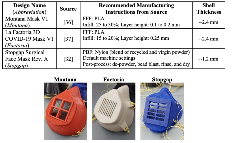
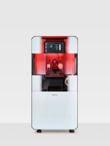
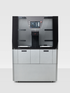
The masks chosen were the Montana mask by Make the Masks, the Factoria mask, and the Stopgap Surgical Face Mask. They printed these masks on a Sinterstation (polymer powder bed fusion), Fortus 400mc (industrial FFF) and an Afinia (desktop FDM). We have made some progress since the venerable Sinterstation and porosity has been reduced in current generation sintering machines with better software and processing so that we would expect less porosity than with a machine that was released in 1998. On the one hand, it’s amazing that these machines last so long, but it is perhaps slightly unfair to use a 22-year-old 3D printer as the industrial sintering system for this important test.

The paper states that, “the PBF models were de-powdered and bead blasted to remove adhered powder and improve surface finish,” but, crucially, it is unclear if “rinse parts with water to remove remaining media and dry parts using compressed air” was done as per the general instruction attached to the file. Also, they state that the powder used was “Nylon-12 (Factoria: 100% recycled; Montana/Stopgap: 50% recycled / 50% virgin).” It’s unclear whose powder it was. Now, its not apparent why they would use different mixes between virgin and recycled powder for different masks but a 100 percent recycled material is not really something I’d recommend. I think it’s also unfair to compare a 100 percent recycled mask to anything.

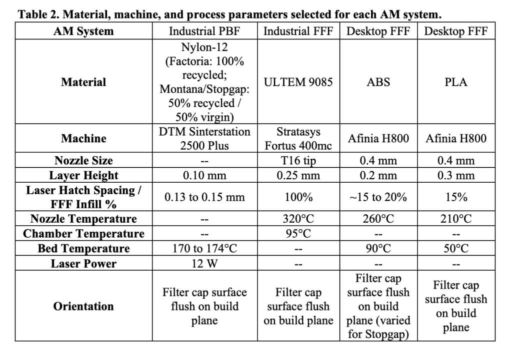
I also have concerns about the filament materials printed. I also thought that ULTEM 9085 printed at 350°C? I’m confused about the ABS print that has 15-20% infill. To me, for a day-to-day use part, I’d use a much higher infill percentage of 30 percent at least. I also don’t understand why the PLA part has 15 percent infill either. I couldn’t find the machine settings or the name of the filament supplier either. There could be a lot of variability in their nozzle temperature as per indicated and actual also. We all know that we can get a lot of layer adhesion differences in prints from speed, material, temperature. So this is one caveat. I’d really like for the Cura profile and the machine settings to be included in this kind of research. If we’re going to be testing parts then we should know how they were made.

This isn’t a gripe specific to this paper however; no papers have this. I personally can’t really get ABS to work at all below a 100°C bed temperature and most recommend 110°C, so that seems low, while 260°C sounds like it could be rather too fume-y. I’d never recommend that you print ABS above 250°C and, most of the time, I’d expect the right temperature to be far lower than that, much lower than 260°C anyway. Also, each test part was only printed once (apart from the stopgap that they tried in two orientations). That to me is putting rather a lot of stock in the five-year-old Afinia’s accuracy and I would have much rather seen a number of parts printed and tested.

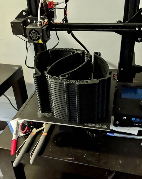
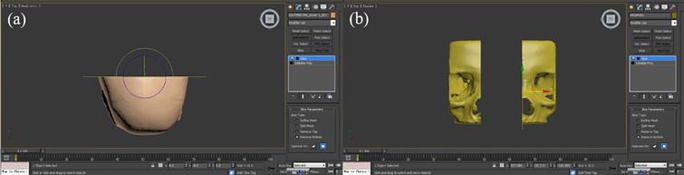
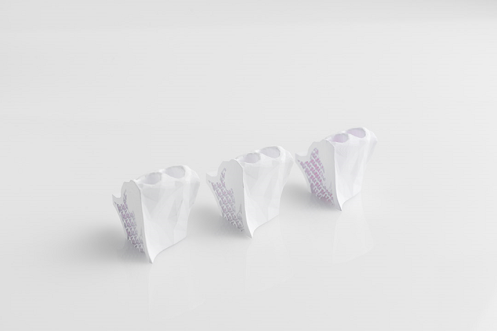
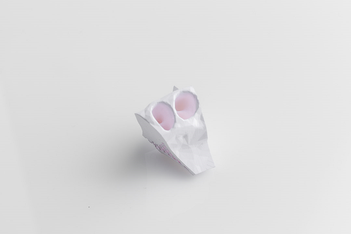
The team then shows us that they had visible defects in the prints.

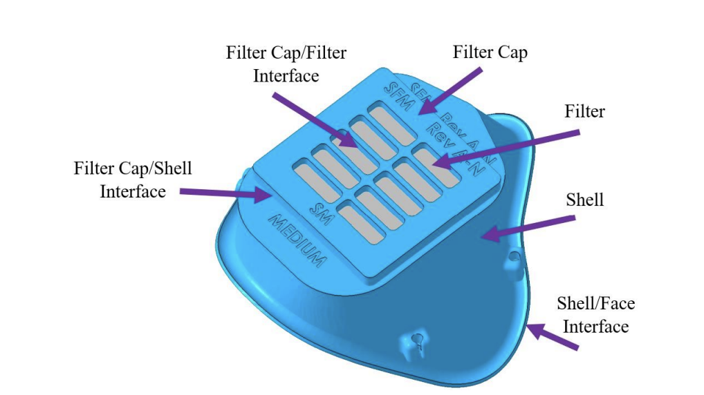
“(a) The Stopgap respirator in ABS oriented with the filter cap face down on the build plane has a few mislaid layers; (b) The Stopgap respirator in ABS in an alternate orientation also suffers from periodic sparsity; (c) The Stopgap respirator in PLA is visibly thin across most surfaces; (d) The Stopgap respirator in ULTEM shows porosity on the surface parallel to the filter.”

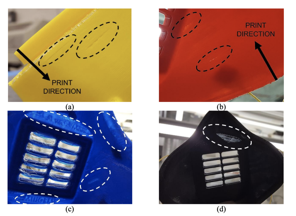
“Figure 4c shows the Stopgap respirator fabricated with PLA held up to a light to enable observation of several regions of thin material along the shell (as in Figure 4a and b), along the seal to the face, and on the surface flush with the filter cap. Figure 4d displays the Stopgap respirator fabricated with ULTEM held up to a light. Macroscale pores across the entire surface flush on the build plane are observed despite this part being printed in 100% infill on an industrial-scale FFF system,” the authors write. The team does say that the Stopgap respirator was made for powder bed fusion ,so that it was not meant to be printed with FFF/FDM. They go on to test the Stopgap FFF/FDM prints and I think that this is rather unfair.

I have a real issue with the authors changing the roll of filament for build orientation prints “a” and “b” and not mentioning that this is a different material. Even if it was from the same vendor and the handling was the same, then the different colorants mean that there is a different optimal print temperature there. It’s strange to me to both change print orientation and material and then compare those prints. Also, the authors say that this is an adhesion issue, but is it? Is it digging by the nozzle? The “c” part is a great example why you should not have letters on your part. The hatched pattern on the “d” print made from ULTEM is very strange. Is that the Sparse Double Density infill pattern? Did it not print because they didn’t support the part well?

The team went on to test the results of the different filter designs:

“The particle analyzer simply counts the frequency of detected nanoparticles; it does not distinguish between nanoparticles resulting from the generated aerosol and residual nanoparticles resulting from stray particulates shed from the shell,”  was an issue that they identified.

They go on to treat the masks, saying that the “FFF respirators were rinsed thoroughly with tap water and dried with compressed air. Since water could cause aggregation among dry powder, the cleaning step for PBF respirators involved additional compressed air followed by the application of two coats of acrylic paint to form a sealant.”

I’m confused about this since I know that water can have effects on porous sintered parts long-term, but am not sure why the researchers didn’t just wash them in water, which would be fine short-term. Also, painting it changes the part and makes it less flexible. I don’t understand the “aggregation among dry powder” part at all really and am not sure why they’d need to paint the model. I especially worry that the coats of paint will effect how the different parts of the mask fit together. I may have read it wrong but why then in the table above do they say that they rinse and dry the PBF parts? Also I’m pretty sure that the PLA models were made more brittle by the water, but perhaps that’s a limitation of the mask that’s good to include.

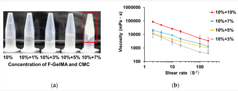
The paper goes on to show that, “none of the printed respirators provided the requisite 95% filtration efficiency.”

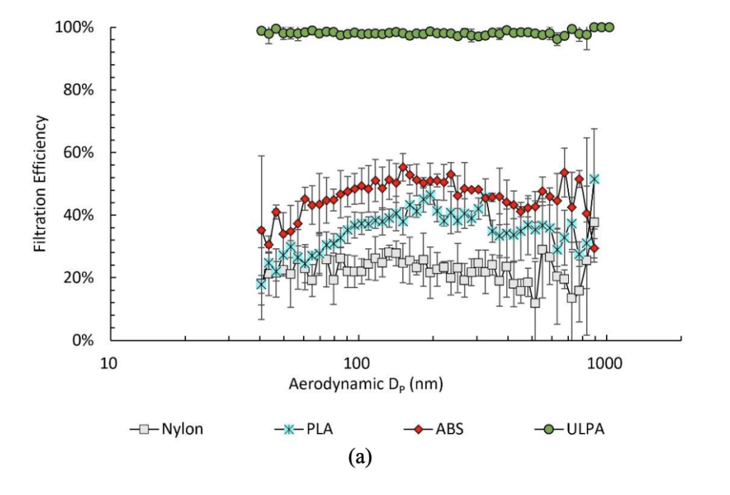
“Montana respirator results (Figure 5a) show filtration efficiency consistently under 60% for the ABS, PLA, and nylon materials, which is far from the baseline performance of the ULPA filter medium. The ULTEM variant of the Montana respirator could not be tested as printed because the filter cap was too loose to adequately secure the filter.”

The team makes the following determination:

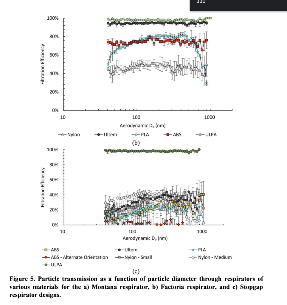
“The Factoria respirator results are provided in Figure 5b. The PLA and ABS respirators filter out more particles than in the Montana respirator design, but both still only protect against ~75% of particles. The ULTEM Factoria respirator provides the highest observed performance, with a filtration efficiency between 90-95%, depending on particle diameter; however, it falls slightly less than the tested ULPA filter (99% efficiency). Similar to the Montana respirator results, the PBF-printed respirator presents the lowest filtration efficiency (~45%).”

“Montana and Factoria respirators are nearly identical in shell design, it is expected that the difference in filter cap design is the cause for the consistently worse performance of the Montana respirator compared to the Factoria respirator. The press-fit cap of the Montana respirator may have allowed particles around the filter (which correlates to the loose-fitting filter cap printed in ULTEM), whereas the larger cap of the Factoria respirator completely encloses the filter.”

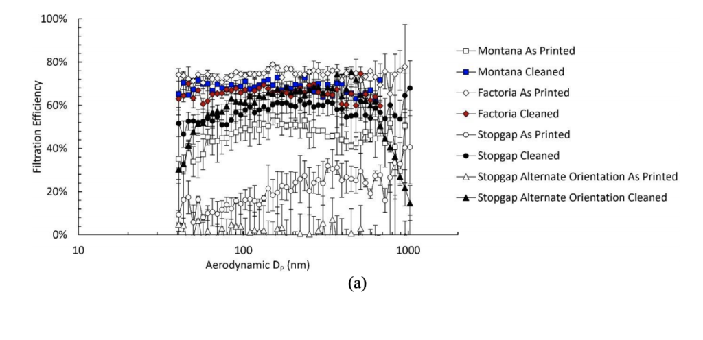
Another thing that I don’t get is this: “It is observed in Figure 6a that cleaning the ABS Montana respirator increases the filtration efficiency measurement by ~20%, but the ABS Factoria measurement decreases in efficiency by ~10%. The ABS Stopgap efficiency measurements significantly improve, with both print orientations offering similar performance once cleaned. In Figure 6b, it is seen that the ULTEM Factoria respirator decreases by ~15% efficiency following cleaning.”

I’m quite surprised that there would be such a huge difference in filtration efficiency just from cleaning the parts? To me, this points to the fact that the testing apparatus is picking up loose powder and particles on the masks themselves from before, or that they are created or released through cleaning. But, I don’t know enough about the filtration side of things to know.

The team concedes, “These results highlight the inherent variability in results due to the testing method and testing conditions, which is why it was critical to use the same respirators for repeat tests. The testing environment was kept as close to the same conditions each time, yet the Factoria respirators somehow declined in filtration efficiency. It is believed that a coupling of the failure modes identified in Section 1.2 could be contributing to the erratic trends.”

They go on to look deeper, “Application of the epoxy sealant to the shell increases efficiency to peak at ~75%. This indicates that the porosity of the PLA material drops filtration efficiency by ~20%.” And “Residual powders from printing, post-process, or handling are likely to blame for the poor performance of the respirators as-printed. This also corroborates the reason why the as-printed nylon Montana and Factoria respirators had such low filtration efficiency. While testing some intermediate modifications were forgone, it is evident that the dominant failure mode is the filter cap/shell interface.”

Their conclusions are the following:

“As printed, most of the respirators performed poorly, with almost all providing less than 60% filtration efficiency (significantly below the requisite 95% efficiency of a N95 respirator). This result is especially discouraging when considering that the testing was done with the approximation of a perfect seal between the respirator and user’s face (a common failure mode for standard N95 textile respirators, and likely a significant failure mode for the rigid printed polymers). When printed in ULTEM on an industrial-scale FFF system, the Factoria respirator provided the best filtration efficiency of those evaluated, consistently exceeding 90% efficiency for all particle sizes.”

They also say that, “For example, while the Factoria respirator in ULTEM reached >90% filtration efficiency in the as-printed state, its measured efficiency was reduced to ~80% following cleaning. No tested design with modifications was able to consistently attain 95% filtration efficiency, although the nylon Stopgap respirator with modifications was able to filter ~85% of particles at the size of 300 nm.”

“The results from this study do not completely discount AM from being appropriate for making an effective N95 respirator,” the authors write. “The ULTEM Factoria’s performance suggests that (i) high quality, repeatable printing technology with (ii) proper process settings, and (iii) tolerancing of the filter cap/shell interface that is aligned with a specific machine/material combination could provide an effective solution.”

Further on they, say, “In the case of the Montana and Stopgap respirators, the as-printed performance falls below that of many simple textile materials. The as-printed Factoria respirators and post-process modified Stopgap respirators provide equivalent protection to these textile materials and surgical masks, with the ULTEM Factoria and modified PBF Stopgap respirators providing slightly enhanced performance to these materials.” This was a result that many of us would actually have been happy with, I believe.

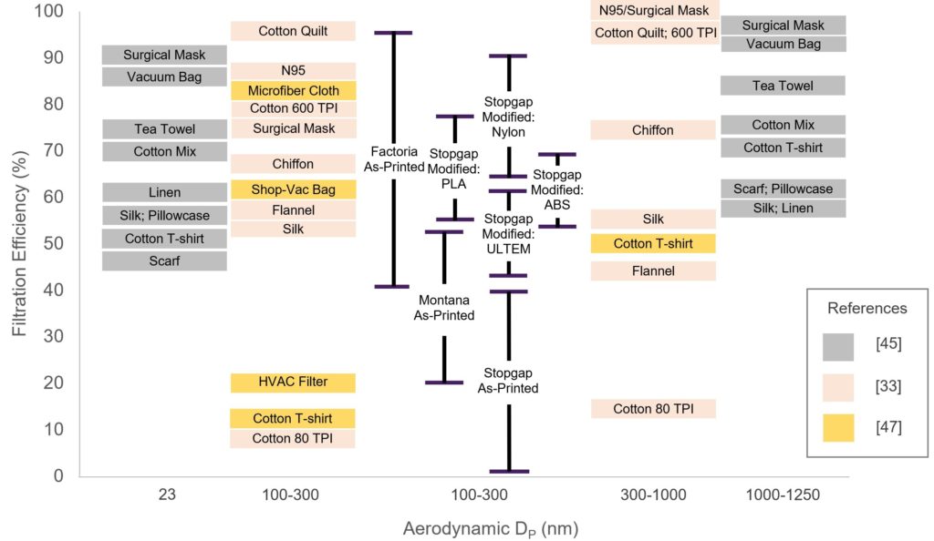
Also, “The modified PBF Stopgap respirators can perform better than the surgical mask, high-threaded cotton, and N95 respirator from the study by Konda [33]. This study shows AM respirators are capable of achieving competitively high filtration efficiency on par with non-medical use masks only when assuming a perfect seal to the face.” This is a very good result however and one that we’d be very happy with. But, as the paper rightfully states, this perfect seal is illusory and is probably not the case for these relatively rigid parts. The inability to make a good seal, especially when compared to a home-sewn mask has always to me been the Achilles heel of 3D-printed respirators.

On the whole, it is very good that this kind of research is being done. I’m a little confused by some of the printing and parameters involved. I would have liked to have seen more consistency there. But assembly and print-related issues in experiments only cause me to consider how such variability precludes us from making respirators. On the whole, we can conclude that it will be difficult to make a respirator that works well with 3D printing. This does not mean that we should be dissuaded from trying to improve these designs but rather that we should welcome scientific rigor and analysis to our endeavors.

The post 3D-Printed Respirator Masks Below N95 Standards, Says Virginia Tech Team appeared first on 3DPrint.com | The Voice of 3D Printing / Additive Manufacturing.

3D-Printed Models for Training & Planning of Endoscopic Pituitary Surgery

A small gland (about the size of a small bean) located at the base of the skull, the pituitary secretes hormones and regulates the other major organs. Also known as the master gland, it is vital to good health, and can be seriously affected by over-production of hormones—or the opposite. When something goes wrong, surgery can be necessary, but tricky; for instance, endoscopic endonasal transsphenoidal pituitary surgery presents numerous challenges and risks as a procedure.

Seeking better solutions for surgeons, researchers came together to perform a recent study, detailing the results in ‘The manufacturing procedure of 3D printed models for endoscopic endonasal transsphenoidal pituitary surgery.’ Honing in on the need to provide better training—especially as cadavers can be hard to come by for both medical students and surgeons to practice on—the researchers examined the further potential of 3D printed models for improved planning.

In removing pituitary tumors, surgeons must work through the nose and sphenoid sinus, using tools to enter the nasal cavity and then actually break the bone until the sphenoid sinus is visible. This is considered to be challenging due to the complex anatomy of the nose, as well as in terms of how the sinus is situated, with the endoscope method most commonly used for surgical removal.

For this study, the authors not only propose that surgeons should be able to 3D print and customize skull models themselves but also use them ‘directly for simulation of the surgery.’ Along with that, their goal was to use a multi-tiered software system to offer better precision in creating the models from CT data.

Overview of the main goals and operations in digital model processing.

“The limited accuracy of CT scanning and threshold segmentation may cause missing features and unexpected holes in the digital model,” explained the authors, moving forward to refine the process with new software.

Set the thresholding value for Bone (CT). We select Menu bar > Segmentation > Thresholding, and set the minimum value as 226 (Bone (CT)) to get the required part of the skull model in this case. The thresholding result is saved as a new mask automatically.

Using Materialise Interactive Medical Image Control System (Mimics), they were able to improve the CT scan by fixing the holes at the base, along with using Geomagic to optimize and repair the skull base and surgical area. Segmentation consisted of extracting the nasal cavity and sellar region.

An unexpected hole on the model.

Fix the hole by drawing slice by slice. We use Menu bar > Segmentation > Edit Masks and select ‘Circle’ with reasonable size and select ‘Draw’ to connect the part.

The hole is fixed.

Comparison of the surgical area model before and after the repair.

3ds Max was used for segmentation and production of molds, correcting the surgical area and base into a polygon shape.

Slicing operations on the skull base and the nasal cavity.

The skull base with supports.

An Ultimaker 2 was used for 3D printing the base and molds, printing with PLA; however, for the surgical area, the researchers switched to binder jetting, using a 3D Systems ProJet 660 Pro with plaster.

“For the practice, the surgical area should be printed by the breakable and low-cost material (like plaster), as the sellar region will be broken during the real surgery,” explained the researchers.

Molds were used for fabrication of the soft tissue due to the expense in 3D printing the material directly. The researchers mixed pigment and silica gel to represent the following:

  • Face
  • Pituitary
  • Optic nerves
  • Internal carotid arteries

To imitate the pituitary tumor properly, the researchers set the model tumor underneath the optic nerve, using an adhesive for proper placement.

Small red dots, which look like capillaries.

The complete model and the face.


Surgical area models with different levels of tumors.

“With the assistance of 3D printed medical models, the surgery can be practiced repeatedly,” concluded the researchers. “The surgical safety can be improved, and the risk of death and morbidity can be reduced. In addition, the 3D printed medical model can be a good tool for the patient or their family members to learn about the disease, the condition and the risk of the surgery, which can promote the communication between the patients and neurosurgeons.”

“The outcomes demonstrate that the 3D printed skull model is able to improve the structure recognition learning. This case proves that the 3D printed anatomical model is worthy of use. Obviously, the model for specific surgery is able to improve the understanding of students or neurosurgeons on the specific or special situations.”

3D printed models are helpful today in a wide array of applications, but within the medical realm they are being used for diagnosing health conditions like tumors, as well as allowing for more streamlined treatment. Even better, such models allow more detailed explanations for patients and their families about ensuing treatments and possible surgical procedures.

Medical students are able to train with 3D printed models, surgeons can prepare for rare procedures—or those which have never been performed before—and such models may also be used as extremely helpful guides in the operating room too. Discuss this article and other 3D printing topics at 3DPrintBoard.com.

[Source / Images: ‘The manufacturing procedure of 3D printed models for endoscopic endonasal transsphenoidal pituitary surgery’]


The post 3D-Printed Models for Training & Planning of Endoscopic Pituitary Surgery appeared first on 3DPrint.com | The Voice of 3D Printing / Additive Manufacturing.

Free Automated Software to Design 3D Printable Cranial Implants

Repairing skull defects with custom cranial implants, otherwise known as a cranioplasty, is expensive and takes a great deal of time, as the the existing process often results in bottlenecks due to long wait times for the implant to be designed, manufactured, and shipped. While 3D printing the implants can help with these issues, a team of researchers from the Graz University of Technology and Medical University of Graz in Austria published a paper, “An Online Platform for Automatic Skull Defect Restoration and Cranial Implant Design,” about an automated system for cranial implant design they’ve devised that can do even better.

“Due to the high requirements for cranial implant design, such as the professional experience required and the commercial software, cranioplasty can result in a costly operation for the health care system,” the researchers wrote. “On top, the current process is a cause of additional suffering for the patient, since a minimum of two surgical operations are involved: the craniotomy, during which the bony structure is removed, and the cranioplasty, during which the defect is restored using the designed implant. When the cranial implant is externally designed by a third-party manufacturer, this process can take several days [1], leaving the patient with an incomplete skull.”

In the case study they cited above, the researchers explained that a professional design center in the UK designed the cranial implant for a patient who lived in Spain. The CT scans had to be transferred from the hospital in Spain to the UK design center, and then a separate UK company 3D printed the titanium implant, which was shipped back to Spain. That’s a lot of unnecessary back and forth.

“Therefore, the optimization of the current workflow in cranioplasty remains an open problem, with implant design as primary bottleneck,” they stated.

“Illustration of In-Operation Room process for cranial implant design and manufacturing. Left: a possible workflow. Right: how the implant should fit with the skull defect in terms of defect boundary and bone thickness.”

One option is developing ad hoc free CAD software for cranial implant design, but the design process still requires expertise and an extended wait.

“In this study, we introduce a fast and fully automatic system for cranial implant design. The system is integrated in a freely accessible online platform,” the team explained. “Furthermore, we discuss how such a system, combined with AM, can be incorporated into the cranioplasty practice to substantially optimize the current clinical routine.”

The system they developed has been integrated in Studierfenster, an open, cloud-based medical image processing platform that, with the help of deep learning algorithms, automatically restores the missing part of a skull. The platform then generates the STL file for a patient-specific implant by subtracting the defective skull from the completed one, and it can be 3D printed on-site.

“Furthermore, thanks to the standard format, the user can thereafter load the model into another application for post-processing whenever necessary,” the researchers wrote. “Multiple additional features have been integrated into the platform since its first release, such as 3D face reconstruction from a 2D image, inpainting and restoration of aortic dissections (ADs) [4], automatic aortic landmark detection and automatic cranial implant design. Most of the algorithms behind these interactive features run on the server side and can be easily accessed by the client using a common browser interface. The server-side computations allow the use of the remote platform also on smaller devices with lower computational capabilities.”

3D printing the implants makes the process faster, and combining it with an automated implant design solutions speeds things up even more. The researchers explained how their optimized workflow could potentially go:

“After a portion of the skull is removed by a surgeon, the skull defect is reconstructed by a software given as input the post-operative head CT of the patient. The software generates the implant by taking the difference between the two skulls. Afterwards, the surface model of the implant is extracted and sent to the 3D printer in the operation room for 3D printing. The implant can therefore be manufactured in loco. The whole process of implant design and manufacturing is done fully automatically and in the operation room.”

The cost decreases, as no experts are required, and the wait time is also reduced, thanks to the automatic implant design software and on-site 3D printing. The patient’s suffering will also decrease, since the cranioplasty can be performed right after removal of the tumor.

“Architecture of automatic cranial implant design system in Studierfenster. The server side is responsible for implant generation and mesh rendering. The browser side is responsible for 3D model visualization and user interaction.”

The team’s algorithm, which processes volumes rather than a 3D mesh model, can directly process high dimensional imaging data, and is accessible to users, and easy to use, through Studierfenster. Another algorithm on the server side of the system converts the volumes of the defective, completed skull, and the implant into 3D surface mesh models. Once they’re rendered, the user can inspect the downloadable models in the browser window.

“An example of automatic skull defect restoration and implant design. First row: the defective skull, the completed skull and the implant. Second row: how the implant fits with the defective skull in term of defect boundary, bone thickness and shape. To differentiate, the implant uses a different color from the skull.”

“The system is currently intended for educational and research use only, but represents the trend of technological development in this field,” the researchers concluded. “As the system is integrated in the open platform Studierfenster, its performance is significantly dependent on the hardware/architecture of the platform. The conversion of the skull volume to a mesh can be slow, as the mesh is usually very dense (e.g., millions of points). This will be improved by introducing better hardware on the server side. Another limiting factor is the client/server based architecture of the platform. The large mesh has to be transferred from server side to browser side in order to be visualized, which can be slow, depending on the quality of the user’s internet connection.”

Discuss this and other 3D printing topics at 3DPrintBoard.com or share your thoughts below.

The post Free Automated Software to Design 3D Printable Cranial Implants appeared first on 3DPrint.com | The Voice of 3D Printing / Additive Manufacturing.

NP Swabs Prove 3D Printing’s Scalability and Speed-to-Market Advantages

A year ago, if you would have asked anyone in our industry what they thought might be a future killer application, it’s highly doubtful that anyone would have replied with, “nasopharyngeal swabs.” Until recently, it was a niche product and the entire market was serviced by a few dominant industry players.

While there are now other protocols, the main test for COVID-19 testing involves gathering virus from deep in a person’s nasal cavity. The SARS-CoV-2 virus is collected using a nasopharyngeal (NP) swab. Traditionally, these swabs are made in two parts, including a polyester handle and a tip with tiny rayon fibers called flock.

A 3D-printed NP swab developed by Carbon. Image courtesy of Carbon.

The two primary companies that make them, Purtian Medical Products Co. and Copan Diagnostics, bulk manufacture them in multiple steps, which include production, assembly, sterilization and packaging, among others. Their process requires customized machinery and a sizable group of relatively skilled people.

When the global pandemic struck, the demand for COVID-19 test kits skyrocketed, far outpacing the combined capacity of these two companies. For several reasons, they had difficulties scaling their businesses. They both produce many other products for the medical industry and adding new equipment is a timely endeavor. To make matters worse, Copan which is located in Italy (a hotspot for the virus) was challenged with maintaining the health of its own workforce.

3D Printing to the Rescue

As it became apparent that the normal suppliers couldn’t fully meet the need, the additive manufacturing industry began working on the problem. Markforged, a manufacturer of filament-based 3D printers, partnered with Neurophotometrics to produce 3D-printed NP swabs made from their Fiberflex Rayon.

Separately, Northwell Health teamed up with the University of South Florida, Tampa General Hospital and Massachusetts-based Formlabs and worked with physicians to design their own NP swab, which Formlabs recently started printing in its FDA-registered, ISO 13485-certified factory in Ohio.

Results from a clinical trial of 3D-printed NP swabs. Image courtesy of Beth Israel Deaconess Medical Center.

Soon after, a consortium of 3D printing companies was codified. Their goal was to deliver clinically tested, FDA-registered, 3D-printed COVID-19 NP test swab designs with superior or equivalent efficacy to flocked swabs, at scale.

Origin Partners with Henkel

Origin, manufacturer of stereolithography (SLA) 3D printers was one of the founding members of the consortium. It began working with several partners to develop what it is now calling the world’s first FDA-compliant, sterile, 3D-printed NP test swab. 

3D printed NP swabs with detailed lattice structure. (Image courtesy Origin.)

In a new announcement, the startup is providing more detail about the process. Origin collaborated with materials company, Henkel and Beth Israel Deaconess Medical Center (BIDMC) on the swab’s development. Working with generative design software, Origin was able to design a one-piece swab that performed as well as the traditional version. Henkel leveraged its own Albert software platform to specify a material that would meet the requirements for an in-body medical device. Together they tested the design’s clinical capabilities, in addition to validating each step in the sterilizations process, and conducting rigorous mechanical testing and packaging certification.

Scalability and Speed-to-Market

Within weeks they were able to bring a product to market that is classified as a sterile device and is considered a finished medical product, which is regulated by the FDA.

It’s a perfect example of two of 3D printing’s biggest benefits: scalability and speed-to-market. As Chris Prucha, Founder and CEO at Origin noted in the press release, “by working collaboratively and utilizing each other’s technologies, we identified, optimized and scaled the manufacturing process to bring an application to market extremely fast.”

Origin’s sterile NP swabs are currently shipping to leading healthcare facilities, government institutions, and independent testing centers in the U.S. and several other countries. They’re also available for purchase on Amazon.

But beyond the opportunity with NP swabs, this collaboration also further substantiates the industry’s growth into functional part production.  

In the press release, Ken Kisner, Head of Innovation for 3D Printing at Henkel said, “From inception, the vision behind Henkel’s Open Materials Platform was to enable collaboration all along additive manufacturing’s value chain. Working together with Origin, we were able to develop a product which is just as effective as its mass-produced counterpart. With the constraints commercial medical suppliers are facing, this presents a significant opportunity for the 3D printing industry to demonstrate its capabilities, beyond prototyping.”

Innovate Globally, Produce Locally

The problem wasn’t just related to the manufacturing of NP swabs. There were constraints all across the medical supply chain. Some of it had to do with the traditional model of centralized manufacturing and logistics. The healthcare industry relies on a relatively small number of producers and distributors. When they’re impeded, all bets are off. Further, the vast number of products, the niche nature of some of them, and shelf life issues make some medical products difficult to stockpile.

Perhaps more than anything else, this application demonstrates the value of a nimble, distributed manufacturing network, where identical parts can be made as close as possible to the point of need. In some cases, it can be financially beneficial, but in others like this decentralizing production provides an insurance policy in the event the unimaginable happens. We know it can, because it has.

About the Author

John Hauer is the Founder and CEO of Get3DSmart, a consulting practice which helps large companies understand and capitalize on opportunities with 3D printing. Prior to that, John co-Founded and served as the CEO of 3DLT. The company worked with retailers and their suppliers, helping them sell 3D printable products, online and in-store.

As a technology journalist, John focuses primarily on the topics of 3D printing, artificial intelligence, virtual reality and automation. His original content has been featured on Forbes, TechCrunch, Futurism, QZ.com, Techfaster.com, 3DPrint.com and Fabbaloo, among others.

Follow John on Twitter @Get3DJohn

The post NP Swabs Prove 3D Printing’s Scalability and Speed-to-Market Advantages appeared first on 3DPrint.com | The Voice of 3D Printing / Additive Manufacturing.

Bioresorbable Ceramics Allow Admatec to Make 3D-Printed Bone Implants

Before I started writing about additive manufacturing a number of years ago, I thought ceramics were only used to make fancy pottery. While it is used for this purpose as well, the hard, non-metallic material is so much more than that: it’s strong in compression, can withstand very high temperatures and chemical erosion, and a SmarTech Analysis report suggests that the ceramics AM market will reach an estimated $4.8 billion in revenue by the year 2030.


Focused on advanced ceramics and metal 3D printing, Dutch company Admatec is now introducing bioresorbable ceramics to its portfolio. CAM Bioceramics, an ISO 13485:2016 certified supplier of medical grade ceramics, is supporting Admatec in its endeavor to introduce new 3D printing opportunities for the material via its Admaflex printers in the medical and dental fields.

“As a supplier for the Orthopaedic and Dental market, CAM Bioceramics acknowledges that 3D Printing will play an increasingly significant role in numerous patient treatment plans. Calcium Phosphate based bone reconstructions will play an important part in this next generation medical device solutions due to its proven biocompatibility,” said C. Hogeboom, CEO of CAM Bioceramics. “As one of the market leaders in this area, CAM Bioceramics actively partners with 3D print experts and promotes the evolution of 3D printed solutions.”

Advanced ceramic materials have a great deal of utility in medical applications, particularly when fabricating surgical instruments and implants. 3D-printed, patient-specific implants made from bioresorbable and biocompatible ceramics are often used in craniomaxillofacial and hemimaxillary surgeries, as they can be absorbed by the human body. Native bone tissue will eventually replace the implant, so it won’t need to be removed later along with autologous bone (harvested from the patient’s own body), which can be painful for patients.

While tricalcium phosphate, or bone ash, is often used in this application, bioactive hydroxyapatite—chemically similar to the mineral component of bones—gives the body more time to heal, as it takes longer for the body to absorb it. By working with CAM Bioceramics, Admatec can now use a commercially available hydroxyapatite slurry material in its Admaflex DLP 3D printers to make patient-specific implants.

Implants that are bioresorbable can be made with very defined pore structures and geometries. Additionally, 3D printing these means that it’s possible to define an optimal geometry for the implant, so it perfectly fits the patient. Bone ingrowth from nearby tissues in the body is facilitated by using both dense and porous structures in bone tissue engineering, and 3D printing fits the bill, as it can produce interconnected scaffolds with well-defined sizes and geometries.

“Using Hydroxyapatite, we can manufacture patient-specific, bioresorbable implants, which have defined pore structures and geometries,” Admatec stated in a press release. “Combinations of functional graded ceramic materials, such as dense and porous structures in a single hemi-maxillary bone-like structure, have been printed successfully with help of the lithography based additive manufacturing technology provided by Admatec.”

Admatec’s suggests that its technology can efficiently print complex channels, geometries, and lattice and honeycomb structures with wall thicknesses and features in the 100 μm range. Its batch-oriented process can print larger implants and its multi-part printing software can help print different geometries simultaneously for smaller implants.

“Engineering know-how, flexibility and ease of use of the Admatec system have convinced CAM Bioceramics to collaborate with Admatec in this development thus enabling other medical device companies to take full advantage of this ‘turnkey’ solution,” said Giuseppe Cama, Head of Innovation & Science at CAM Bioceramics. “Customization to optimize patient outcomes can easily be achieved, supported by the physical proximity of the two companies.”

With its newly available bioresorbable ceramics, Admatec can offer medical device manufacturers even more customized solutions. For instance, the company is now working on multimaterial DLP 3D printing, where a combination of zirconia and hydroxyapatite is used to print combination implants for the lower jaw.

Large bone defects resulting from tumors or severe trauma can’t heal on their own without assistance, and one potential solution is a cage, 3D printed out of high-strength zirconia or another ceramic material, that can support the area while it’s healing. The cage would be paired with an implant, featuring an inner volume of bioresorbable ceramic, like hydroxyapatite, and because they’re biocompatible, both could be left in the body.

Discuss this story and other 3D printing topics at 3DPrintBoard.com or share your thoughts in the Facebook comments below.

(Source/Images: Admatec)

The post Bioresorbable Ceramics Allow Admatec to Make 3D-Printed Bone Implants appeared first on 3DPrint.com | The Voice of 3D Printing / Additive Manufacturing.

3DHEALS 2020 Virtual Medical Summit: 3D-Printed Materials in Healthcare

There were a multitude of sessions and sub-sessions to follow at 3DHEALS 2020 (running from June 5-6), with over 70 speakers and four workshops, covering many topics on complex design, and patient-specific treatment. Here at 3DPrint.com, we have covered many stories on materials, as researchers and manufacturers delve into their uses in other major applications too like automotive, aerospace, construction, and so much more.

At the “Material Science in Healthcare 3D Printing” session, medical applications were discussed in detail by Balaji Prahbu (Director of Strategic Marketing for Medical Device Solutions/Evonik Industries), Steve Kranz (Lab Manager and Senior Scientist at Origin), Sean Dsilva (Medical Marketing Segment Head for 3D Printing Henkel) Adhesive Technologies Division), and Mike Vasquez (Founder and CEO, 3Degrees). Topics covered included the importance of bioresorbable materials, biocompatible materials, those used to create devices and tools during the COVID-19 pandemic, as well as workflow systems designed by materials engineers.

Balaji Prahbu opened the presentation with statistics on osteoporosis, a condition that causes millions of fractures in patients around the world. Currently, Evonik uses a variety of polymers to create materials and implants that assist in treating and healing and reconstructing human bone. While some materials dissolve in the body over time, other types of materials created by Evonik can also be used permanently.

The Germany-based company is also developing other new powders and filaments for patient-specific implants that can be produced in just a few hours and completely suited to the individual needs of patients—evidence of some of the greatest benefits in using 3D printing technology.

Sean Dsilva offered information regarding Henkel’s biocompatible materials, explaining more too about the keys to developing high-quality, high-performance UV resins—all tied together with effective workflow processes. The $20 billion company specializes in adhesives technology, including that for the medical field. Currently, Henkel offers four different levels of biocompatible materials:

Henkel’s materials can be used for an extremely diverse number of applications, from auditory devices like hearing aids, to prosthetics such as orthotics, bionics, and more. 3D-printed models can also be created, offering a host of benefits like better diagnostics, treatment, and education for patients and their families. Not only that, 3D-printed medical models allow for better training of medical students and allow surgeons to prepare for delicate procedures too.Henkel’s materials are also used to fabricate other industrial components like jigs, fractures, and devices.

Steve Kranz definitely offered some of the most interesting information regarding materials, and Origin’s recent transformation from software developer and 3D printer manufacturer to a ‘swab factory’ in response to the coronavirus pandemic. The San Francisco-headquartered company began manufacturing a variety of different 3D-printed swabs for testing purposes, as well as originally designing shields and other personal protection equipment, to include adapting snorkels to be transformed into N-95-style face masks.

Kranz began by explaining, however, that while Origin has—previous to the COVID-19 pandemic—been centered around software and hardware endeavors, they do not develop materials; instead, they rely on experts like Henkel, BASF, DSM and others.

“When COVID-19 hit, things changed for us. It was a lot different, so we had to adapt to survive,” explained Kranz. “We transformed ourselves from a platform that allowed other people to do 3D printing to becoming a factory, to printing parts ourselves.”

While the Origin team did initially begin creating other types of COVID-19 devices, Kranz stated that they quickly realized they could offer the most important contribution by 3D printing nasopharyngeal swabs. They began collaborating with nTopology, drafting a flexible, effective design.

While the two companies were able to work together in creating the actual swab, there were numerous obstacles. Some supplies were difficult to attain, such as isopropyl alcohol, gloves, and paper products. They were also challenged in scaling up with more inventory and other resources, dealing with waste, and hiring additional staff to work a lot of long, and “sometimes crazy” hours.

“That’s been Origin’s journey for the past couple of months. It has been very intense, challenging, strange at some times, but also really rewarding and I feel like we have learned a lot. We’ve kind of put ourselves right in the fire in terms of testing out our own production, our own capabilities, and we have learned a lot that is going to help improve our own printers in the future,” said Kranz.

Mike Vasquez opened by explaining that, as a materials engineer, he realizes that additive manufacturing is “fundamentally driven by materials, but it is complicated.” This is due to a lack of accessibility in many cases, an “opaque and often confusing” supply landscape, and limited standards. Material properties may be an issue as well, as they often do not match up with what users are expecting or needing for specific projects.

Because there can be so many challenges—and so much data—involved with creating a 3D-printed part, the 3Degrees team developed the TRACE process for 3D printing workflow management. In creating TRACE, they spoke with over 50 additive manufacturing users, auditors, manufacturers, and standards organizations. The workflow management tool, complete with comprehensive analytics is meant to be customized for different projects.

During the fabrication of 3D-printed medical devices, TRACE can be used to keep track of variables like data inputs, specifications for materials and machine processes, post-processing, and inspections.

Although originally set for ‘the heart of San Francisco’ as a venue, this year’s 3DHEALS Global Healthcare 3D printing conference became a virtual—and inspiring—event. Focusing on the continued impacts to the field of medicine, rather than cancel the annual event due to the COVID-19 restrictions, founder and CEO, Dr. Jenny Chen, committed to an online format, and along with seeing every speaker conform to the changes, she was even able to able 25 percent more in programming.

What do you think of this news? Let us know your thoughts! Join the discussion of this and other 3D printing topics at 3DPrintBoard.com.

[Source / Images: 3DHeals 2020 – from the ‘Material Science in Healthcare 3D Printing’ session)

The post 3DHEALS 2020 Virtual Medical Summit: 3D-Printed Materials in Healthcare appeared first on 3DPrint.com | The Voice of 3D Printing / Additive Manufacturing.