Free Automated Software to Design 3D Printable Cranial Implants

Repairing skull defects with custom cranial implants, otherwise known as a cranioplasty, is expensive and takes a great deal of time, as the the existing process often results in bottlenecks due to long wait times for the implant to be designed, manufactured, and shipped. While 3D printing the implants can help with these issues, a team of researchers from the Graz University of Technology and Medical University of Graz in Austria published a paper, “An Online Platform for Automatic Skull Defect Restoration and Cranial Implant Design,” about an automated system for cranial implant design they’ve devised that can do even better.

“Due to the high requirements for cranial implant design, such as the professional experience required and the commercial software, cranioplasty can result in a costly operation for the health care system,” the researchers wrote. “On top, the current process is a cause of additional suffering for the patient, since a minimum of two surgical operations are involved: the craniotomy, during which the bony structure is removed, and the cranioplasty, during which the defect is restored using the designed implant. When the cranial implant is externally designed by a third-party manufacturer, this process can take several days [1], leaving the patient with an incomplete skull.”

In the case study they cited above, the researchers explained that a professional design center in the UK designed the cranial implant for a patient who lived in Spain. The CT scans had to be transferred from the hospital in Spain to the UK design center, and then a separate UK company 3D printed the titanium implant, which was shipped back to Spain. That’s a lot of unnecessary back and forth.

“Therefore, the optimization of the current workflow in cranioplasty remains an open problem, with implant design as primary bottleneck,” they stated.

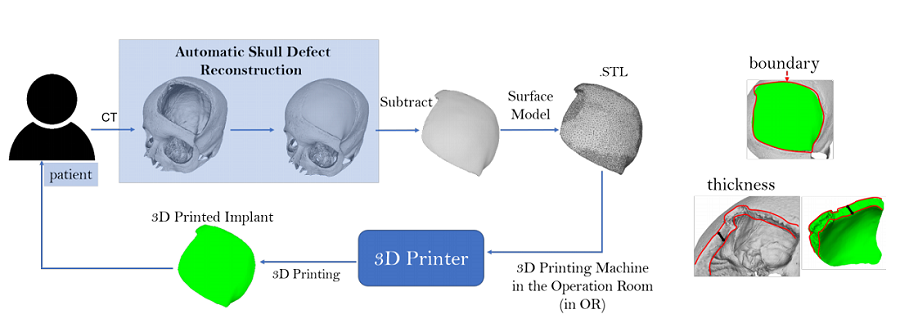
“Illustration of In-Operation Room process for cranial implant design and manufacturing. Left: a possible workflow. Right: how the implant should fit with the skull defect in terms of defect boundary and bone thickness.”

One option is developing ad hoc free CAD software for cranial implant design, but the design process still requires expertise and an extended wait.

“In this study, we introduce a fast and fully automatic system for cranial implant design. The system is integrated in a freely accessible online platform,” the team explained. “Furthermore, we discuss how such a system, combined with AM, can be incorporated into the cranioplasty practice to substantially optimize the current clinical routine.”

The system they developed has been integrated in Studierfenster, an open, cloud-based medical image processing platform that, with the help of deep learning algorithms, automatically restores the missing part of a skull. The platform then generates the STL file for a patient-specific implant by subtracting the defective skull from the completed one, and it can be 3D printed on-site.

“Furthermore, thanks to the standard format, the user can thereafter load the model into another application for post-processing whenever necessary,” the researchers wrote. “Multiple additional features have been integrated into the platform since its first release, such as 3D face reconstruction from a 2D image, inpainting and restoration of aortic dissections (ADs) [4], automatic aortic landmark detection and automatic cranial implant design. Most of the algorithms behind these interactive features run on the server side and can be easily accessed by the client using a common browser interface. The server-side computations allow the use of the remote platform also on smaller devices with lower computational capabilities.”

3D printing the implants makes the process faster, and combining it with an automated implant design solutions speeds things up even more. The researchers explained how their optimized workflow could potentially go:

“After a portion of the skull is removed by a surgeon, the skull defect is reconstructed by a software given as input the post-operative head CT of the patient. The software generates the implant by taking the difference between the two skulls. Afterwards, the surface model of the implant is extracted and sent to the 3D printer in the operation room for 3D printing. The implant can therefore be manufactured in loco. The whole process of implant design and manufacturing is done fully automatically and in the operation room.”

The cost decreases, as no experts are required, and the wait time is also reduced, thanks to the automatic implant design software and on-site 3D printing. The patient’s suffering will also decrease, since the cranioplasty can be performed right after removal of the tumor.

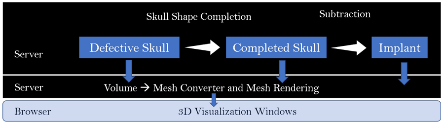
“Architecture of automatic cranial implant design system in Studierfenster. The server side is responsible for implant generation and mesh rendering. The browser side is responsible for 3D model visualization and user interaction.”

The team’s algorithm, which processes volumes rather than a 3D mesh model, can directly process high dimensional imaging data, and is accessible to users, and easy to use, through Studierfenster. Another algorithm on the server side of the system converts the volumes of the defective, completed skull, and the implant into 3D surface mesh models. Once they’re rendered, the user can inspect the downloadable models in the browser window.

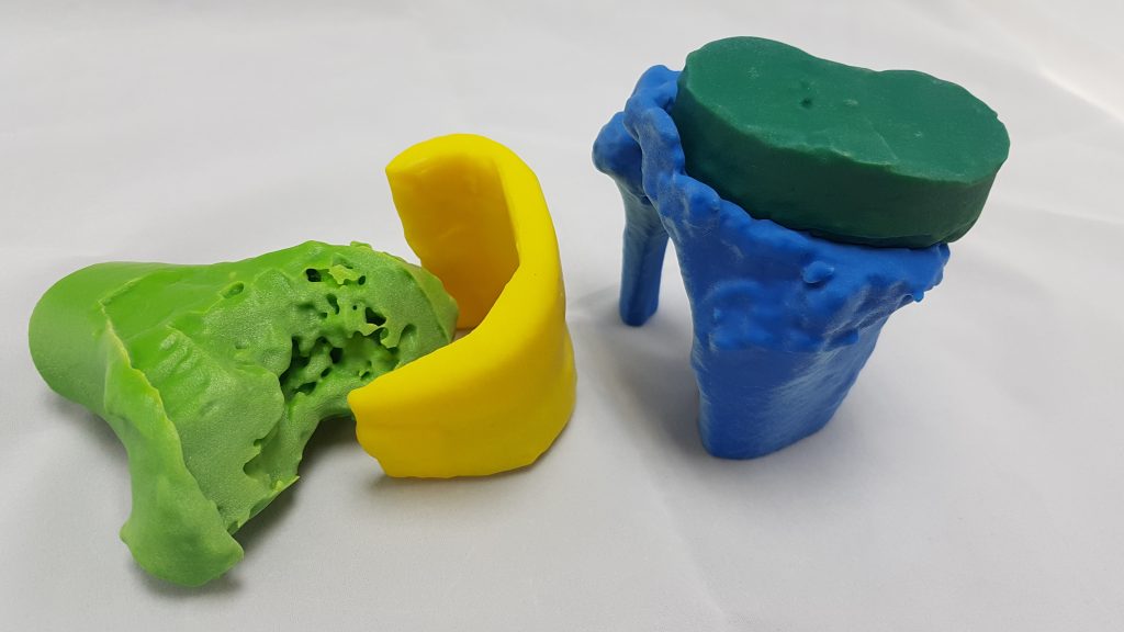
“An example of automatic skull defect restoration and implant design. First row: the defective skull, the completed skull and the implant. Second row: how the implant fits with the defective skull in term of defect boundary, bone thickness and shape. To differentiate, the implant uses a different color from the skull.”

“The system is currently intended for educational and research use only, but represents the trend of technological development in this field,” the researchers concluded. “As the system is integrated in the open platform Studierfenster, its performance is significantly dependent on the hardware/architecture of the platform. The conversion of the skull volume to a mesh can be slow, as the mesh is usually very dense (e.g., millions of points). This will be improved by introducing better hardware on the server side. Another limiting factor is the client/server based architecture of the platform. The large mesh has to be transferred from server side to browser side in order to be visualized, which can be slow, depending on the quality of the user’s internet connection.”

Discuss this and other 3D printing topics at 3DPrintBoard.com or share your thoughts below.

The post Free Automated Software to Design 3D Printable Cranial Implants appeared first on 3DPrint.com | The Voice of 3D Printing / Additive Manufacturing.

Creatz3D Medical Service Bureau To Offer Medical 3D Printed Models

We’ve written about the enterprising Singapore based Creatz3D before. They made a robot arm, we interviewed them on their expansion in the region and we’ve mentioned their work in News Briefs before. We’re interested in them because they are a reseller of ceramics, metals, bioprinting and plastic systems but also offer software and services. There is currently a huge shift underway in the 3D printing market where people are moving towards enterprise solutions and acting more like systems integrators than box movers. Creatz3D is a great example of a firm that is broadening its offering and moving into more high value applications. A great example of this is their service bureau for medical models. We interviewed Nigel Yap, Accounts Manager for the Creatz3D Medical Service Bureau to find out more.

What is Creatz3D Medical?

Creatz3D Medical Service Bureau pride ourselves as a solution provider in working together with our customers to provide the perfect medical solution for their needs. Being a service bureau that is focused entirely on medical applications, our seamless end-to-end solution guarantees a hassle-free experience in the conversion of CT/MRI images into 3D printed medical models.

What kinds of parts do you produce?

We recreate 3D printed medical models based on patients’ scan such as CT and/or MRI imaging.

Biological models are 3D printed medical models that are solely converted from patient scans (CT/MRI). These are used by surgeons in the interpretation of complex surgical conditions and helps in uncovering new surgical procedures (such as minimally invasive alternatives) that helps in the reduction of surgical time, recovery time and overall risk to the patient.

Educational models meanwhile are used for procedural skills training and simulation. As opposed to operating on human, animal cadavers or medical simulators, these models allow a pathological approach to the practice of rare and complex surgical procedure that would traditionally be taught through 2-dimensional illustrations.

What kinds of materials do you use?

Using the multi-material capabilities of our in-house PolyJet 3D Printer, we can work with a range of materials from rigid to clear and rubber-like properties. These materials have been tested with surgical practitioners and their feedback are that the materials react quite similarly as cortical and cancellous bone structures as well as the soft textural feeling of various human organs. The multi-material capabilities also help in separating anatomical features through color differentiation.

What printers?

We have 2 in-house machines that are used for various purposes, namely the Fortus 450mc as well as the Stratasys J750.

If I wanted to make a medical implant what do I have to take into account?

That is something that we do not offer as the materials that we print with are non-biocompatible for prolonged usage.

Who would you like to partner with?

We are always open to medical device companies who are looking at developing their own range of simulators, that can be used in their training or research and development purposes.

How do you take MRI scans and turn them into printable files?

Imagine the individual slices of the CT/ MRI scans as pages in a book. What our software does is that we take these individual pages and assemble them together back into a book, placing successive layers on top of each other in the creation of this book. After we have stacked the images in successive order, we clean the data to remove unwanted anatomies.

For instance, for the creation of a 3D printed heart model, with a CT scan of the chest cavity, we will remove the lungs and liver and just focus on the heart.

Thereafter, the file is cleaned up in a post-processing software that helps in ensuring that the file is printable and subsequently exported as an STL file that is then read by the 3D printer.

Do you do medical models today? What would they cost?

We currently provide the service of converting CT/MRI images into 3D medical models. The costing of each part varies depending on the anatomy in question. As such, it is difficult for us to provide any figure off hand. It would be best to have a consultation with the customer/ doctor in understanding their requirements before we are able to provide them with an ideal solution.

How can 3D prints help in surgical planning?

There are a number of ways that 3D Printing can help in surgical planning, these includes:

  • Better pre-operative planning through enhanced visuospatial appreciation of the defect in a relationship with other anatomical structures

  • Better pre-operative planning reduces overall surgical time, thus reducing time under general anesthesia, and improving the recovery rate

  • Pre-planned cuts and angles can be done in a customized surgical guide that will reduce surgical time

  • New minimally invasive procedures can be practiced prior to surgery to enable faster surgical time

  • Sizing of implants can be done on the 3D printed medical model instead of traditional methodology such as X-Ray templating on knee implant that has been proven to be inaccurate

How can they help educate doctors?

Medical education has traditionally been going through the concept of “see once, do once, teach once”. This form of education has proven to be effective but it is labour and time intensive. In addition, rare and complex conditions do not normally go through the above concept because of its rarity. This makes it a challenge to enable the right opportunity to be taught to junior doctors.

Many of these rare and complex conditions are thus traditionally taught through 2-dimensional representations. These are however unable to provide a 3D spatial appreciation of the anatomy and do not allow for simulated surgery.

Cadaveric training is also increasingly difficult to organize due to specimen shortages. Even if there is a steady supply, the specimens do not reflect the pathological features that would be required in the teaching of rare and complex conditions.

With 3D printed medical models, medical educators can show rare and complex conditions in a tangible form. They are also able to create customized simulation models where junior doctors can practice upon them in a controlled and risk-free environment. The repeatability nature of 3D printed medical models allows for multiple practice session, akin to being exposed to a real-life simulator for high-risk scenarios, before being allowed to operate.

What kinds of customers do you have?

There are mainly 3 types of customers that we work with for varying applications:

  1. Surgical Practitioners (Doctors)

    1. Pre-surgical planning models
    2. Pre-surgical simulation models
  2. Medical Educators

    1. Medical education models
    2. Procedural skills training simulation models
  3. Medical Device Companies

    1. Training models

    2. Research and development models