Medtronic to Acquire French Spinal Surgery Maker Medicrea, Strengthening 3D Printed Implants

As part of medical device maker Medtronic‘s push toward a fully integrated solution for surgical planning, the company announced its intent to acquire Medicrea, a French pioneer in innovative surgical technologies for the treatment of complex spinal pathologies, in a transaction valued at €7 ($8) per share. The all-cash agreement, set to purchase all of Medicrea’s outstanding shares, had unanimous approval by both companies and is expected to close by the end of 2020, subject to regulatory approvals and other customary closing conditions from both France and the United States.

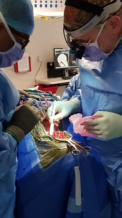
Medtronic treats the first U.S. patients with spinal surgery robot (Image courtesy of Medtronic)

“Combining Medtronic’s innovative portfolio of spine implants, robotics, navigation, and 3D imaging technology with Medicrea’s capabilities and solutions in data analytics, artificial intelligence, and personalized implants, would enhance Medtronic’s fully-integrated procedural solution for surgical planning and delivery. This marks another important step in furthering our commitment to improving outcomes in spine care,” said Jacob Paul, senior vice president and president of the Cranial and Spinal Technologies division, which is part of the Restorative Therapies Group at Medtronic, headquartered in Ireland. “Medtronic will become the first company to be able to offer an integrated solution including artificial intelligence-driven surgical planning, personalized spinal implants and robotic-assisted surgical delivery, which will significantly benefit our customers and their patients.”

Following news of the deal, Medicrea shares jumped by 20% in regular trading, most likely due to the premium the acquiring company was set to pay on the target’s share price, in this case, 22 percent over the closing price of Medicrea shares on 14 July 2020.

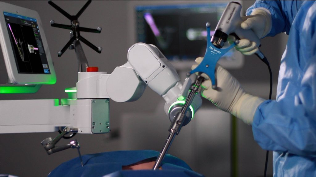
Medicrea’s UNiD technology (Image courtesy of Medicrea)

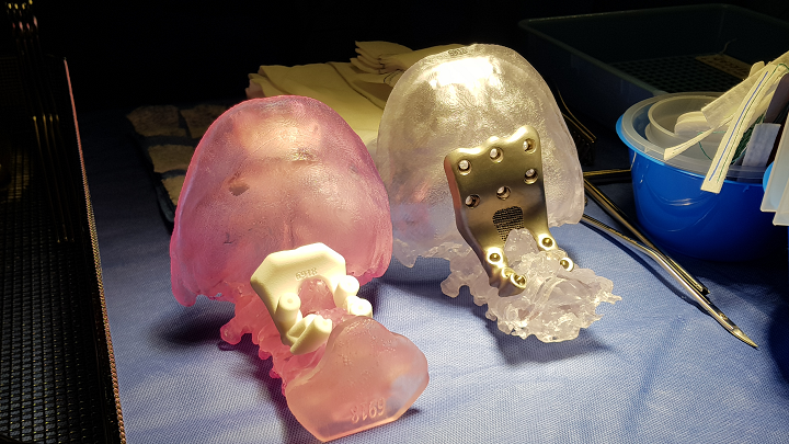
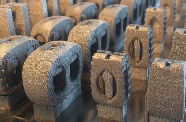
The deal will allow Medtronic to incorporate Medicrea’s latest innovations, which include the UNiD ASI (Adaptive Spine Intelligence) technology, designed to support surgeon workflow in pre-operative planning and incorporating 3D printing processes to create personalized implant solutions for surgery. The company’s portfolio also consists of artificial intelligence-driven surgical planning using predictive modeling and sophisticated algorithms that measure and digitally reconstruct the spine to its optimal profile. As well as an ultra-modern manufacturing facility in Lyon, France housing the development and production of 3D printed titanium patient-specific implants.

“Spine surgery is one of the more complex procedures in healthcare because of the high number of different parameters to take into consideration. It is impossible for the human brain to compute all of them for one single patient,” said Denys Sournac, founder, chairman and CEO of Medicrea. “The medical world has been waiting for the arrival of customization in spinal surgery. With scientific progress in understanding sagittal balance and spinal injury, combined with the advent of new digital technologies, it is now possible to offer spinal patients entirely customized implants. We are thrilled to be joining forces with Medtronic because we share a similar mission to restore the long-term quality of life for patients. Now, together, we can help more patients in more places benefit from consistently high-quality surgical care.”

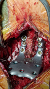
3D-printed spinal implants from Medicrea (Image courtesy of Medicrea)

The news comes amid expectations of an eventual recovery from the coronavirus pandemic and as Medtronic’s stock bounces back from a significant fall in the early months after COVID-19 emerged. The overall decline in procedures and supply chain disruptions have been among the key causes of concern for Medtronic, as well as impacted sales generated from China.

Medtronic said in a statement that the completion of the deal was subject to Medtronic getting at least 66.67% of Medicrea’s share capital. Up until now, Medtronic has entered into agreements with Medicrea shareholders totaling 44.4% of the company’s current outstanding share capital. The tender offer is expected to be filed with the French Markets Authority (AMF) in September 2020 and will be opened once the foreign investment approval in France and the merger control clearance in the United States are finalized.

Over the last seven decades, Medtronic has introduced a wide range of products to treat up to 70 health conditions, from cardiac devices and surgical tools to cranial and spine robotics, even insulin pumps, and patient monitoring systems. In the last few years, teams of scientists and engineers at the company have been working on new possibilities for personalized medicine using 3D printing technology, like its titanium 3D printing platform for spinal surgery implants. At the company’s facility, seven 3D printers work around the clock filling orders for rapid prototyping and medical models that allow doctors to practice procedures on life-like simulations. Additionally, researchers from Medtronic teamed up with academia to create a new operating room system powered by personalized 3D images, to give neurosurgeons better tools to remove brain tumors.

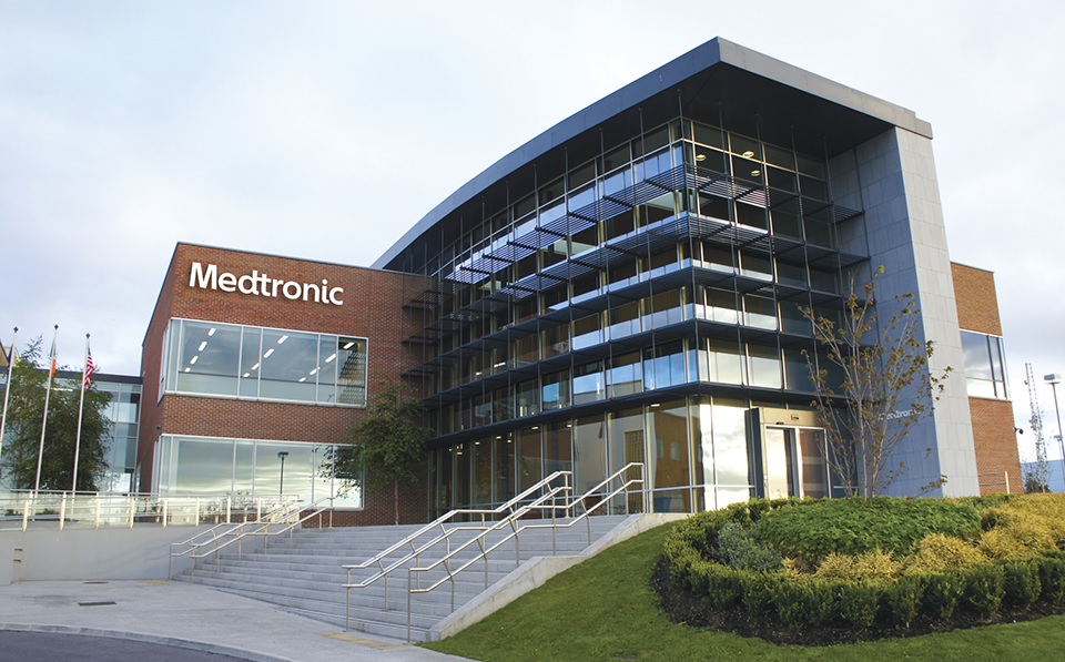
Medtronic headquarters in Dublin, Ireland (Image courtesy of Medtronic)

As of 2017, Medtronic was the leader in the U.S. market for spinal implants with a share of over one third. Once the acquisition is complete, the company will be able to expand and strengthen its position as a global innovator in further enabling technologies and solutions for spine surgery.

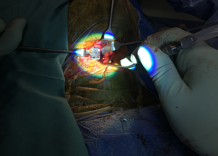
Spinal procedures are considered by experts as one of the most painful in neurosurgery and orthopedics, with over 1.62 million instrumented interventions performed every year. ResearchMoz analysts predicted the global spine surgery products market to hit $16.7 billion by 2025, mainly due to an increase in spine disorder cases among the geriatric population. The demand for innovative, minimally invasive solutions to this problem is critical for patient healthcare, which is why Medtronic is looking towards the predictive medicine opportunity that Medicrea has been developing, by collecting an unprecedented amount of data to develop its proprietary predictive models and employing disruptive technologies in every step of the way. Overall, the combination of the companies’ technical know-how would probably improve the clinical experience for patients and strengthen the future of spinal health.

The post Medtronic to Acquire French Spinal Surgery Maker Medicrea, Strengthening 3D Printed Implants appeared first on 3DPrint.com | The Voice of 3D Printing / Additive Manufacturing.

China: 3D Printed Vertebral Body Used to Reconstruct Upper Cervical Spine of 9 Patients

Primary osseous spinal tumors make up roughly 5% of all primary bone tumors, and reconstruction is required to restore the spine’s integrity and stability. However, it’s hard to reconstruct this complex section, which is responsible for transitioning the axial loading force from the cranium to the spinal column, and subpar implants can result in complications like migration and nonfusion.

3D printing can be used to fabricate patient-specific porous implants for fixing these bone defects. A group of researchers from Beijing published a study, “Upper cervical spine reconstruction using customized 3D-printed vertebral body in 9 patients with primary tumors involving C2,” where they described “the clinical outcomes of upper cervical spine reconstruction using customized 3D-printed vertebral body,” with “a mean follow-up of 28.6 months” for the patients.

“Patients with primary tumors involving C2 who were treated in our institution between July 2014 and November 2018 were enrolled,” the team stated.

“Nine patients (2 males and 7 females) were included in the study with a mean age of 31.4 years (12 to 59 years). Seven patients demonstrated tumors located in C2 and 2 showed involvement of C2 and C3.”

The nine patients initially complained of “aggravating pain,” with two suffering neurological impairment, and average duration since the onset of these symptoms was almost three months. Here’s the tumor breakdown for the patients, established using a CT-guided biopsy:

  • 4 giant cell tumors (GCT)
  • 2 chordoma
  • 1 Ewing sarcoma
  • 1 paraganglioma
  • 1 aggressive hemangioendothelioma

Fig. 1: Imaging studies for patient #3. The achievement of osseointegration was defined when new bone formation was observed around the bone-implant interface on X-ray (B) and CT (D) during the follow-up compared to that of immediately postoperative (A,C). The postoperative segment vertebral height was measured on the midsagittal reconstruction CT from atlas anterior tubercle to the midpoint of the adjacent lower endplate (C).

Making the implants was a 7-day process. First, CT scans were performed on the patients’ spines, and the DICOM data was imported into Materialise Mimics 15.0 software, where a CAD model for the implant was designed. Ti6Al4V powder was used to print the porous metal scaffold implants with Arcam EBM’s electron beam melting technology.

“Based on our previous studies, the parameters set for the trabecular structure and the size of the uniform micro-pores were determined to generate the optimized biomechanical and osteoinductive properties (,,). The upper contact surface morphology of the implant coincided with the inferior articular surfaces of C1, while the lower contact surface morphology coincided with the upper endplate of the caudal vertebra,” they wrote.

Fig. 2: The 3D printed artificial vertebral body with porous scaffold fabricated out of titanium alloy powder.

A two-stage intralesional spondylectomy was performed on each patient, and the 3D printed vertebral body was used to accomplish anterior reconstruction, without the use of a bone graft.

“The average interval between the posterior and anterior procedures was 14.4 days,” the researchers said.

“In the first 4 cases in this series, occipitocervical fixation was performed (Figure 1). Subsequently, with more confidence in the stability of the 3D-printed anterior construct, we were able to preserve the atlanto-occipital joint in the next 5 cases.”

If you’re interested in the rest of the nitty-gritty surgical details, check out the full research paper.

Table 1: The details of the 9 patients

In the table above, you can see the details of the patients, who all had follow-up appointments after the 3D printed vertebral body was implanted. All nine received postoperative radiotherapy, while two also received chemotherapy.

“Patient one died of systemic metastases 15 months postoperatively without signs of local recurrence. Patient seven had tumor local recurrence. The others were alive and functional in their daily livings at the last follow-up without evidence of disease. At their final follow-ups, the neurological status of all alive patients was ASIA E, and the average VAS score was 0.9. Three patients had ECOG 1, while 5 patients had ECOG 0 for their general well-being and activities of daily life,” they stated.

Fig. 3: Imaging studies for patient #3.

Through radiograph and CT examinations, the researchers observed new bone formation around the bone to implant contact surfaces, “which provided the evidence of osseointegration,” and they found that all of the 3D printed vertebral bodies were stable, without any signs of ” implant displacement or subsidence.” Additionally, none of the screws had come loose, and there was no rod breakage in the posterior instrumentation systems.

The researchers found several advantages to using 3D printing for this reconstruction rather than traditional methods of manufacturing, such as the implant offering “reliable primary immediate postoperative stability.” A patient-specific implant provides a better match to bony surfaces and a larger contact area, and because screw tracks are actually integrated directly into the artificial vertebra, “self-stabilization” occurs.

Fig. 4: Imaging studies for patient #6 showing fusion process. Compared to the immediate postoperative X-ray (A) and CT (D), regenerated osseous tissue can be seen to have gradually grown along the implant 12 months (B,E) and 24 months (C,F) post-op (arrow).

“Secondly, the anatomical design of the contact surface of the curved porous endplate and its biocompatibility provided reliable mid-long-term stability. The porous bone-contacting surface of the 3D-printed vertebral body is conducive to bone in-growth into the trabecular pores to achieve firm osseointegration, which was supported by evidence from previous basic research and in vivo studies (,,),” they explained.

Additionally, post-op radiotherapy may not affect the 3D printed vertebral body as much, so long as osseointegration on two ends occur, “because solid combination was accomplished.” Conversely, this treatment can lead to instrumentation failure with conventionally manufactured implants.

“In our study, the progress of osseointegration is evident on follow-up with imaging studies. On lateral radiography, regenerated osseous tissue was seen adhering to the 3D-printed vertebral body (Figures 1B,4B,C1B,4B,C).),” the researchers noted. “Sagittal CT revealed new bone tissue crawling and growing around the ends of the 3D-printed vertebral body from the upper and lower vertebra (Figures 1D,4E,F1D,4E,F).). All patients were capable of resuming normal activity without mechanical pain associated with spinal instability at 12-month follow-up.”

Finally, a 3D printed vertebral body could mean there’s less of a need for transoral (direct access through the mouth) or transmandibular surgical approaches. For example, as noted above, this research team used the posterior-anterior approach to perform C2 spondylectomy, which made it easier and safer to isolate the vertebral arteries.

“Our study suggests that 3D-printed implant may be a good option in upper cervical reconstruction, the tailored shape matching with the contact surfaces and the porous structure conductive to osseointegration provide both short- and long-term stability to the implant,” the researchers concluded. “However, a higher level of evidence is still needed.”

Discuss this and other 3D printing topics at 3DPrintBoard.com or share your thoughts below.

The post China: 3D Printed Vertebral Body Used to Reconstruct Upper Cervical Spine of 9 Patients appeared first on 3DPrint.com | The Voice of 3D Printing / Additive Manufacturing.

Mayo Clinic is Using 3D Printing to Teach the Next Generation of Spine Surgeons

Resident life is hard. After years of living, studying, and breathing medicine, they are finally one step closer to officially being able to practice. Medical residencies can take three years for primary care physicians, but some surgical specialties require even more training time to develop new procedures without risking lives, sometimes up to seven years. And did we mention that training is not a breeze? Its hard work and some of it is not for the queasy.

Residents need cadavers for educational purposes, but these are difficult to come by. In the United States, only 20,000 bodies are donated to science every year, that’s barely enough to satisfy a population of more than 27,000 residents. Plus you need to add the costs of purchasing them, according to an article by The New York Times, delivery of an intact cadaver costs as little as $1,000, but different specialists seek out specific pieces of anatomy for their work, and individual parts can be expensive, such as a torso in good conditions which can go for $5,000, or a spine for $3,500. With residents needing more than one cadaver to practice on and clinics usually having only limited resources, there is a sensical need to find new ways to educate the next generation of doctors. For William Clifton, Neurosurgery Resident at the Mayo Clinic in Florida, it seemed there had to be another way to train surgeons, so he consulted with an engineer, who was also a friend, and realized that creating 3D printed models for medicine and simulators for resident training could be the solve-all solution.

Clifton had an idea which involved buying an Ultimaker S5 FDM desktop printer with a personal loan, and finding room at home to begin printing out models in his free time, not an entirely easy job considering the young doctor and his wife have four children. But Clifton, one of the first recipients of the Mayo Clinic John H. and Carolyn O. Sonnentag Neurosurgery Residency in Florida, was on a mission, one he became very passionate about, using 3D printing technology to build simulators that can be used to train residents for surgery. Along his path, he ran into Aaron Damon, a researcher and lab specialist at the Simulation Center at Mayo Clinic, and immediately discovered that they shared a desire to change the future of resident training.

His early designs helped him build the training models used today. Since last March, they have developed hundreds of their original Biomimetic Human Tissue Simulators, more than 30 peer-reviewed publications, as well as several patent submissions for neurosurgical devices. The Department of Neurologic Surgery also became heavily engaged in planning for dedicated neurosurgery 3D printing space and there is now a materials science laboratory based on the ideas and infrastructure Clifton helped create. Today their lab houses three FDM printers: two Ultimaker S5 and one Raise3D Pro for bigger volumes, such as when they need to print cases of scoliosis (a medical condition in which a person’s spine has a sideways curve) to help residents that need to deal with pediatric scoliosis. 

“We are using 3D printed patient-specific models in a certain way to characterize the material properties of actual bone, soft tissue, muscle, ligaments, tendons, and more, exactly how it is in vivo,” said Clifton in an interview with 3DPrint.com. “The big innovation is that our models exhibit the same biomechanics as the human spine or the human skull so that the surgeon can see where the structures are in realtion to each other, bend and move them, as well as watch how the dynamic relations change.” 

According to Mayo Clinic, the 3D printing process comprises stripping down information from a patient’s CT scan and converting the image to stereolithography. Based on spatial information, Mayo Clinic can program the printer to create patient-specific models, such as anatomically precise vertebrae. Physician-scientists use these surgical training models in tandem with resources provided by the J. Wayne and Delores Barr Weaver Simulation Center at Mayo Clinic’s campus in Florida. The Weaver Simulation Center partnered with Clifton to share his 3D training models with surgical learners.

The real innovation was simulating human tissue and creating a new polymer for FDM 3D printing of anatomical models that will be used for practicing pre-surgical operations using electrocautery, something that has never been done before. The innovative new polymer chemistry and the 3D printed film should be commercially available soon as Clifton and Damon are in talks with multiple companies through Mayo Clinic Ventures (the business office at Mayo that handles this type of negotiations and commercialization).

3D printed phantom for teaching neurosurgical trainees the freehand technique of C2 laminar screw placement

“The faculty is ecstatic about this project because the skill of the residents has gone up considerably. Traditionally interns learn on cadavers or during the residency in living people. For example, surgical residents have to learn to place screws into the C2 vertabrae and this is a complex procedure because they are learning to navigate between the vertebral artery and the spinal cord; but with our models they can practice puting in a couple of hundred C2 pedicle screws so that when they go to the Operating Room (OR) they know exactly what to do, because they have done it so many times on such reallistic models,” claimed Clifton, whose specialty as a neurosurgeon leads him to perform surgery on the brain, spinal cord, and peripheral nerves.

Mayo Clinic is all about research, education and patient care, which is why Damon and Clifton are printing their way into teaching future doctors how to take care of the next generation of patients. The duo gets along extremely well, understanding that cooperation and collaboration are one of the keys of their success, which will shortly be used in the other two Mayo Clinics, in Rochester and Arizona. In the short eight months since their adventure began, they have created 14 different models for spine pathologies alone, and now they will begin developing brain models. Damon indicated that they have a brain tumor resection model in the works, “that residents can use to practice the removal of a brain tumor, coagulation of vessels, measuring blood loss, brain manipulation, and even brain mapping, meaning that now we are going into the microsurgery skill set, and not just spinal surgeries.” 

“We can print a lot of different pathologies, wereas you are kind of limited with a cadaver. With 3D prinitng you can model specific pathologies after a patient, as well as complex and rare pathologies that residents might not see that often in the OR,” explained Damon. “This is a very specific and universal development at the same time, because we hope that other clinics around the world that barely have any access to cadavers for resident training, can use them and eventually this will improve outcomes and lives all over the world.” 

Each simulator costs approximately $50, whereas a cadaver normally purchased for this type of surgery training can run into the thousands, helping Mayo Clinic save thousands of dollars as well. The medical center, which ranks as the best clinic for neurosurgery worldwide, will still get cadavers of course; but Clifton recalled that he spent over $50,000 in just three months, on cadaveric tissue for research and education in 2018. That experience and the reality of the burden of costs behind his training is one of the reasons that sparked the interest in developing a more cost-effective way to practice.

“One of the distinct advantages of FDM 3D printing specifically is that it allows us to build models made from different material compositions and properties. We do this to simulate different disease processes for resident training, and to approach patient care and surgical planning in an individualized way,” said Clifton.

Clifton and Damon want to take this technology to the point where it is streamlined so that residents will have dedicated simulation training. The key is that even before they hit the OR, they have done the procedure at least 10 times, and not just the simplest parts of the procedure, but also all the complex elements. Their hope for the future is that this will become a standardized model in the US.

Damon suggested that a big part of their work has been refining and validating their models until they achieved a high fidelity that is comparable to cadavers. “With these simulators, residents are able to acquire the same skill level as surgeons who have been in the field for many years by high fidelity and cost-effective repeated practice. The support we get from Mayo Clinic also allows our creativity to grow and continue to push the boundaries of innovation in 3D printing.”

Damon suggested that “even in our lab at Mayo, there were up to five surgical residents sharing one cadaveric torso, but now each person can have their own model and countless amount of experience, which is invaluable.” The two experts claim that training on cadavers gives residents one chance of getting a procedure right, while models can be 3D printed hundreds of times. 

Three of Clifton’s patients recently benefited from his applications in 3D printing. The unique models contributed to surgical planning sessions, helped to pinpoint precise diagnoses and guided postoperative management choices. The first patient had a congenital spinal deformity, the next had a large metastatic tumor near the lumbar spine, and the third had a rare cyst compressing her brainstem. All three patients have recovered from their successful surgeries because of the combined forces of innovation and surgical skills.

“We are really looking into the education component, trying to obtain a better, safer and cheaper solution for the future. My goal is to teach the next generation of residents to become great spine surgeons.”

[Image credit: Mayo Clinic]

The post Mayo Clinic is Using 3D Printing to Teach the Next Generation of Spine Surgeons appeared first on 3DPrint.com | The Voice of 3D Printing / Additive Manufacturing.

Indonesian Researchers 4D Print Spacers for Minimally Invasive Transforaminal Lumbar Interbody Fusion (MI-TLIF)

Researchers at Universitas Indonesia are investigating the potential of 4D printed spacers for spinal surgeries, outlining their recent, published findings in ‘Modelling the shape memory properties of 4D printed polylactic acid (PLA) for application of disk spacer in minimally invasive spinal fusion.’ Authors Nindya Aprilia Alief, Sugeng Supriadi, and Yudan Whulanza created a disk spacer able to morph according to its environment.

Using PLA as the material for the disk spacer, the goal was to make a patient-specific implant that can help to stabilize the lumbar segment during a surgical treatment called Minimally Invasive Transforaminal Lumbar Interbody Fusion (MI-TLIF). This procedure is less aggressive and less traumatic to the body and to the patient, but a disk spacer is required for a successful outcome.

Pattern of disk spacer model with hollow spaces. a) Uniformed vertical hollow spaces in 1 mm width. b) Uniformed vertical hollow spaces in 2 mm width. c) Non-uniformed vertical hollow spaces varies from 1 to 3 mm width.

“In order to obtain the right shape transformation, the expansion and thermal distribution of 4D printed polylactic acid model were investigated,” stated the researchers. “It is indicated that specific PLA structure possesses thermal shape memory behavior that can be thermomechanically trained into temporary shape and return to their permanent shape when heated.”

The researchers created several models of the disk spacer, designing a pattern for the temporary shape. The disk spacers were 50 mm long, 18 mm wide, with two identical parts, partitioned. Once the structure is created, hollow areas are added, with the three models varying in detail. PLA was chosen because of its clarity and rigidity, and most importantly, its biocompatibility.

Representative of the PLA model deformation after 3 seconds. a) Solid disk spacer model. b) Uniformed vertical hollow spaces in 1 mm width. c) Uniformed vertical hollow spaces in 2 mm width. d) Non-uniformed vertical hollow spaces varies from 1 to 3 mm width.

After 3D printing, a beaker was filled with 300 ml of hot water, with each model immersed at 60 °C, the glass temperature of PLA.

“… the time needed for a PLA pattern to expand has an average of 0.0513 second. The model (a) needs 0.064 seconds, model (b) needs 0.07 seconds, and model (c) needs 0.02 seconds. Afterwards, the model has reached the final expansion. Models are expanded towards x-axis and y-axis paralleled to the shape of the hollow spaces. Each model has adjacent value of maximum total displacement. Subsequently, the model (a) has maximum total displacement as much as 0.94 mm, the model (b) has 0.94 mm, and the model (c) has 0.95 mm, which all of the values are located on the tip of the pattern.

“Moreover, the time needed for a pattern to completely reach the external temperature has an average of 0.052 seconds. Hence, the model (a) needs 0.058 seconds, model (b) needs 0.058 seconds, and model (c) needs 0.04 seconds.”

Side by side comparison before and after adding stimulus. Left: a PLA model before adding stimulus and right: after adding the stimulus.

The authors noted that non-uniformed hollow spaces were superior in the amount of time required for thermal expansion, explaining that less time required for deformation, the better, as that means there would be less valuable time needed to affix the 4D planted structure during the medical procedure.

“This paper presents a study of thermal shape memory behavior of the PLA pattern model. The time needed for a model to expand is less than one second. The pattern expands toward x-axis and y-axis and the expansion happens gradually until it completely expands,” concluded the researchers. “The expansion value between simulation and experimental result are much the same yet the values from the experiment are on the different direction. However, based on the simulation result, nonuniformed hollow spaces pattern has favorable result that could be a reference for the future research in order to design the suitable pattern for the 4D PLA model.

“The current pattern that has been developed also need to combine with other different pattern in order to achieve desired transformation.”

The impacts that 3D and 4D printing are making in the medical field are stunning in many cases, allowing researchers to bioprint brain tumors to understand them better, 3D print splints to open up breathing airways, and fabricate mandibular implants to improve chewing, facial structure, and much more. Find out more about the potential for 4D technology in creating disk spacers for spinal fusion surgery here. What do you think of this news? Let us know your thoughts! Join the discussion of this and other 3D printing topics at 3DPrintBoard.com.

[Source / Images: Modelling the shape memory properties of 4D printed polylactic acid (PLA) for application of disk spacer in minimally invasive spinal fusion]

Osseus Fusion Systems achieves FDA clearance for 3D printed spinal implants

Texan medical device company Osseus Fusion Systems, has received FDA clearance for its family of 3D printed spinal implants known as Aries. The implants are designed to help surgeons ease back pain and shorten spinal recovery time. They are the first example of Osseus’ new line of 3D printed products, with several other 3D printed […]

Breakthrough 3D Printed Neural Scaffold Could Help Patients with Spinal Cord Injuries Regain Some Functions

Right now, 285,000 people in the US suffer from spinal cord injuries, with roughly 17,000 new injuries each year. 3D printed spinal implants have been shown to help patients recover more easily, and a team of engineers and medical researchers from the University of Minnesota (UMN) have spent the last two years developing an innovative new 3D printed medical device that could help long-term spinal cord injury patients regain some function in the future.

“This is a very exciting first step in developing a treatment to help people with spinal cord injuries. Currently, there aren’t any good, precise treatments for those with long-term spinal cord injuries,” said Ann Parr, MD, PhD, a UMN Medical School Assistant Professor in the Department of Neurosurgery and Stem Cell Institute.

The method involves a 3D printed silicone guide, which acts as a scaffold for special stem cells that are bioprinted directly on top of it. The aim is to surgically implant the guide into the injured part of the spinal cord, and it should act as a bridge between living nerve cells both above and below the area, which could help alleviate pain for patients, in addition to helping them gain control over functions like bladder, bowel, and muscle control again.

“We’ve found that relaying any signals across the injury could improve functions for the patients. There’s a perception that people with spinal cord injuries will only be happy if they can walk again. In reality, most want simple things like bladder control or to be able to stop uncontrollable movements of their legs,” Parr explained. “These simple improvements in function could greatly improve their lives.”

Spinal cord scaffold assembly process: 3D bioprinting cells on silicone scaffolds
allows for in vitro culture of sNPCs and OPCs. (a) Silicone scaffolds are printed with
channels, and (b) cells are dispensed inside the channels. (c) A layer of silicone covers the channels, and (d) scaffolds are placed inside a dish and cultured for 7 days.

The team recently published a paper on their potentially life-changing work, titled “3D Printed Stem-Cell Derived Neural Progenitors Generate Spinal Cord Scaffolds,” in the peer-reviewed scientific journal Advanced Functional Materials.

Fluorescence images of 3D printed cell-laden Matrigel (50%) matrices cultured for 0 (3 hours), 1, and 4 days. Timescale images show (a) sNPCs extending axons, and (b) OPCs exhibiting bi-polar processes.

The abstract reads, “A bioengineered spinal cord is fabricated via extrusion‐based multimaterial 3D bioprinting, in which clusters of induced pluripotent stem cell (iPSC)‐derived spinal neuronal progenitor cells (sNPCs) and oligodendrocyte progenitor cells (OPCs) are placed in precise positions within 3D printed biocompatible scaffolds during assembly. The location of a cluster of cells, of a single type or multiple types, is controlled using a point‐dispensing printing method with a 200 µm center‐to‐center spacing within 150 µm wide channels. The bioprinted sNPCs differentiate and extend axons throughout microscale scaffold channels, and the activity of these neuronal networks is confirmed by physiological spontaneous calcium flux studies. Successful bioprinting of OPCs in combination with sNPCs demonstrates a multicellular neural tissue engineering approach, where the ability to direct the patterning and combination of transplanted neuronal and glial cells can be beneficial in rebuilding functional axonal connections across areas of central nervous system (CNS) tissue damage. This platform can be used to prepare novel biomimetic, hydrogel‐based scaffolds modeling complex CNS tissue architecture in vitro and harnessed to develop new clinical approaches to treat neurological diseases, including spinal cord injury.”

The process begins with any type of adult stem cell, be it blood or skin, and medical researchers use the latest bioengineering techniques to reprogram these into neuronal stem cells. These cells are then 3D printed onto a silicone guide with a unique extrusion-based technology, which can print both the cells and the guide from the same 3D printer.

Michael McAlpine, PhD, UMN Benjamin Mayhugh Associate Professor of Mechanical Engineering in the University’s College of Science and Engineering, said, “This is the first time anyone has been able to directly 3D print neuronal stem cells derived from adult human cells on a 3D-printed guide and have the cells differentiate into active nerve cells in the lab.”

Photograph of customized 3D bioprinting setup.

The 3D printed silicone guide keeps the stem cells alive, so they can change into neurons.

“Everything came together at the right time. We were able to use the latest cell bioengineering techniques developed in just the last few years and combine that with cutting-edge 3D-printing techniques,” said Parr.

The researchers created a prototype implantable guide to help connect the living cells on each side of a damaged spinal cord area, though this task was not without its difficulties.

“3D printing such delicate cells was very difficult. The hard part is keeping the cells happy and alive,” explained McAlpine. “We tested several different recipes in the printing process. The fact that we were able to keep about 75 percent of the cells alive during the 3D-printing process and then have them turn into healthy neurons is pretty amazing.”

With any luck, the team’s next steps in the process will be successful, which should provide some hope for the future to patients with long-term spinal cord injuries.

Co-authors of the paper are Daeha Joung, Vincent Truong, Colin C. Neitzke, Shuang-Zhuang Guo, Patrick J. Walsh, Joseph R. Monat, Fanben Meng, Sung Hyun Park, James R. Dutton, Parr, and McAlpine.

Discuss this and other 3D printing topics at 3DPrintBoard.com or share your thoughts below.

Patient-Specific 3D Printed Spinal Solutions by Anatomics

3D printed anterior cervical cage

Australian medical device company Anatomics has a very prolific 3D printing portfolio in the healthcare industry, from assisting with a 3D printed heel bone to 3D printing the first titanium sternum and set of ribs and vertebrae. Anatomics founder Paul D’urso, MD, a neurosurgeon at Epworth Healthcare, began his medical 3D printing research way back in 1991, four years before starting the not-for-profit Anatomics, which has since helped over 5,000 patients with its custom 3D printed medical solutions.

D’Urso said, “Anatomics has lead the world in custom 3D printed spine technology for over 20 years and is proud to have developed numerous world first applications.”

3D printed occipito-cervical plate

D’Urso has a particular interest in developing spinal applications for biomodeling, and at the recent 3DHEALS conference in San Francisco, reported that he’s used 3D printing in nearly 700 spinal fusion procedures, including what D’Urso tells us is “the world’s first custom occipito cervical plate.”

“In the future Anatomics plans to create disruptive Spinal Solution Centres that will enable Community Based Personalised Healthcare allowing surgeons and hospitals to 3D print a range of spinal implants and share designs through-out the world,” D’Urso told 3DPrint.com.

The success of some of the innovative 3D printed solutions that D’Urso and Anatomics have developed, including 3D printed custom spinal implants, have recently been described in articles and research papers printed in various publications, such as the European Spine Journal and the Journal of Clinical Neuroscience.

The latter paper, titled “Designing patient-specific 3D printed devices for posterior atlantoaxial transarticular fixation surgery,” discussed how biomedelling and 3D printing are both useful tools for pre-surgical planning, developing titanium implants and patient-specific tools, and intraoperative stereotaxy – a minimally invasive surgical procedure which uses a 3D coordinate system to locate small targets inside the body and then perform an action, like an ablation, biopsy, injection, or implantation, on them.

The abstract reads, “Atlantoaxial transarticular screw fixation is an effective technique for arthrodesis. Surgical accuracy is critical due to the unique anatomy of the atlantoaxial region. Intraoperative aids such as computer-assisted navigation and drilling templates offer trajectory guidance but do not eliminate screw malposition. This study reports the operative and clinical performance of a novel process utilising biomodelling and 3D printing to develop patient specific solutions for posterior transarticular atlantoaxial fixation surgery. Software models and 3D printed 1:1 scale biomodels of the patient’s bony atlantoaxial spine were developed from computed tomography data for surgical planning. The surgeon collaborated with a local medical device manufacturer using AnatomicsC3D to design patient specific titanium posterior atlantoaxial fixation implants using transarticular and posterior C1 arch screws. Software enabled the surgeon to specify screw trajectories, screw sizes, and simulate corrected atlantoaxial alignment allowing patient specific stereotactic drill guides and titanium posterior fixation implants to be manufactured using 3D printing. Three female patients with unilateral atlantoaxial osteoarthritis were treated using patient specific implants. Transarticular screws were placed using a percutaneous technique with fluoroscopy and neural monitoring. No screw malposition and no neural or vascular injuries were observed. Average operating and fluoroscopy times were 126.0 ± 4.1 min and 36.7 ± 11.5 s respectively. Blood loss was <50 ml per patient and length of stay was 4–6 days. Clinical and radiographic follow up data indicate satisfactory outcomes in all patients. This study demonstrates a safe, accurate, efficient, and relatively inexpensive process to stabilise the atlantoaxial spine using transarticular screws.


The paper also explained how operative ergonomics and the placement of atlantoaxial transarticular screws can both be simplified using 3D printing. Authors include Ganesha K. Thayaparan, with Epworth’s Department of Neurosciences, and Anatomics’ Mark G. Owbridge, Robert G. Thompson, and D’Urso.

Discuss this and other 3D printing topics at 3DPrintBoard.com or share your thoughts in the comments below. 

[Images provided by Anatomics]