Interview with Jason Chuen: Shaping Australia’s Medical 3D Printing Environment

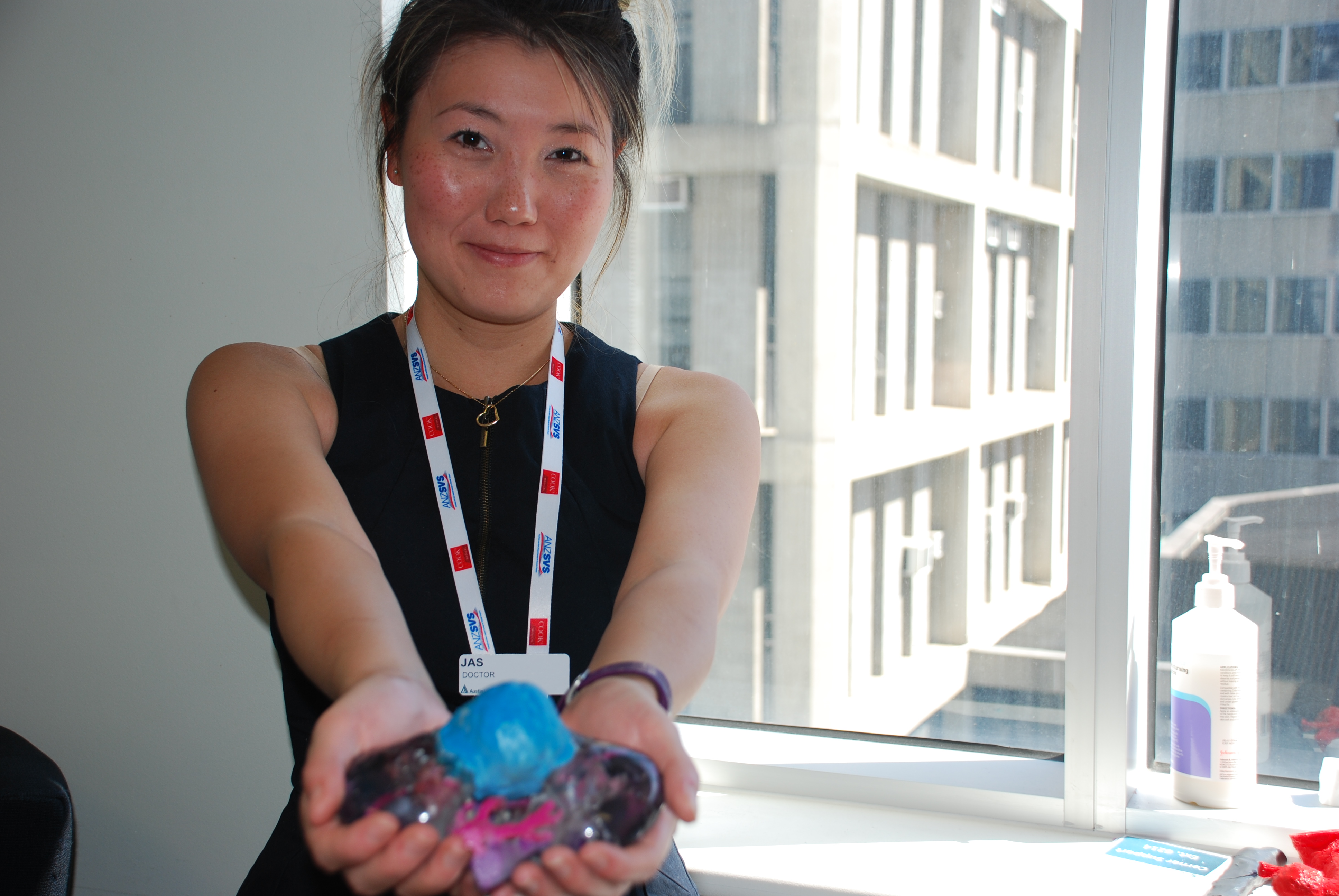
In Australia, vascular surgeon, Jason Chuen understands that 3D printing is the exciting next step in personalized medicine, which is why he uses 3D scans and 3D printing to deliver anatomical models. During an interview with 3DPrint.com, Chuen, who is also the Director of Vascular Surgery at Austin Health and Austin Health’s 3D Medical Printing Laboratory (3D Med Lab), suggested that “there is a lot of interest because the field of 3D printing in medicine is growing; we are seeing the doctors and researchers involved more than ever, as well as more application development originating from clinicians.”

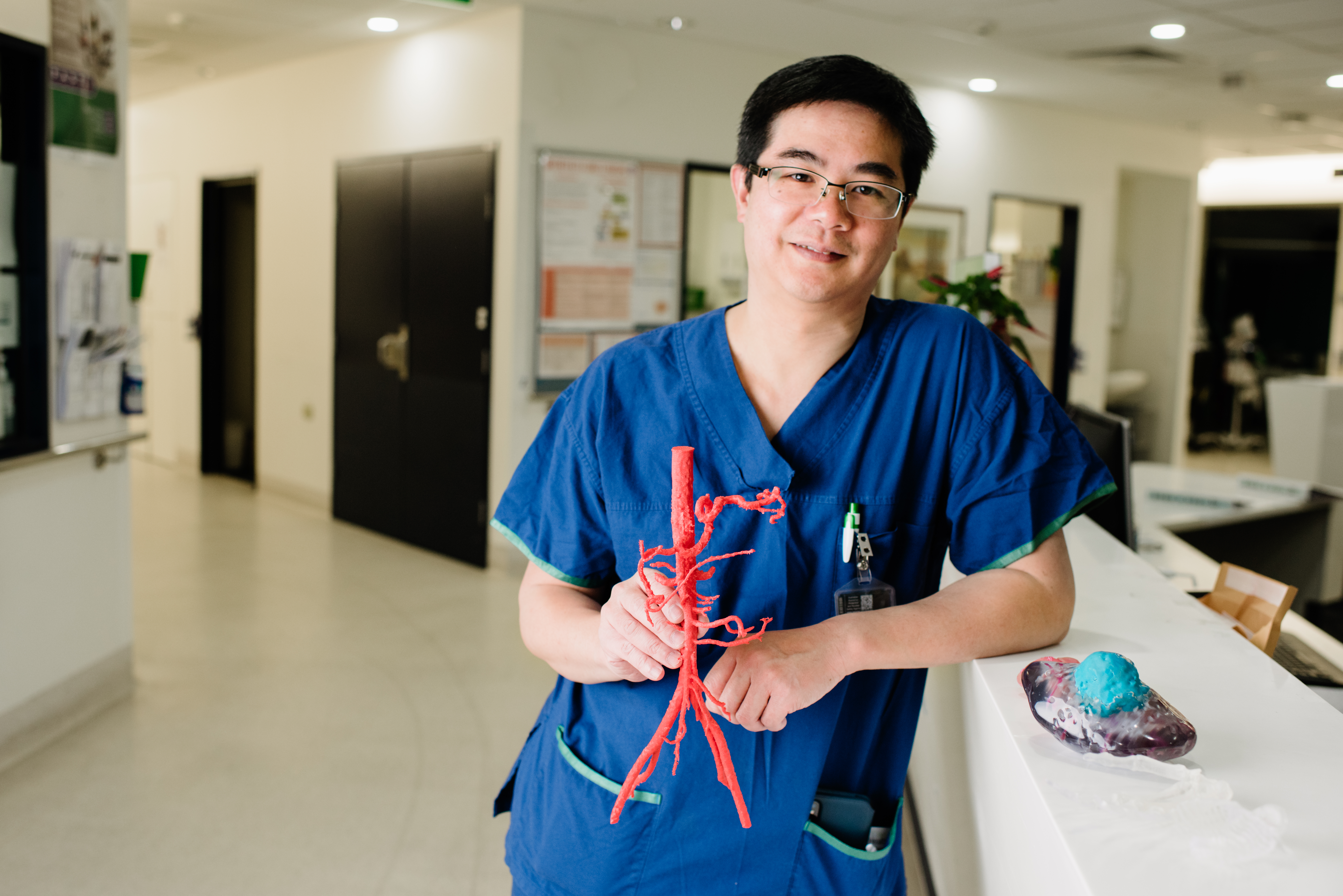
At The University of Melbourne, in Australia, the 3D Med Lab supports 3D printing for clinical applications and runs an active research program exploring how it can be used for teaching, procedural simulation, patient education, surgical planning, and prosthetic implants. The first facility of its kind in Australia, 3D Med Lab, frequently prints models of diseased aortas to perform a “practice-run” of surgery. What makes this lab unique is that it is hospital-based, and works with many different specialties. Chuen has been looking into the landscape of medical 3D printing for many years and earlier this month along with his colleague Jasamine Coles-Black, a Doctor and Vascular Researcher at the Department of Vascular Surgery at Austin Health and the 3D Med Lab, organized the fifth annual 3D Med Australia Conference, which he claims is the only meeting of its kind in Australasia, with only one or two more around the world of a similar nature, like Materialise‘s medical 3D printing meetup in Belgium.

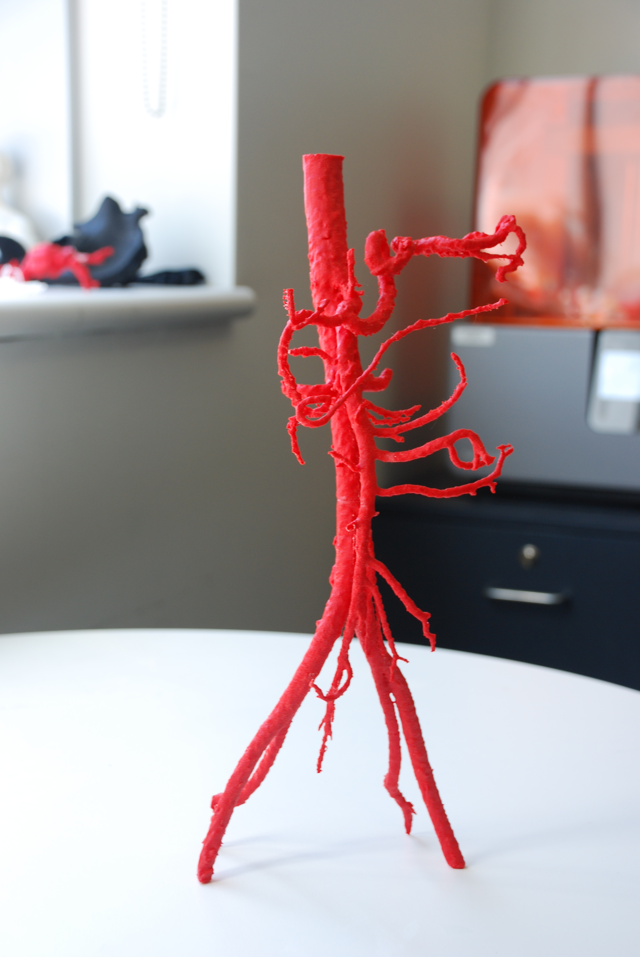
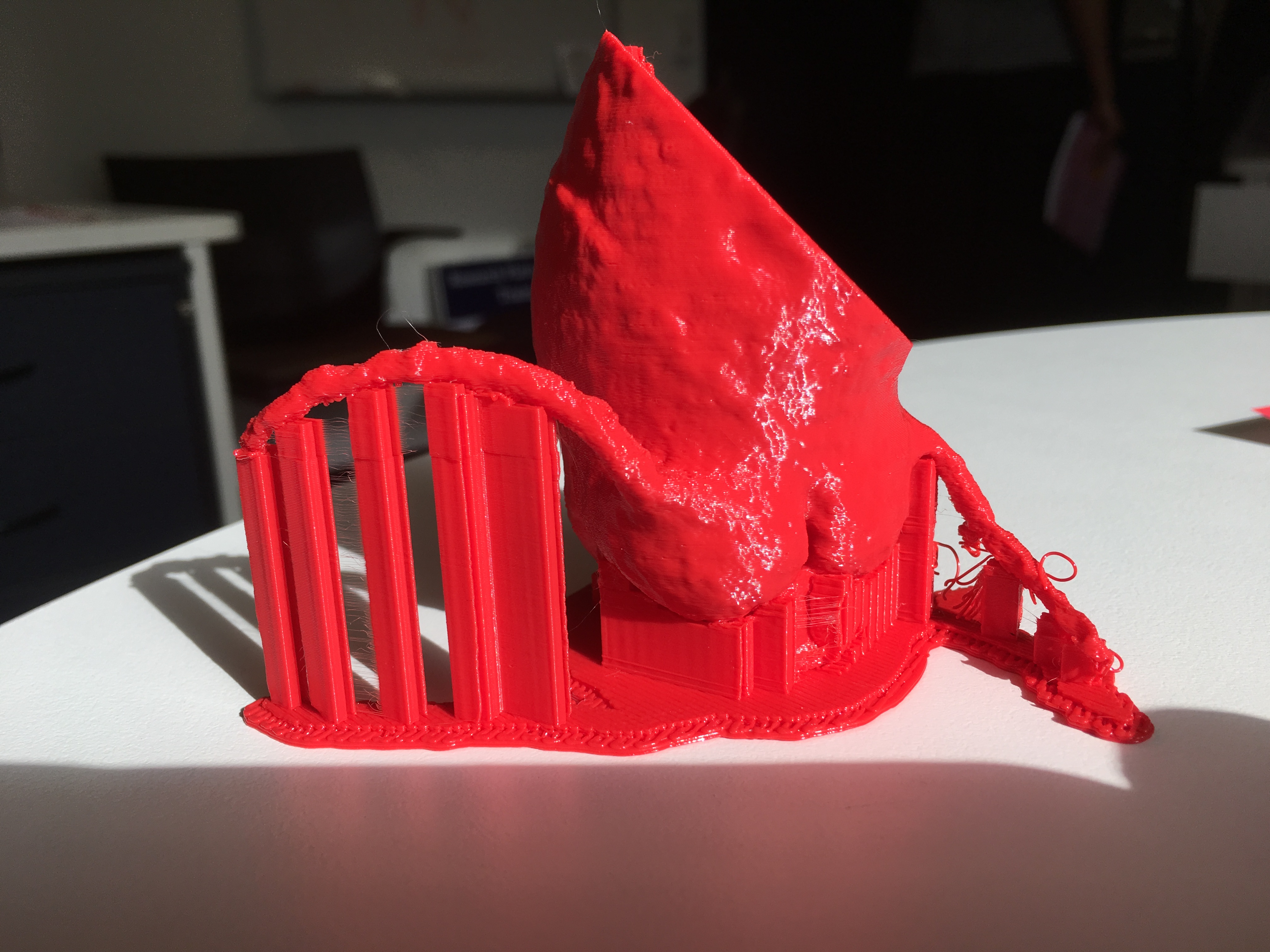
Normal anatomical branches on an abdominal aortic model 3D printed on MakerBot Replicator 2X FDM

Chuen and Coles-Black even begun printing out copies of patient kidneys to help surgeons at Austin Health plan the removal of kidney tumors. Moreover, Chuen understands that the immediate challenge in medical 3D printing is ensuring that medical professionals themselves are up to speed with the technology because it is their clinical experience that will drive new applications and projects. 

During our interview, Chuen asserted that the conference has once again proved that Australia is leading the way with cross institution development cooperations, ethical issues surrounding 3D printing and he looks forward to many exciting possibilities of the technology for the future.

Why was the 3D Med Conference so important to the region?

We noticed there were a lot of groups that existed previously that didn’t know about each other and the meeting has become a really good focal point for people to find out about what others are researching and selling. So rather than working on their own and almost in secret, they can join together and create projects that cross different institutions, specialties and disciplines. During the conference, at every corner I encountered groups of people from different universities and cities gathering to hatch a project, proving that there was a very cooperative atmosphere. They all clearly had common interests and discovered that they can work outside of their own space with others. 

What was so unique about the 3D Med Conference?

Because there really aren’t many meetings like this, the areas of interest are still growing, anyone who is working with these technologies have applications in different areas so that is why we have a lot of crossover between the fields. The strength of the confreence comes from encouraging people to have an overview of what was happening in the field, so rather than just understanding technical aspects of technology, everyone started to become knowledgeable about the whole landscape, for example, why we need to care about ethics and regulation, or considering the useful implications of applying techniques from a different area of science and research. 

One of the biggest challenges for 3D printing is?

One of the big problems in customized medical devices and the 3D printing space is that there is uncertainty about what will happen in the future. Apart from the guidance of the US Food and Drug Administration (FDA), there hasn’t been a lot of resources for manufacturers and researchers on how 3D printing and customized medical devices will be regulated. Australia’s own Therapeutic Goods Administration (TGA) representation in the International Medical Device Regulators Forum (IMDRF) has been very strong,particularly around 3D printing and customized medical devices. During the conference John Skerritt, Deputy Secretary of the Australian Department of Health, outlined the broad framework around the field and has engaged in a consultation process with the medical 3D printing community (and we have provided some proposals for the final documentation that will be ready soon.)

Distributed production will present new risks for ensuring the quality control of end products. It will need a fundamental shift in responsibility from the supplier to wherever the medicines or devices are manufactured. That represents a huge change and we have to work out how it could work. But if we get the regulation right then it will transform access to medical products.

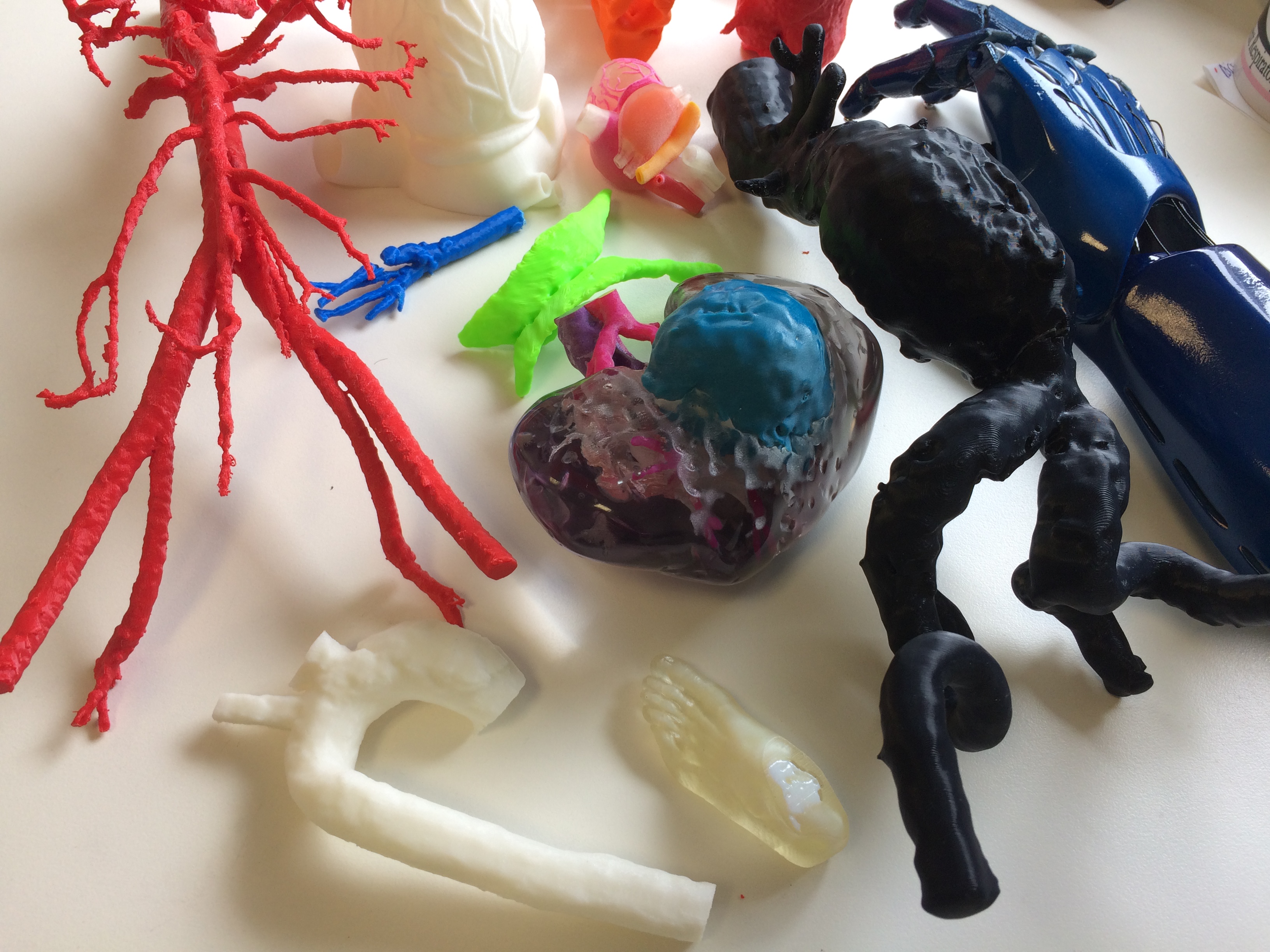
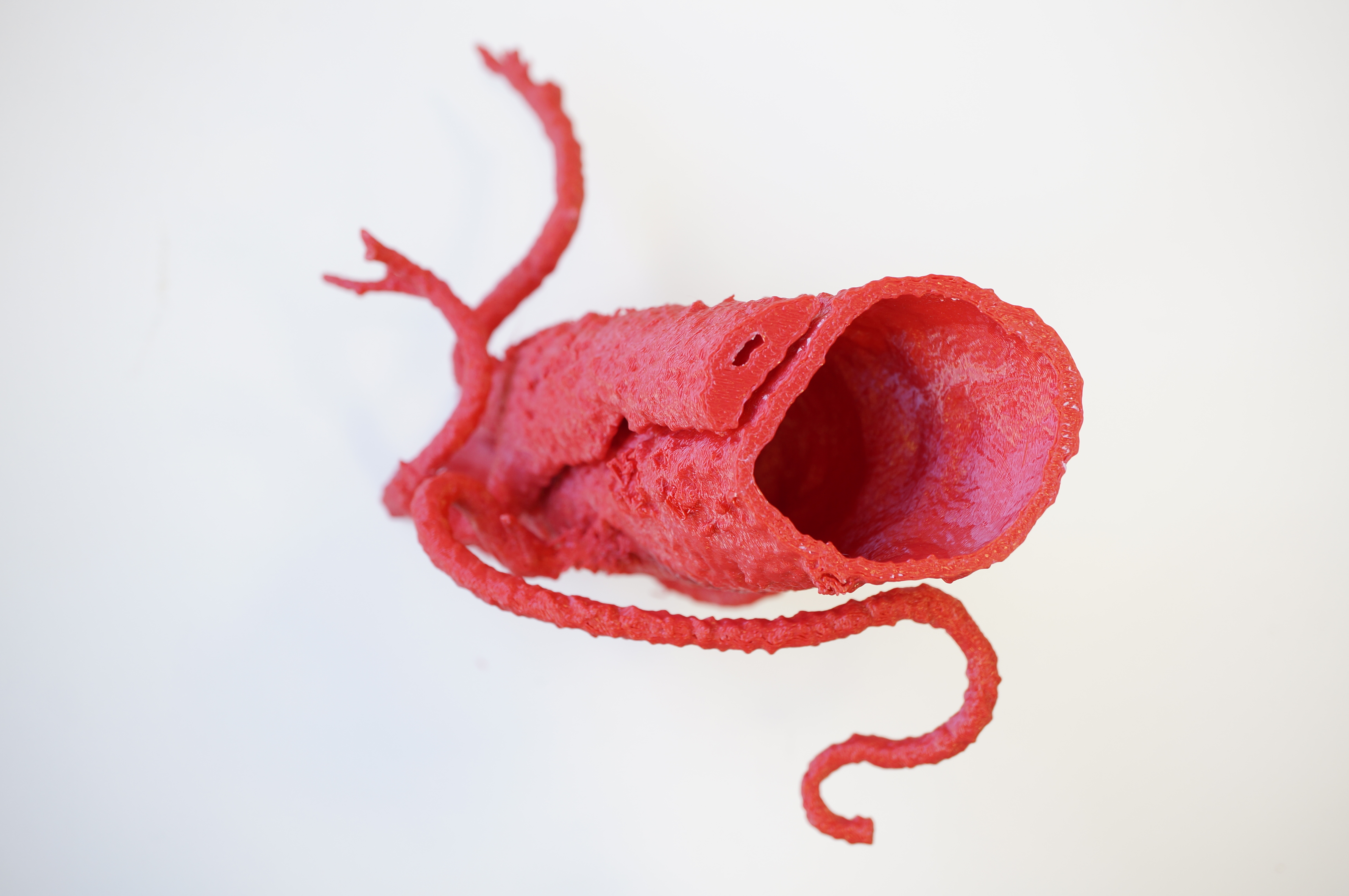
Collection of 3D printed objects

What does the future of 3D printing in medicine look like?

The whole point of what we do is improve patient care, so we have to think very carefully about our next steps and analyze whether it is helpful or not. For patients, anatomical models help them see and understand the condition or surgery they plan for. We have done projects and have some conclusive evidence that patient understanding is improved with anatomical 3D printed models. 

Patients are interested to know what will happen in the future, especially with 3D printed kidneys and stents. But the truth is that that technology is very far away. We may never be able to 3D print an organ, not at least the way we imagine it to be. Realistically, if we are talking about an organ for transplantation, we have to think that no matter what the organ looks like, the question is: does it do the job? For example, if we were thinking about bioprinting in order to replace a kidney, as long as it performs the function of the kidney, it doesn’t matter what shape it comes in. And for that, we have to be able to reproduce a structure. This could be in shapes, rather than in one block, or it could be a composition of an external and an internal device, meaning we would be looking into something that is assembled. Today the technology to have the replacement kidney is available, it is a dialysis machine, yet you wouldn’t expect a dialysis machine to look like a kidney. The same is going to happen with 3D printed organs, where we need to separate the appearance and structure of the organ from the function. In the end, the function is what matters.

As such, if we were to imagine what a 3D printed heart would look like, we would need to go into the field of soft robotics or mimicking natural structures, all of that changes fundamentally how we think about organs for the human body.

How can your particular medical field benefit from 3D printing?

As a vascular surgeon, I’m also looking at 3D printed stents, and there is quite some work around that. Mainly it is based on printing something that looks like a stent, but it is very difficult to reproduce the mechanical properties of a stent using 3D printing. The benefits revolve around the different materials that could be used with 3D printing, for example, if you could reproduce a stent in a bioabsorbable plastic it would allow surgeons to deploy it with embedded drugs (like antibiotics and pain medication) that get released at a set time. There are a lot of options in terms of using multi material technology in customized implant production, as well as great precision, and that is an area where 3D printing helps. 

Ideally, we need to understand the technology to know where the errors can happen. But in general, it is improving, both in hardware and software, the challenge will be about making it accessible. We have done randomized trials around anatomical models for teaching, education and simulation. There are already some 3D printed medical devices, such as for joints and implants. It would be ideal to have assessments of the economics to determine whether the anatomical models will be worthwhile. 

How is Australia changing the paradigm of medical 3D printing?

Australia has world leading technology, but in terms of the way we have collaborated and worked together, we are quite unique. Even globally one of the big problems is finding the groups that are doing this kind of work. We have been in touch with research groups in Poland, Boston, and Toronto, even engaging with large centers like the Mayo Clinic, in Minnesota. Key collaboration between international centers are great and we are keeping an eye out for other major hubs of activity, like in China, South Korea, and Europe. We need to link up all the international groups, that’s where we see things are going!

[Image credit: 3dMedLab, Austin Health]

The post Interview with Jason Chuen: Shaping Australia’s Medical 3D Printing Environment appeared first on 3DPrint.com | The Voice of 3D Printing / Additive Manufacturing.

Interview with Jasamine Coles-Black: Benefits of 3D Printed Models in Vascular Surgery

Seven years ago, the World Health Organization estimated the total global volume of operations to be 312.9 million. That could probably mean that in our lifetime, a lot of us will go under the surgeon’s knife, and because it’s not as scary anymore as perhaps the early 1900’s (if you saw the Cinemax hit show The Knick, you know what I mean), we should be thankful to live in the 21st. century. Surgery has become safer, and cosmetic surgery, microsurgery and many other elective-type surgeries are quite common and often painless procedures. Still, some of the more complex surgeries still have high death rates. In an attempt to take advantage of the ongoing technological advances, many medical specialists have noticed that 3D printing can improve surgical planning and training, which will directly benefit surgical procedures.

Last year, Nova Scotia surgeons used a 3D printed brain model in surgical planning for the first time, while 60 specialists at the Mayo Clinic dedicated three and a half years and more than 50 Saturdays to practicing a very complex face transplant, thanks to 3D imaging and virtual surgical procedures. In Melbourne, Australia, the Austin Health 3D Printing Laboratory supports 3D printing for clinical applications and runs an active research program exploring how 3D printing can be used for teaching, procedural simulation, patient education, surgical planning, and prosthetic implants. The first facility of its kind in the southernmost city of the country, 3dMedLab came about after discussions for a more coordinated and organised approach to 3D Printing Resources throughout The University of Melbourne and in particular the availability of this technology to Clinician-Researchers at hospitals associated with the Faculty of Medicine, Dentistry and Health Sciences. Since then they have expanded the number of medical specialties and researchers that they work with across multiple Melbourne hospitals as well as developing nationwide and international partnerships.

3DPrint.com spoke to Jasamine Coles-Black, doctor and vascular researcher at the Department of Vascular Surgery at Austin Health, in Melbourne to understand how 3D printing can improve surgical planning in complex endovascular procedures to treat the arteries and veins of the body and training for medical residents.

Dr. Jasamine Coles-Black with kidney tumor model

“3D printing is the exciting next step in personalized medicine. The anatomical models are custom-made using scans that we have acquired as part of a patient’s medical care. From these scans, we can highlight the parts of the patient that we are interested in operating on and use these images to make a 3D model. From there, it can be printed using any 3D printer. The lab frequently prints models of diseased aortas to perform a ‘practice-run’ surgery. We have found that these 3D printed models are of greatest value when we are confronted with challenging cases. In addition to using them for surgical simulation, they can also be used to teach trainee surgeons about the anatomy and approach in difficult cases. These same models can be used to explain to patients their disease, and the procedure we are proposing they undergo. We have found that we are better able to communicate with patients about their disease when they can hold their own anatomy in their hands,” explained Coles-Black, who is Co-Founder of 3DMedLab along with Jason Chuen, the Director of the Lab.

Abdominal Aortic Model 3D printed on MakerBot Replicator 2X FDM printer in ABS, used for anatomical teaching of surgical trainees

At the 3DMedLab experts have also 3D printed internal carotid arteries (the main blood vessel in the neck supplying the brain) to help visualise them before the team performs operations aimed at preventing strokes, or before they operate on tumours in the neck involving these arteries. Tracie Barber, assistant professor and a collaborator for the University of New South Wales, 3D prints fistulas created by vascular surgeons for dialysis access as an educational tool for patients and nurses. Furthermore, the very first anatomical model 3D printed at the lab was to plan a particularly complex abdominal aortic aneurysm that would have been life-threatening to the patient if left untreated. The operation was succesfully performed by Chuen, and according to Coles-Black, the patient has even taken home the 3D printed replica of his aneurysm, where it has been given a name!

Hospital patients will benefit with new research that shows surgery is far superior if doctors do 3D printouts of the relevant body parts, so that people requiring operation will get customized surgery, making procedures quicker, patients wont be under anaesthetics for as long, allowing the production of efficient simulators for endovascular training, improving residents’ surgical performance and self-confidence. Coles-Black claims that 3D-printed models may help in understanding the behaviour of the endovascular material in three dimensions, inside a specific anatomy and can be directly manipulated and inspected, which can help identify some details that might not have been noticed on a CT scan. Furthermore, 3D printing allows a patient-specific simulation, which is more efficient than a generic one. Today, with online 3D printing services, like Shapeways or Materialise, taking care of any bespoke 3D project, it’s easier for doctors who don’t have access to 3D printers to outsource their anatomical printed models.

But although 3D printing improves surgical outcomes, as a cutting-edge field, there are still gaps in the literature that need to be filled in order to validate the benefits of these anatomical models in surgery. Scientific articles published to date have shown that surgeons find 3D printed models a beneficial advancement in surgical planning, and quite useful to train junior surgeons. Also, studies have shown that 3D printed anatomical models enhance patient safety by reducing a patient’s time under anesthesia, reducing operation times, recovery times, and even blood loss. The applications of producing life-size 3D models can have great benefits as well to a patient’s understanding of their disease and lessens their anxiety about the upcoming procedure. Personalised 3D printed models have been associated with increased patient understanding of basic anatomy, physiology, and the planned surgical procedure. This in turn helps the patient be more satisfied with their care resulting in a better engagement with the treating doctor.

Hollow aorta demonstrating type B aortic dissection

The 3DMedLab is fully equipped with many 3D printers which are used depending on the function required of the 3D printed anatomical model. Including printers from FormLabs, the Form2 which Coles-Black claims was very useful in the creation of transparent, flexible, and autoclavable models which can be sterilised and applied in the operative field, and the Form Cure and Form Wash. While for inexpensive initial prototypes and to print bony models such as for orthopaedic or maxillofacial surgery, they use an Ultimaker S5, for intricate structures such as the delicate bones of the face, Coles-Black and her team uses Ultimaker PVA (polyvinyl alcohol) a water-soluble support material for multi-extrusion 3D printing. To produce Orthopaedic models to size prostheses and simulate the reaming of bone, they rely on an Objet Scholar. And also have access to a Connex 3, Makerbots, and a metal SLS printer through the University of Melbourne. 

So how much time does pre-surgical simulation take? As Coles-Black told 3DPrint.com, it depends very much on the procedure that is being planned:

“The process can be as quick as bending a metal plate for facial reconstructive surgery against a 3D printed replica of a patient’s jaw in order to save time during surgery, to doing an entire “practice-run” of a procedure, such as introducing a needle and wire into a life-sized 3D printed aorta, navigating its anatomy, and deploying a stent into it as you would in a real patient. In essence, the strength of 3D printed anatomical models is that it allows us to create a life-sized replica of a patient’s anatomy. Doing a ”practice-run” before performing surgery on a real patient allows us to better plan for and anticipate challenging scenarios. Every individual’s anatomy is unique, and having the opportunity to practice the procedure beforehand allows us to make a plan to deal with potential problems before we operate on a patient. This improves the speed and the safety of our operations. Sometimes doing a “practice-run” on an exact replica of a patient’s anatomy also changes the techniques and equipment used.”

Model of Aortic Valve 3D printed in Formlabs Form2 SLA Printer

With research involving 3D printing hollow, diseased patient-specific abdominal aortic aneurysms, dilatation of the abdominal part of the main artery in the body which has a 70 to 80% mortality rate when ruptured, Coles-Black is very interested in introducing 3D printing technology to her work. Using 3D printed models help visualise and physically hold the patient’s individual anatomy in their hands, as well as practice minimally invasive repairs before performing them on the actual patient.

“These replicas of patient’s aortas also help to guide our selection of technique and equipment,” she stressed.

In order to improve the realism of her models, the expert is searching for a material that “most closely mimics a real aorta.”  Every month, doctors at the 3DMedLab print dozens of anatomical models to help with the pre-surgical planning of Cardiothoracic Surgery, Orthopaedic Surgery, Plastic Surgery, Maxillofacial Surgery, Ear Nose and Throat Surgery, Urology, General Surgery, Neurosurgery, and of course Vascular Surgery. They even produce 3D printed anatomical models to help Vets with their surgical planning too; something many veterinarians have been attempting to do.

Hollow aortic root model showing aortic valve, used for planning prior to cardiac surgery

“In addition to this, we 3D print training models used by medical professionals to teach and upkeep their procedural skills,” Coles-Black said. “For example, we have just 3D printed a large batch of airway training models requested by a paramedic at a base hospital in the Middle East. We also receive requests from research labs hoping to quickly and cheaply replace equipment parts. 3D printing technology has been around for a while, but a few years ago, doctors and other health professionals began to make use of it as part of patient care. Five years ago, when Jason and I founded 3D Med Lab I was still a medical student, so I have always envisioned 3D printing as part of my practice. In the past few years we have noticed an increase in the uptake of this technology amongst medical professionals. Certainly, the number of requests our lab receives seems to reflect this. 3D Med Lab is open to the medical space and offers clinicians everywhere a chance to get started in 3D printing.” 

Dr. Jason Chuen, Director of Vascular Surgery and Austin 3D Medical Printing Laboratory

The 3D Med Lab supports doctors and scientists interested in this technology by providing a central hub for the exchange of knowledge and ideas. Thanks to their workshops they teach other interested clinicians in using 3D printing as part of their practice. Their next annual Australian 3D printing in medicine conference (#3dMed19) will take place November 14 through 16, in Melbourne. 

As Coles-Black says, it is a small but growing field, getting a lot of interest from enthusiastic research students. Nonetheless, to really disrupt the medical industry, some of these models will require even more human-like properties, so it might be a few more years before advances in 3D printing technology allow printing of multi-material products to achieve the mechanical properties required, using cost-effective methods. But this is a great start for many research centers, universities and hospitals around the world that are getting a noticeable advantage through the use of additive manufacturing, and certainly, more patients would benefit with its widespread use.

[Images: 3dMedLab, Austin Health]