Researchers Create 3D Printed Bacterial Cellulose Material for Wound Healing

When it comes to medical applications, we’ve seen 3D printing used in the past for healing and repairing wounds, whether through the use of 3D printed bandages, 3D printed blood platelets, or bio-based materials, like nanocellulose. Researchers Dr. Mohamed M. Kanjou, Hassan Abdulhakim, Gabriel Molina de Olyveira, and Pierre Basmaji published a paper, titled “3-D Print Celulose Nanoskin: Future Diabetic Wound Healing,” about using bacterial cellulose for the purposes of wound healing.

“Most 3D printers use heat to melt the plastic or metal to be printed, and biobased materials are degraded,” the team wrote. “But cellulose nanofibrils have a solution to this problem: the printing paste is wet and dries out to a solid material. In this work, it was showed recent wound healing in Vinous Ulcer with kidney and other health complications using bacterial cellulose 3D print membranes.”

[Image: American Process Inc.]

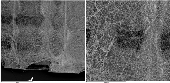
Cellulose nanofibrils, also known as nanocellulose, are made from wood or bacteria, and are the smallest fibers into which cellulose can be decomposed. They can contain up to 50% water, and this viscosity makes it ideal for a 3D printing paste, which can produce strong, biodegradable materials once they’ve dried out. By manipulating the cross-links between the fibrils, the properties can be modified, which allows for the fabrication of strong, porous, and flexible structures.

“Nanocellulose increases the opportunities for creating new materials in wound healing therapy. But this development still requires moisture tests to develops 3D printing with cellulose nanofibrils for medical and biotechnology applications,” the researchers explained.

“Several articles were published by our group since 2015 using Nanoskin membranes for wound healing treatment with successful results in diabetic ulcers, car and other accidents, amputation required ulcers [4] [5] [6]. In this work, it was showed recent wound healing in Vinous Ulcer with kidney and other health complications using bacterial cellulose 3D print.”

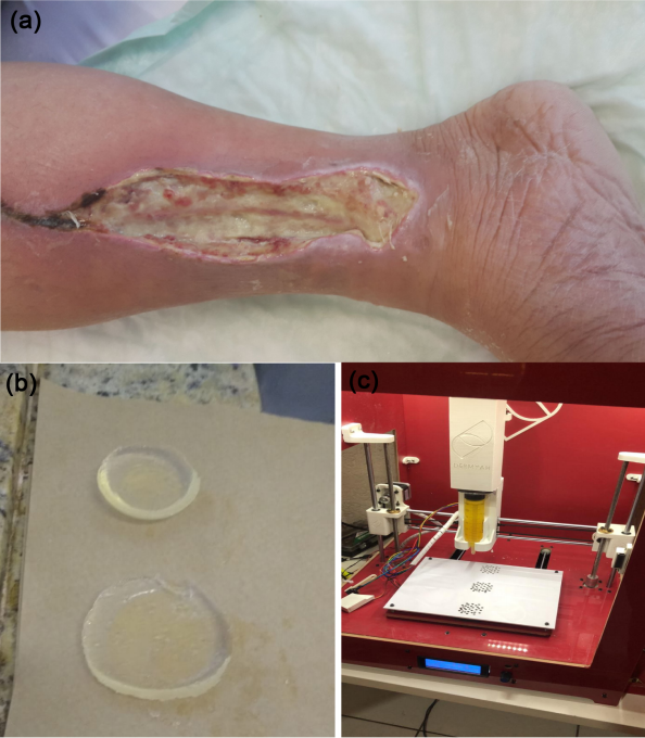
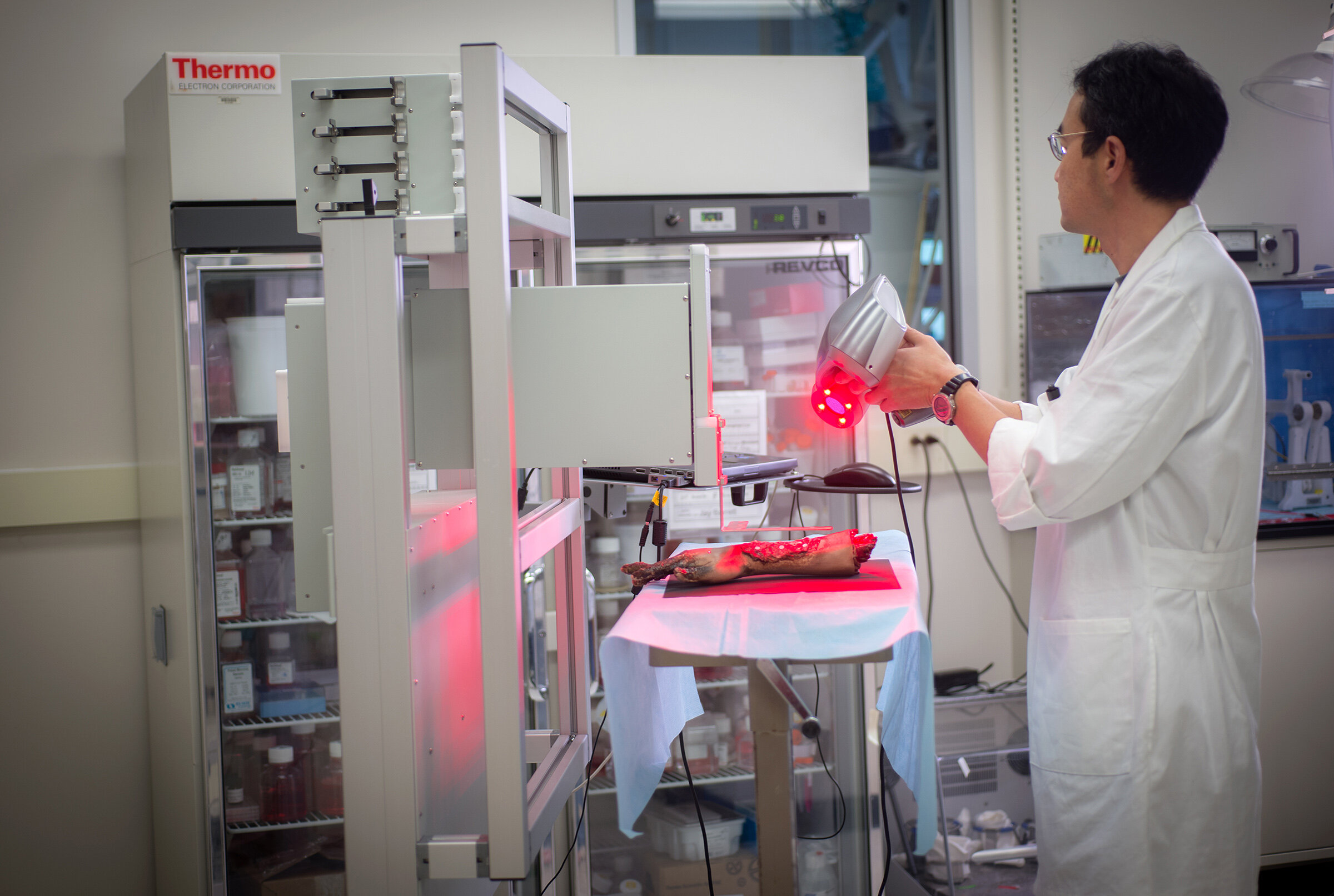
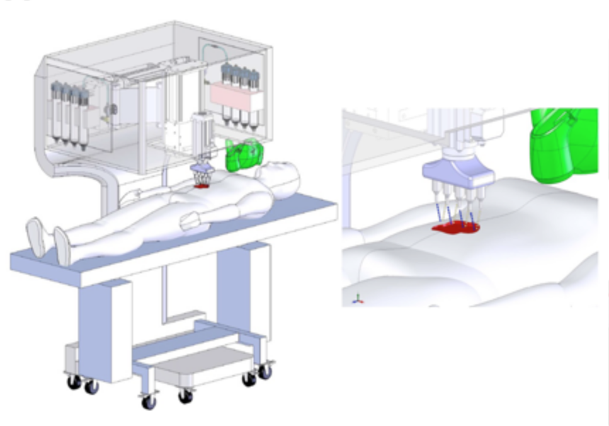
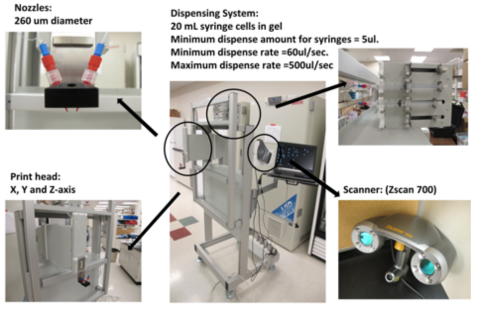
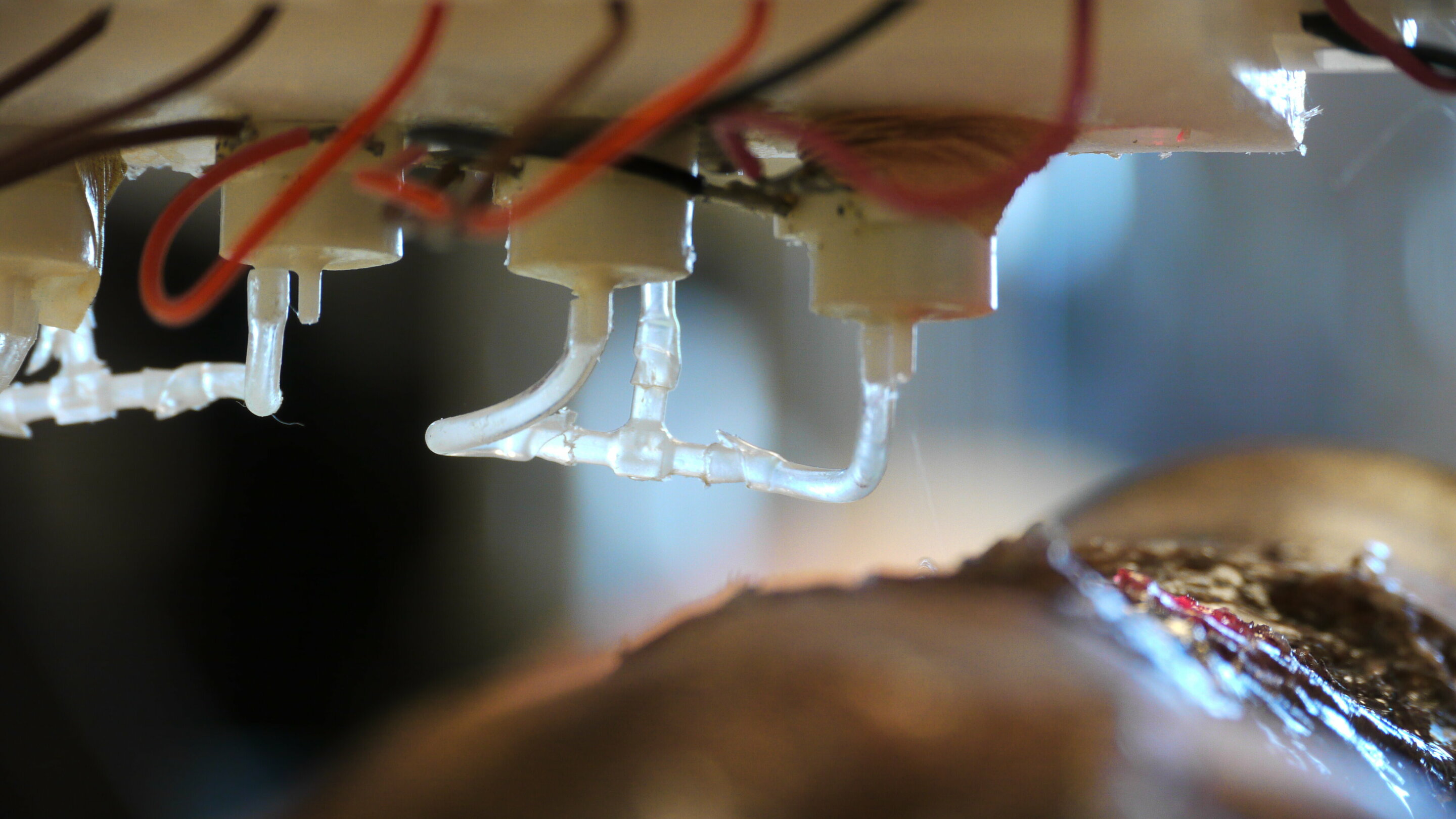
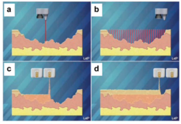
Wound healing treated with 3D bacterial cellulose-biological wound dressing (a); developed membrane (b) and Nanoskin developed equipament (c).

This time, the team explored a novel biomaterial and prepared a variety of different bacterial cellulose nanocomposites, such as BC/chondroitin sulfate and hyaluronic acid cross linked with sodium alginate and calcium chloride. They also synthesized bacterial cellulose and bacterial cellulose/chondroitin sulfate/hyaluronic acid.

“The acetic fermentation process was achieved by using glucose as a carbohydrate source,” the researchers explained. “Results of this process were vinegar and a nanobiocellulose biomass. The modifying process was based on the addition of hyaluronic acid and chondroitin sulfate (1% w/w) to the culture medium before bacteria inoculation. Bacterial cellulose (BC) was produced by Gram-negative bacteria Gluconacetobacter xylinus, which could be obtained from the culture medium in the pure 3-D structure, consisting of an ultra fine network of cellulose nanofibers.”

Dr. Kanjou and Abdulhakim supervised the completion of an in vivo analysis – the model was a 60-year-old patient diagnosed with a diabetic foot wound.

Here’s your warning – more icky wound pictures are coming.

Wound healing evolution in 1 month and 3D bacterial cellulose impact use with biological wound dressing.

When the patient, also suffering from kidney failure, arrived at Sheikh Khalifa Hospital, the wound was infected and had accumulated a lot of slough tissue. A classic silver dressing did not show any progress, so the researchers began treating the patient’s wound with 3D printed bacterial cellulose membranes.

For one month, the 3D printed bacterial cellulose material was used on alternating days, to some excellent results – the edge and bottom of the wound were starting to heal, and the wound area was reduced.

Additionally, the slough tissue was easy to remove, and healthy red granulation tissue was starting to grow, which you can see in the below image.

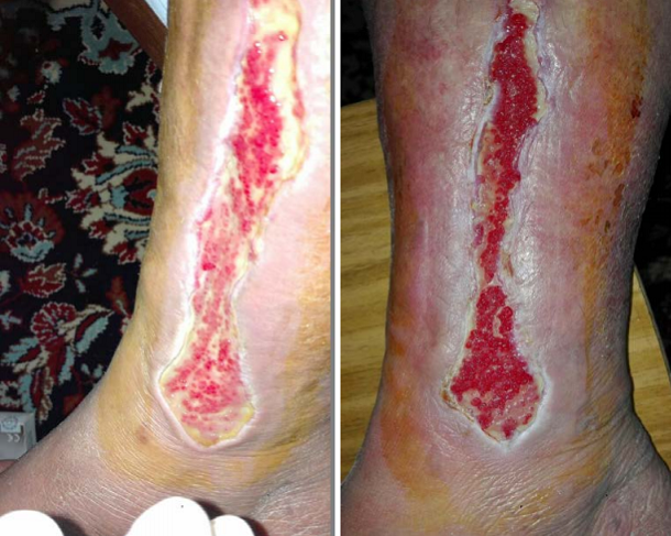
Wound healing evolution in 2 months, 3D print Bacterial cellulose impact use in biological wound dressing.

“Then, after more 1 month, almost all slough tissue is removed by treating with 3-D print Bacterial cellulose only; granulation and building up of healthy tissue is coming up with approximation of skin and the wound is closing,” the researchers wrote.

“Finally, after 4 months of treatment, there is complete healing with minimizing the scar in wound area and able to decrease with time.”

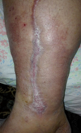
Figure 4. Complete wound healing evolution in 4 months and impact use of biological wound dressing 3D print of bacterial cellulose.

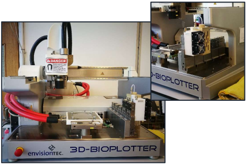
The researchers were able to successfully modify bacterial cellulose by “changing the fermentation medium with hyaluronic acid, chondroitin sulfate, besides of crosslinked with alginate sodium and calcium chloride.” In so doing, they were able to fabricate promising 3D printed scaffolds out of the bio-based material. In addition, the team developed new equipment for carrying out its work.

“In conclusion, 3-D print bacterial cellulose membranes apply to diabetic ulcers, with significant lesions and wound healing requirement; furthermore, natural membranes applications are for all population with different age.”

Discuss this research and other 3D printing topics at 3DPrintBoard.com or share your thoughts in the Facebook comments below.

The post Researchers Create 3D Printed Bacterial Cellulose Material for Wound Healing appeared first on 3DPrint.com | The Voice of 3D Printing / Additive Manufacturing.

Adding Lignin & Curcumin to PLA for 3D Printed Wound Healing Meshes

As innovation in materials grows into a vast science—especially within the 3D and 4D printing realms, medical patients of today and the future can look forward to improved, patient-specific care. Researchers from Queen’s University Belfast study the implications and potential with enhanced PLA in ‘Antioxidant PLA Composites Containing Lignin for 3D Printing Applications: A Potential Material for Healthcare Applications.’

Lignin (LIG) is a natural biopolymer containing antioxidants. To see if these properties would carry through after serving as a coating for PLA pellets and then being 3D printed, the researchers placed the material into an extruder at 200 ◦C. Their suppositions proved correct as not only did the filament work successfully, but it passed on antioxidants.

“A wound healing model compound, curcumin (CUR), was applied in the surface of the mesh and its diffusion was studied,” stated the researchers. “It was observed that the dimensions of the meshes affected the permeation rate of CUR. Accordingly, the design of the mesh could be modified according to the patient’s needs.”

Photographs of: PLA and PLA coated pellets (A); LIG and TC containing PLA filaments (B); LIG and TC containing 1 cm × 1 cm squares prepared using 3D printing (C); and different shapes printed using the filament containing 2% (w/w) LIG (D).

The use of PLA is popular for many reasons, beginning with its percieved biodegradability factor, and lack of toxicity. Suitable for FFF 3D printing, the vegetable-based filament can be combined with other molecules and has shown increasing merit for medical applications, especially in accelerating healing of wounds. This type of study has not been expansive previously, however, harnessing the power of lignin’s antioxidant and antimicrobial properties. Lignin is of interest as an extremely abundant polymer that the researchers contend is highly unexploited. It is an affordable material to acquire and use, and useful in a variety of other applications currently.

Scheme of the different meshes produced using FFF.

The researchers used different types of mesh with a 2 percent combination of LIG in the PLA, along with curcumin (CUR) applied in the material and diffused. They discovered better effectiveness with the meshes when using a size of 1mm. The research team also found that the release rate was delayed if they used both the mesh and a soluble PVA film, printed with the mesh on an FDM 3D printer with a dual extruder. The PVA film may also function in dual capacity as it not only delays the release of CUR, but also keeps the wound moist.

“A potential scenario for this material is as a wound dressing material due to the antioxidant activity of the composite material that can contribute to wound closure. Due to the low price of 3D printing equipment and its versatility, these materials can be used in hospitals to print wound dressings for patients on demand,” concluded the researchers.

“Due to the enhanced cell proliferation on antioxidant materials [16], these materials can be used for tissue culture applications or even for regenerative medicine. Due to the versatility of FFF, complex geometries can be prepared such as scaffolds. However, before this type of materials can be implanted into humans, the safety of lignin-based materials should be evaluated. It has been reported before that LIG-based materials are biocompatible [45] but more studies should be performed.”

3D printing continues to make substantial impacts in the medical arena, innovating for better ways to heal wounds, along with improving drug-delivery systems, and assisting in tissue regeneration. What do you think of this news? Let us know your thoughts! Join the discussion of this and other 3D printing topics at 3DPrintBoard.com.

Experimental setup used to measure drug diffusion trough the 3D printed meshes (A);
photographs of the 3D printed meshes made of PLA and 2% (w/w) LIG (B); and CUR release through
1.5 mm (C) and 1 mm (D) 3D printed meshes (n = 3).

[Source / Images: Antioxidant PLA Composites Containing Lignin for 3D Printing Applications: A Potential Material for Healthcare Applications]

Wake Forest: In Situ 3D Printing of Tailored Skin Grafts, a Possible Step Forward in Tissue Engineering

In recent years bioprinting has proved to be an important tool for tissue engineering applications and it holds great promise towards the fabrication of skin tissue. A team from Wake Forest Institute for Regenerative Medicine (WFIRM) has brought progress in this direction by developing a proof of concept validation of a mobile skin bioprinting system that provides rapid on-site management of full-thickness wounds. The prototype is intended to be used for wound healing and the coverage of large wound areas directly on the patient. The experiments were conducted on mice and pigskin, so this experiment is years away from being trialed in humans. For now, it is great news if you’re trying to repair a ham. Nonetheless, this study represents a significant advance in potential personalized wound treatment approaches as it is similar and goes further than other approaches in bioprinting skin directly onto patients.

It also represents a very strategic Intellectual Property move by Wake Forest to claim, potentially patent and exploit a logical path to large scale wound treatment. Bioprinting gradients, correct vascularization, actually implementing patient specific bioprinting, the combination of scanning and printing, and in situ printing of skin are all pivotal areas. This is especially relevant since Wake Forest has in the past been quick to press release and slow to publish when compared to others working in the field.   

picture os a man using the bioprinter on a limb

A member of WFIRM team operates with the bioprinter

This is salient since this idea could represent a substantial improvement in health care. Large, nonhealing or chronic wounds as the ones derived from burn injuries, diabetes or venous and pressure ulcers represent a burden for patients and mean large costs on health services. Only in the United States, over 7 million patients suffer each year from this issue, representing an expenditure of $25 billion each year. There is no need to explain then the importance of the improvement in regenerative medicine for our society.

The bioprinting system developed by the scientist at WFIRM offers a step forward to the improvement of this issue. It is a bedside machine that can tailor skin tissue directly on the patient.

Illustration of functioning of bioprinting machine

A schematic demonstrating scale of the skin bioprinter via WFIRM

The main components of the skin bioprinter system consist of a hand-held 3D scanner and a printing head with an XYZ movement system containing eight 260 µm diameter nozzles, each driven by an independent dispensing motor. The ZScanner Z700 scanner offers the ability to capture the entire wound in a continuous scan and it creates a wound map for printing.

All components are mounted on a frame small enough to be mobile inside the operating room.

Directly from the abstract:

“Once the scan is complete, the wound data is compiled to form a model of the wound. The scanned wound area is then processed using Geomagic Studio software orient the model to standard coordinates so the import into Artcam is ideal. Scanned data is in the form of an STL file that has been outputted from Geomagic and inputted to Artcam 3D software to obtain the full volume and the nozzle path needed to print the fill volume.”

Picture of detail of bioprinter nozzle

Detail of the bioprinter nozzle

Once the data is taken, it can be modified to better fit the structure of the wound to treat:

“The user can define a series of bitmap images where each non-black pixel corresponds to a cell drop, and the color of the pixel corresponds to the cell type to print. This method allows the user to create complex structures using any cell type in any configuration. Finally, a list of available cell types loaded in the printer is provided to the user by the print-head configuration.”

Once ready, it is used to generate the fill volume and the path points for nozzle head to print. The delivery system is similar to the ones used in traditional inkjet printing. It has several cartridges, each contains different biomaterials and every single nozzle is connected to a separate cartridge. It 3D prints directly on the wound a double layered skin substitute consisting dermal fibroblasts and epidermal keratinocytes cells that exactly match the patient’s wound.

Illustration of how 3D skin printing works

Skin bioprinting concept bia WFIRM

“The wound depth is then split into layers on the Z axis to determine which layers correspond to the dermis and epidermis. Each layer is overlaid with a series of XY lines that cover the entire wound area. These lines are used in conjunction with the cartridge-based delivery system to determine a path for the printer. The skin cell delivery system is controlled by a custom software employing a three-tiered architecture design based on the Microsoft.NET Framework 2.0 and written in C++.”

What makes this technology special is that while being mobile, the skin bioprinter has the ability of 3D print customize skin tissue directly on a patient by scanning and measuring the patient’s injury. Through the compilation and manipulation of data, it makes possible to manage extensive wound surface and deposit with precision customize biomaterial that simulates skin.

In contrast with the existing technologies used to heal wounds such as using a manually seeded matrix or cell spraying, this has possible advantages. The bioprinted skin may prove to be the most adequate due to its ability to place specific cell types into needed position and creating a complex skin structure that improves wound healing with better aesthetic properties. From an intellectual property standpoint, Wake Forest has really made a very timely disclosure towards what is the most straightforward path to personalized in situ 3D printing of skin tissue.  

At the same time, this technique has shown a positive outcome when applied on mice and pigskin wounds. The future of its success lies in the ability of this approach in conducting a successful clinical trial in humans with prosperous results, which we all hope for.

3D Printing and Electrospinning PCL for Dressings and Wound Repair

When the body is functioning as it should, wounds repair themselves naturally. Sometimes, however, wounds do not heal, due to conditions such as diabetes or other chronic diseases. This puts the patient at risk of infection and other complications, at worst even requiring amputation. Chronic wounds affect 37 million patients around the world, and there is a great need for better treatment of these wounds.

In her Master’s Degree thesis, Politecnico di Torino student Viola Sgarminato discusses how 3D bioprinting and electrospinning can create scaffolds that actually promote wound repair. Using a combination of electrospinning and 3D printing with an EnvisionTEC 3D-Bioplotter, Sgarminato developed scaffolds that would promote healing by electrically stimulating skin cells.

“To this aim hierarchical scaffolds of polycaprolactone (PCL) and piezoelectric barium titanate (BaTiO3) nanoparticles were fabricated using 3D-bioprinting and the electrospinning technique,” Sgarminato explains.

Electrospinning is a technique that has been used for decades, and it involves the use of an electric charge to spin nanometer threads from a polymer solution. Lately, it’s been frequently used in conjunction with 3D printing, particularly bioprinting. In one part of the study, Sgarminato used the technique to create fibrous patches. She also used a 3D-Bioplotter to 3D print scaffolds for skin regeneration.

“In this work the bio-plotting technique was used to produce polycaprolactone (PCL) scaffolds with and without barium titanate nanoparticles,” she says. “Several sample thicknesses, porosities and geometries were tested to obtain optimized scaffolds for skin regeneration. Low and high temperature processes were performed to produce piezoelectric and nonpiezoelectric scaffolds, respectively. Indeed, to print scaffolds with homogenously distributed BTNPs, a solution of PCL and consequently a low temperature process are needed.”

The scaffolds were seeded with cells, which were then evaluated 24 and 72 hours later.

For the electrospinning portion of the project, one solution, in particular, worked better at producing defect-free mats of fibers – PCL was the key ingredient. The 3D printed scaffolds were examined to test their porosity, and to compare those that were produced through high temperature and low-temperature 3D printing. The composite wound dressings were also examined using a scanning electron microscope to verify the adhesion of the fibers to the scaffold, and good results were shown: even if subjected to mechanical stretching, the fibers remained attached to the substrate.

SEM images of electrospun fibers deposited onto a composite scaffold.

“The results that have been measured in 3T3 cells after 24h from seeding demonstrate that the cellular adhesion is comparable for each substrate,” says Sgarminato. “By considering the absorbance values after 72h, the highest increment of proliferation occurs for samples 4,5 and 6 that correspond to the PCL nanofibers…Indeed, PCL electrospun nanofibers represent a suitable substrate for growth and proliferation of NIH 3T3 cells. The small differences between PCL nanofibers and composites (samples 7 and 8) suggest a good cytocompatibility of the composite wound dressings. On the contrary, as expected, the printed scaffolds without nanofibers (samples 2 and 3) are not appropriate substrates for cell proliferation because of the limited culture surface.”

You can read the full thesis, entitled “Composite scaffolds with porosity over multiple length scale for skin regeneration,” here. The paper demonstrates that electrospinning and 3D bioprinting are effective methods of creating wound dressings that can treat chronic wounds and promote healing. This is an important application of bioprinting; while many people excitedly await the day that it’s possible to 3D print and transplant working human organs, it’s applications like these that are more immediate and can save lives just as effectively as a new heart.

Discuss this and other 3D printing topics at 3DPrintBoard.com or share your thoughts below.